

The DOI matches its Crossref record; 35 of the 35 references it cites were found in a registry or an index.
Elevated Levels of G-Quadruplex Formation in Human Stomach and Liver Cancer Tissues
Open accessAbstract
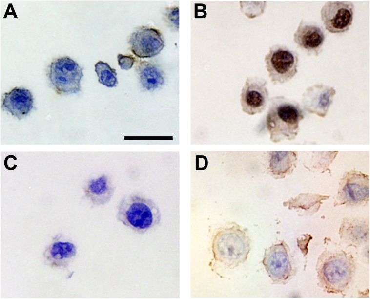
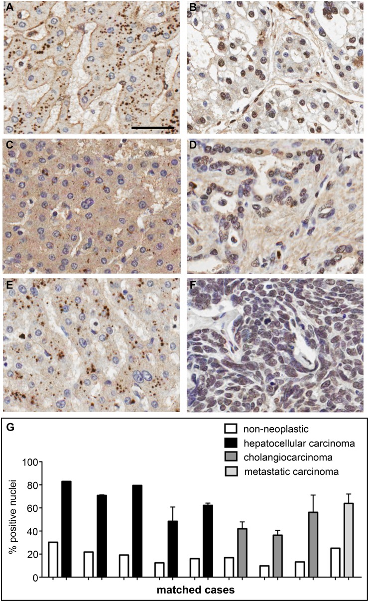
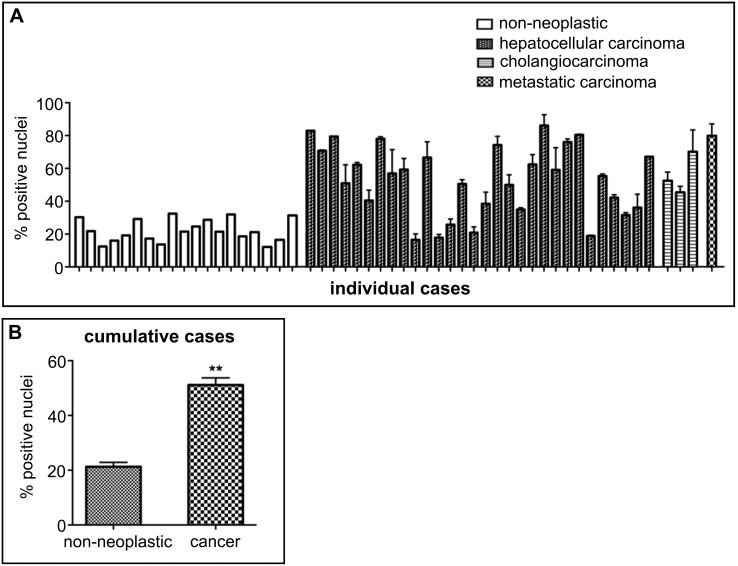
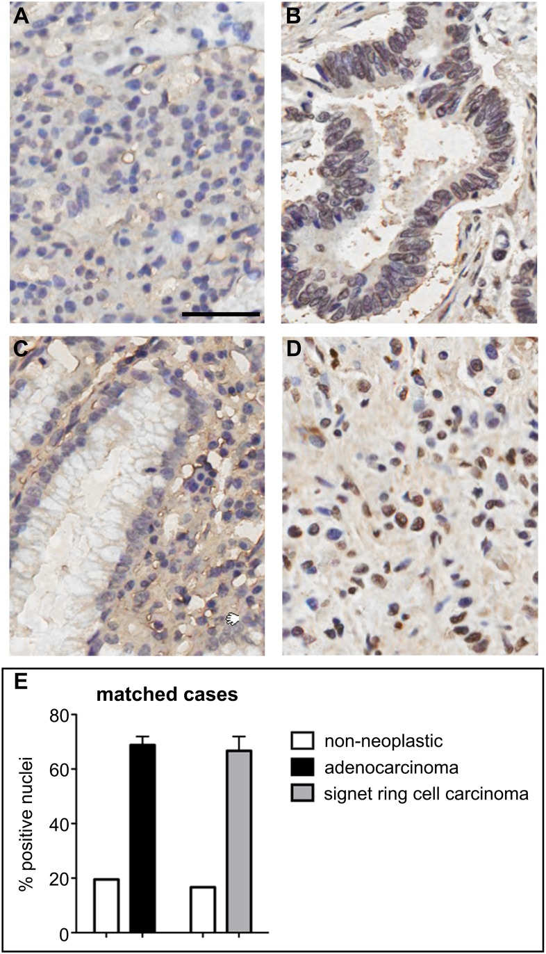
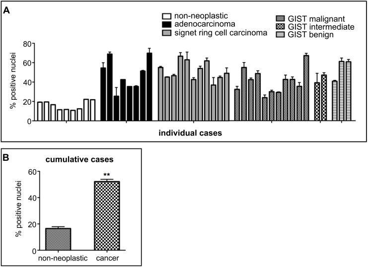
Four-stranded G-quadruplex DNA secondary structures have recently been visualized in the nuclei of human cultured cells. Here, we show that BG4, a G-quadruplex-specific antibody, can be used to stain DNA G-quadruplex structures in patient-derived tissues using immunohistochemistry. We observe a significantly elevated number of G-quadruplex-positive nuclei in human cancers of the liver and stomach as compared to background non-neoplastic tissue. Our results suggest that G-quadruplex formation can be detected and measured in patient-derived material and that elevated G-quadruplex formation may be a characteristic of some cancers.
Overview
The claims and statistics of this paper haven’t been checked yet.
Run the check — free, no account needed. It reads the open-access text and checks the sources it cites.
Integrity signals
Registry
Cites
Statistics
Figures
Verification
Claims the paper makes
The claims and statistics of this paper haven’t been checked yet.
Run the check — free, no account needed. It reads the open-access text and checks the sources it cites.
Claims other papers make about this one
Each is checked against this paper’s own text: does it say what the citing paper says it does?
The source backs part of the claim, not all of it — or backs it for a different population, condition or comparison.
In the citing paper
“Additionally, G-rich sequences, like G4 motifs, are prone to oxidative DNA damage, which can lead to mutations if not repaired properly (76).”
From G-quadruplex-mediated genomic instability drives SNVs in cancer (2024) · cites this paper as [76]
Experimental — accuracy not yet measuredThe cited source says what the claim attributes to it.
In the citing paper
“This suggests that elevated G-quadruplex formation might be a characteristic of some cancers and supports the hypothesis that G4s could contribute to genomic instability in cancer patients (76).”
From G-quadruplex-mediated genomic instability drives SNVs in cancer (2024) · cites this paper as [76]
The cited source says what the claim attributes to it.
In the citing paper
“recent studies have shown that G4s can be detected in patient-derived tissues using immunohistochemistry, with a significantly elevated number of G4-positive nuclei observed in human cancers of the liver and stomach compared to non-neoplastic tissue (76).”
From G-quadruplex-mediated genomic instability drives SNVs in cancer (2024) · cites this paper as [76]
Self-check
Percentages, sums and reported differences re-computed from the paper’s own numbers.
The self-check needs the paper’s full text.
Citations
Cites
Hallmarks of Cancer: The Next Generation
Hanahan, D., & Weinberg, R. A. (2011) · Cell · doi:10.1016/j.cell.2011.02.013 · Cited by 66,590 · cited 1×
“For example, Gquadruplex structures, if not resolved during DNA replication, may represent fragile sites that promote genome instability, which is a well-known hallmark of cancer [24].”
BackgroundSiddiqui-Jain, A., Grand, C. L., Bearss, D. J., & Hurley, L. H. (2002) · Proceedings of the National Academy of Sciences · doi:10.1073/pnas.182256799 · Cited by 2,274 · cited 1×
“For example, G-quadruplex stabilization by a small molecule can repress transcription of certain oncogenes and/or induce DNA damage at telomeres and oncogenes leading to replication defects and cell death [10–17].”
BackgroundQuantitative visualization of DNA G-quadruplex structures in human cells
Biffi, G., Tannahill, D., McCafferty, J., & Balasubramanian, S. (2013) · Nature Chemistry · doi:10.1038/nchem.1548 · Cited by 2,173 · cited 5×
“To determine the suitability of BG4 for IHC, we first tested this antibody in a model system using sections from FFPE cell pellets of MDA-MB-231 breast cancer cells that were previously observed to display nuclear G-quadruplex foci by IF microscopy [9].”
BackgroundMethodResultInfluentialSelf-citationSen, D., & Gilbert, W. (1988) · Nature · doi:10.1038/334364a0 · Cited by 1,866 · cited 1×
“G-quadruplexes are four-stranded structures that comprise stacks of tetrads formed through Hoogsteen hydrogen-bonding of four guanines coordinated by a central monovalent cation [3].”
BackgroundPrevalence of quadruplexes in the human genome
Huppert, J. L., & Balasubramanian, S. (2005) · Nucleic Acids Research · doi:10.1093/nar/gki609 · Cited by 1,795 · cited 1×
“Non-canonical G-quadruplex DNA secondary structures can be formed from guanine-rich DNA sequences of the type G3+NL1 G3+NL2 G3+NL3 G3+(N= any base and L= loop) and such sequence motifs are prevalent throughout the human genome [1,2].”
BackgroundSelf-citationG‐Quartets 40 Years Later: From 5′‐GMP to Molecular Biology and Supramolecular Chemistry
Davis, J. T. (2004) · Angewandte Chemie International Edition · doi:10.1002/anie.200300589 · Cited by 1,640
G-quadruplexes in promoters throughout the human genome
Huppert, J. L., & Balasubramanian, S. (2006) · Nucleic Acids Research · doi:10.1093/nar/gkl1057 · Cited by 1,392 · cited 1×
“The predicted location of G-quadruplex sequence motifs in regulatory regions of the genome, such as promoters and telomeres, suggests that Gquadruplex structures may have important roles in genome function [5–7].”
BackgroundSelf-citationHighly prevalent putative quadruplex sequence motifs in human DNA
Todd, A. K. (2005) · Nucleic Acids Research · doi:10.1093/nar/gki553 · Cited by 1,002 · cited 1×
“Non-canonical G-quadruplex DNA secondary structures can be formed from guanine-rich DNA sequences of the type G3+NL1 G3+NL2 G3+NL3 G3+(N= any base and L= loop) and such sequence motifs are prevalent throughout the human genome [1,2].”
BackgroundSmall-molecule–induced DNA damage identifies alternative DNA structures in human genes
Rodriguez, R., Miller, K. M., Forment, J. V., Bradshaw, C. R., Nikan, M., Britton, S., Oelschlaegel, T., Xhemalce, B., Balasubramanian, S., & Jackson, S. P. (2012) · Nature Chemical Biology · doi:10.1038/nchembio.780 · Cited by 741 · cited 1×
“For example, G-quadruplex stabilization by a small molecule can repress transcription of certain oncogenes and/or induce DNA damage at telomeres and oncogenes leading to replication defects and cell death [10–17].”
Self-citationDefective Telomere Lagging Strand Synthesis in Cells Lacking WRN Helicase Activity
Crabbé, L., Verdún, R. E., Haggblom, C., & Karlseder, J. (2004) · Science · doi:10.1126/science.1103619 · Cited by 630
Fanconi anaemia and the repair of Watson and Crick DNA crosslinks
Kottemann, M. C., & Smogorzewska, A. (2013) · Nature · doi:10.1038/nature11863 · Cited by 624
Burger, A. M., Dai, F., Schultes, C. M., Reszka, A. P., Moore, M. J., Double, J. A., & Neidle, S. (2005) · Cancer Research · doi:10.1158/0008-5472.can-04-2910 · Cited by 609
The Bloom's Syndrome Helicase Unwinds G4 DNA
Sun, H., Karow, J., Hickson, I. D., & Maizels, N. (1998) · Journal of Biological Chemistry · doi:10.1074/jbc.273.42.27587 · Cited by 554 · cited 1×
“For example, Fanconi anaemia, Bloom’s and Werner’s syndromes, all display genome instability with a predisposition to cancer [27– 29] and result from mutations in DNA helicase enzymes that have G-quadruplex-resolving activity [30–32].”
BackgroundRodriguez, R., Müller, S., Yeoman, J. A., Trentesaux, C., Riou, J.-F., & Balasubramanian, S. (2008) · Journal of the American Chemical Society · doi:10.1021/ja805615w · Cited by 517 · cited 1×
“For example, G-quadruplex stabilization by a small molecule can repress transcription of certain oncogenes and/or induce DNA damage at telomeres and oncogenes leading to replication defects and cell death [10–17].”
BackgroundSelf-citationPif1 family helicases suppress genome instability at G-quadruplex motifs
Paeschke, K., Bochman, M. L., Garcia, P. D., Ćejka, P., Friedman, K. L., Kowalczykowski, S. C., & Zakian, V. A. (2013) · Nature · doi:10.1038/nature12149 · Cited by 492
Marinoni, I., Kurrer, A. M. S., Vassella, E., Dettmer, M. S., Rudolph, T. J., Banz, V. M., Hunger, F., Pasquinelli, S., Speel, E. M., & Perren, A. A. (2013) · Gastroenterology · doi:10.1053/j.gastro.2013.10.020 · Cited by 473 · cited 1×
“neuroendocrine tumors [33], and XPD mutations disrupt nucleotide excision DNA repair that leads to cancer-prone diseases such”
BackgroundLaw, M. J., Lower, K. M., Voon, H. P. J., Hughes, J. R., Garrick, D., Viprakasit, V., Mitson, M., Gobbi, M. D., Marra, M. A., Morris, A. P., et al. (2010) · Cell · doi:10.1016/j.cell.2010.09.023 · Cited by 461
Excess of rare cancers in Werner syndrome (adult progeria).
Goto, M., Miller, R. W., Ishikawa, Y., & Sugano, H. (1996) · PubMed · Cited by 431
Wu, Y., Shin-ya, K., & Brosh, R. M. (2008) · Molecular and Cellular Biology · doi:10.1128/mcb.02210-07 · Cited by 428 · cited 1×
“For example, Fanconi anaemia, Bloom’s and Werner’s syndromes, all display genome instability with a predisposition to cancer [27– 29] and result from mutations in DNA helicase enzymes that have G-quadruplex-resolving activity [30–32].”
BackgroundFan, L., Fuss, J. O., Cheng, Q. J., Arvai, A. S., Hammel, M., Roberts, V. A., Cooper, P. K., & Tainer, J. A. (2008) · Cell · doi:10.1016/j.cell.2008.04.030 · Cited by 369
Friedberg, E. (2008) · doi:10.3410/f.1112038.571082 · Cited by 367 · cited 1×
“as Xeroderma pigmentosum [34].”
Existence and consequences of G-quadruplex structures in DNA
Murat, P., & Balasubramanian, S. (2014) · Current Opinion in Genetics & Development · doi:10.1016/j.gde.2013.10.012 · Cited by 346
Self-citationEddy, J., & Maizels, N. (2008) · Nucleic Acids Research · doi:10.1093/nar/gkm1138 · Cited by 289 · cited 1×
“The predicted location of G-quadruplex sequence motifs in regulatory regions of the genome, such as promoters and telomeres, suggests that Gquadruplex structures may have important roles in genome function [5–7].”
BackgroundTelomere damage induced by the G-quadruplex ligand RHPS4 has an antitumor effect
Salvati, E., Leonetti, C., Rizzo, A., Scarsella, M., Mottolese, M., Galati, R., Sperduti, I., Stevens, M. F. G., D’Incalci, M., Blasco, M., et al. (2007) · Journal of Clinical Investigation · doi:10.1172/jci32461 · Cited by 266 · cited 1×
“For example, G-quadruplex stabilization by a small molecule can repress transcription of certain oncogenes and/or induce DNA damage at telomeres and oncogenes leading to replication defects and cell death [10–17].”
BackgroundG‐quadruplex nucleic acids and human disease
Wu, Y., & Brosh, R. M. (2010) · FEBS Journal · doi:10.1111/j.1742-4658.2010.07760.x · Cited by 265 · cited 1×
“Helicases that resolve G-quadruplexes are also found localized at sites of genome instability [20–22], and diseases such as Bloom’s and Werner’s syndromes, which show increased levels of genome instability and a predisposition to cancer, have contributing mutations in G-quadruplex-resolving helicases [23].”
BackgroundDNA secondary structures and epigenetic determinants of cancer genome evolution
De, S., & Michor, F. (2011) · Nature Structural & Molecular Biology · doi:10.1038/nsmb.2089 · Cited by 252 · cited 1×
“G-quadruplex structures may also be linked to genome instability and G-quadruplex sequence motifs have been found proximal to DNA breakpoints and sites of somatic copy number changes seen in several cancers [18,19].”
BackgroundVerma, A., Halder, K., Halder, R., Yadav, V. K., Rawal, P., Thakur, R. K., Mohd, F., Sharma, A., & Chowdhury, S. (2008) · Journal of Medicinal Chemistry · doi:10.1021/jm800448a · Cited by 234 · cited 1×
“The predicted location of G-quadruplex sequence motifs in regulatory regions of the genome, such as promoters and telomeres, suggests that Gquadruplex structures may have important roles in genome function [5–7].”
BackgroundG quadruplexes are genomewide targets of transcriptional helicases XPB and XPD
Graybuck, L. T., Vallur, A. C., Eddy, J., & Maizels, N. (2014) · Nature Chemical Biology · doi:10.1038/nchembio.1475 · Cited by 217 · cited 2×
“Recent genome-wide studies have also highlighted the recognition of G-quadruplex sequences by additional resolving helicases such as ATRX, PIF1 and XPB/D [20–22].”
BackgroundGómez, D., O’Donohue, M.-F., Wenner, T., Douarre, C., Macadré, J., Koebel, P., Giraud-Panis, M.-J. E., Kaplan, H., Kolkes, A., Shin‐ya, K., et al. (2006) · Cancer Research · doi:10.1158/0008-5472.can-06-1581 · Cited by 212 · cited 1×
“For example, G-quadruplex stabilization by a small molecule can repress transcription of certain oncogenes and/or induce DNA damage at telomeres and oncogenes leading to replication defects and cell death [10–17].”
BackgroundPhatak, P., Cookson, J. C., Dai, F., Smith, V., Gartenhaus, R. B., Stevens, M. F. G., & Burger, A. M. (2007) · British Journal of Cancer · doi:10.1038/sj.bjc.6603691 · Cited by 201 · cited 1×
“For example, G-quadruplex stabilization by a small molecule can repress transcription of certain oncogenes and/or induce DNA damage at telomeres and oncogenes leading to replication defects and cell death [10–17].”
BackgroundNambiar, M., Goldsmith, G., Moorthy, B. T., Lieber, M. R., Joshi, M. V., Choudhary, B., Hosur, R. V., & Raghavan, S. C. (2010) · Nucleic Acids Research · doi:10.1093/nar/gkq824 · Cited by 121 · cited 1×
“Indeed, G-quadruplex sequence motifs are found at DNA breakpoints that lead to translocations within the BCL2 gene in lymphomas and within the HOX11 gene in T-cell leukemias [25,26].”
BackgroundBloom syndrome, genomic instability and cancer: the SOS-like hypothesis
Amor-Guéret, M. (2005) · Cancer Letters · doi:10.1016/j.canlet.2005.04.023 · Cited by 109 · cited 1×
“For example, Fanconi anaemia, Bloom’s and Werner’s syndromes, all display genome instability with a predisposition to cancer [27– 29] and result from mutations in DNA helicase enzymes that have G-quadruplex-resolving activity [30–32].”
BackgroundKatapadi, V. K., Nambiar, M., & Raghavan, S. C. (2012) · Genomics · doi:10.1016/j.ygeno.2012.05.008 · Cited by 90 · cited 2×
“Computational analyses have also suggested possible associations of G-quadruplexes and breakpoint regions in various cancers [19].”
BackgroundTargeting the c-Kit Promoter G-quadruplexes with 6-Substituted Indenoisoquinolines
Bejugam, M., Gunaratnam, M., Müller, S., Sanders, D. A., Sewitz, S. A., Fletcher, J. A., Neidle, S., & Balasubramanian, S. (2010) · ACS Medicinal Chemistry Letters · doi:10.1021/ml100062z · Cited by 70
Self-citationNambiar, M., Srivastava, M., Gopalakrishnan, V., Sankaran, S. K., & Raghavan, S. C. (2013) · Molecular and Cellular Biology · doi:10.1128/mcb.00540-13 · Cited by 56 · cited 1×
“Indeed, G-quadruplex sequence motifs are found at DNA breakpoints that lead to translocations within the BCL2 gene in lymphomas and within the HOX11 gene in T-cell leukemias [25,26].”
Background
Cited by
Sorted by citations
High-throughput sequencing of DNA G-quadruplex structures in the human genome
Chambers, V. S., Marsico, G., Boutell, J. M., Antonio, M. D., Smith, G. P., & Balasubramanian, S. (2015) · Nature Biotechnology · doi:10.1038/nbt.3295 · Cited by 1,261
Self-citationThe regulation and functions of DNA and RNA G-quadruplexes
Varshney, D., Spiegel, J., Zyner, K. G., Tannahill, D., & Balasubramanian, S. (2020) · Nature Reviews Molecular Cell Biology · doi:10.1038/s41580-020-0236-x · Cited by 1,292
Self-citationG-quadruplex structures mark human regulatory chromatin
Hänsel-Hertsch, R., Beraldi, D., Lensing, S. V., Marsico, G., Zyner, K., Parry, A., Di Antonio, M., Pike, J., Kimura, H., Narita, M., et al. (2016) · Nature Genetics · doi:10.1038/ng.3662 · Cited by 964
Self-citationThe Structure and Function of DNA G-Quadruplexes
Spiegel, J., Adhikari, S., & Balasubramanian, S. (2020) · Trends in Chemistry · doi:10.1016/j.trechm.2019.07.002 · Cited by 920
“Subsequently, G4s were visualised in human cells [41–44] and cancer tissue [45].”
BackgroundSelf-citationDNA G-quadruplexes in the human genome: detection, functions and therapeutic potential
Hänsel‐Hertsch, R., Antonio, M. D., & Balasubramanian, S. (2017) · Nature Reviews Molecular Cell Biology · doi:10.1038/nrm.2017.3 · Cited by 882
Self-citation
Metadata
- Authors
- Biffi, G., Tannahill, D., Miller, J., Howat, W. J., & Balasubramanian, S.
- Affiliations
- University of Cambridge (GB)
- Corresponding author
- Balasubramanian, Shankar
- Venue
- PLoS ONE
- Publisher
- Public Library of Science (PLoS)
- Published
- 2014-07-17
- Volume
- 9
- Issue
- 7
- Pages
- e102711
- ISSN
- 1932-6203 (ISSN-L)
- PMC
- PMC4102534
- OpenAlex
- W2100966371
- Cited by
- 223
- References
- 34
- Citations by year
- 2026: 8 · 2025: 17 · 2024: 27 · 2023: 31 · 2022: 20 · 2021: 30 · 2020: 25 · 2019: 15 · 2018: 14 · 2017: 17 · 2016: 7 · 2015: 10 · 2014: 2
- FWCI
- 4.92
- Type
- journal article
- Language
- en
- Open access
- gold
- doi.org
- Locations
- PLoS ONE (publishedVersion, cc-by)
- Europe PMC (PubMed Central) (publishedVersion, cc-by)
- PubMed Central (publishedVersion, cc-by)
- PubMed (publishedVersion)
- DOAJ (DOAJ: Directory of Open Access Journals) (submittedVersion, cc-by-sa)
- eScholarship (California Digital Library) (submittedVersion)
- eScholarship (California Digital Library) (submittedVersion, cc-by)
- Figshare (submittedVersion, cc-by)
- Funders
- Cancer Research UK (11961, A11961, 12488, 22310, C9681/A11961)
- Topics
- Life Sciences › Biochemistry, Genetics and Molecular Biology › Molecular Biology › DNA and Nucleic Acid Chemistry
- Life Sciences › Biochemistry, Genetics and Molecular Biology › Molecular Biology › Advanced biosensing and bioanalysis techniques
- Life Sciences › Biochemistry, Genetics and Molecular Biology › Cell Biology › Telomeres, Telomerase, and Senescence
- Keywords
- G-quadruplex, G-quadruplex formation, immunohistochemistry, gastric cancer, liver cancer, tumor markers, DNA secondary structure, patient-derived tissue
- MeSH terms
- DNA, Gastric Mucosa, Humans, Immunohistochemistry, Liver, Liver Neoplasms, Stomach Neoplasms, Cell Line, Tumor, G-Quadruplexes
About these checks
Registry, retraction, Cites and statistics results are computed, not judged, with one exception: a printed reference that no index lists is read once by a model before it counts as a citation error. Claims are extracted by a model and kept only when their quote appears verbatim in the text; each cited source is then checked, with a model’s help, for whether it says what the claim relies on. Those verdicts can be wrong — read the claim’s quote and the cited source before relying on one.
Statistics and arithmetic are recomputed, not judged.
Confidence bands are shown only for fields calibrated on our annotated test set. None is calibrated yet, so no band is shown.
What counts as a claim ›How we measure confidence ›How we check this ›
Are you an author, or do you see a problem with this page? Request a review
