

The DOI matches its Crossref record; 30 of the 30 references it cites were found in a registry or an index.
Quantitative Computed Tomographic Descriptors Associate Tumor Shape Complexity and Intratumor Heterogeneity with Prognosis in Lung Adenocarcinoma
Open accessAbstract
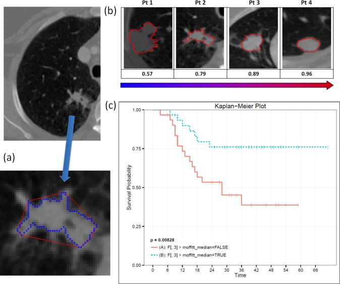
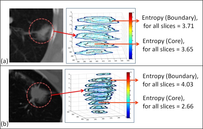
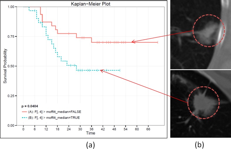
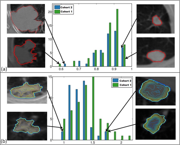
Two CT features were developed to quantitatively describe lung adenocarcinomas by scoring tumor shape complexity (feature 1: convexity) and intratumor density variation (feature 2: entropy ratio) in routinely obtained diagnostic CT scans. The developed quantitative features were analyzed in two independent cohorts (cohort 1: n = 61; cohort 2: n = 47) of patients diagnosed with primary lung adenocarcinoma, retrospectively curated to include imaging and clinical data. Preoperative chest CTs were segmented semi-automatically. Segmented tumor regions were further subdivided into core and boundary sub-regions, to quantify intensity variations across the tumor. Reproducibility of the features was evaluated in an independent test-retest dataset of 32 patients. The proposed metrics showed high degree of reproducibility in a repeated experiment (concordance, CCC≥0.897; dynamic range, DR≥0.92). Association with overall survival was evaluated by Cox proportional hazard regression, Kaplan-Meier survival curves, and the log-rank test. Both features were associated with overall survival (convexity: p = 0.008; entropy ratio: p = 0.04) in Cohort 1 but not in Cohort 2 (convexity: p = 0.7; entropy ratio: p = 0.8). In both cohorts, these features were found to be descriptive and demonstrated the link between imaging characteristics and patient survival in lung adenocarcinoma.
Overview
The claims and statistics of this paper haven’t been checked yet.
Run the check — free, no account needed. It reads the open-access text and checks the sources it cites.
Integrity signals
Registry
Cites
Statistics
Figures
Verification
Claims the paper makes
The claims and statistics of this paper haven’t been checked yet.
Run the check — free, no account needed. It reads the open-access text and checks the sources it cites.
Claims other papers make about this one
Each is checked against this paper’s own text: does it say what the citing paper says it does?
In the citing paper
“Grove et al found these measures to be strong prognostic indicators in patients with early-stage lung cancer (P < .01) (56).”
From Radiomics: Images Are More than Pictures, They Are Data (2016) · cites this paper as [56]
Self-check
Percentages, sums and reported differences re-computed from the paper’s own numbers.
The self-check needs the paper’s full text.
Citations
Cites
Siegel, R., Naishadham, D., & Jemal, A. (2013) · CA A Cancer Journal for Clinicians · doi:10.3322/caac.21166 · Cited by 11,825
A concordance correlation coefficient to evaluate reproducibility.
Lin, L. I.-K. (1989) · Biometrics · doi:10.2307/2532051 · Cited by 8,785
Radiomics: extracting more information from medical images using advanced feature analysis
Lambin, P., Rios-Velazquez, E., Leijenaar, R., Carvalho, S., van Stiphout, R. G. P. M., Granton, P., Zegers, C. M. L., Gillies, R., Boellard, R., Dekker, A., et al. (2012) · European Journal of Cancer · doi:10.1016/j.ejca.2011.11.036 · Cited by 6,084
Self-citationThe quickhull algorithm for convex hulls
Barber, C. B., Dobkin, D., & Huhdanpaa, H. T. (1996) · ACM Transactions on Mathematical Software · doi:10.1145/235815.235821 · Cited by 5,562
Somatic mutations affect key pathways in lung adenocarcinoma
Ding, L., Getz, G., Wheeler, D. A., Mardis, E. R., McLellan, M. D., Cibulskis, K., Sougnez, C., Greulich, H., Muzny, D. M., Morgan, M. B., et al. (2008) · Nature · doi:10.1038/nature07423 · Cited by 2,681
Integrative genome analyses identify key somatic driver mutations of small-cell lung cancer
Peifer, M., Fernandez‐Cuesta, L., Sos, M. L., George, J., Seidel, D., Kasper, L. H., Plenker, D., Leenders, F., Sun, R., Zander, T., et al. (2012) · Nature Genetics · doi:10.1038/ng.2396 · Cited by 1,404
Wojnar, L. (1998) · Cited by 912
Decoding global gene expression programs in liver cancer by noninvasive imaging
Segal, E., Sirlin, C. B., Ooi, C., Adler, A. S., Gollub, J., Chen, X., Chan, B. K., Matcuk, G. R., Barry, C. T., Chang, H. Y., et al. (2007) · Nature Biotechnology · doi:10.1038/nbt1306 · Cited by 629
Quantitative Imaging in Cancer Evolution and Ecology
Gatenby, R. A., Grove, O., & Gillies, R. J. (2013) · Radiology · doi:10.1148/radiol.13122697 · Cited by 515
Self-citationGaneshan, B., Panayiotou, E., Burnand, K., Dizdarevic, S., & Miles, K. A. (2011) · European Radiology · doi:10.1007/s00330-011-2319-8 · Cited by 504
Cited by
Sorted by citations
Radiomics: Images Are More than Pictures, They Are Data
Gillies, R. J., Kinahan, P. E., & Hricak, H. (2016) · Radiology · doi:10.1148/radiol.2015151169 · Cited by 8,421
Self-citationPredicting cancer outcomes with radiomics and artificial intelligence in radiology
Bera, K., Braman, N., Gupta, A., Velcheti, V., & Madabhushi, A. (2022) · Nature Reviews Clinical Oncology · doi:10.1038/s41571-021-00560-7 · Cited by 819
Innovative approaches for cancer treatment: current perspectives and new challenges
Pucci, C., Martinelli, C., & Ciofani, G. (2019) · ecancermedicalscience · doi:10.3332/ecancer.2019.961 · Cited by 864
Limkin, E. J., Sun, R., Dercle, L., Zacharaki, E. I., Robert, C., Reuzé, S., Schernberg, A., Nikos Paragios, A., Deutsch, É., & Ferté, C. (2017) · Annals of Oncology · doi:10.1093/annonc/mdx034 · Cited by 756
Radiomics and radiogenomics in lung cancer: A review for the clinician
Thawani, R., McLane, M. J., Beig, N. G., Ghose, S., Prasanna, P., Velcheti, V., & Madabhushi, A. (2017) · Lung Cancer · doi:10.1016/j.lungcan.2017.10.015 · Cited by 459
Radiomics in breast cancer classification and prediction
Conti, A., Duggento, A., Indovina, I., Guerrisi, M., & Toschi, N. (2020) · Seminars in Cancer Biology · doi:10.1016/j.semcancer.2020.04.002 · Cited by 411
Defining the biological basis of radiomic phenotypes in lung cancer
Grossmann, P., Stringfield, O., El-Hachem, N., Bui, M. M., Rios Velazquez, E., Parmar, C., Leijenaar, R. T., Haibe-Kains, B., Lambin, P., Gillies, R. J., et al. (2017) · eLife · doi:10.7554/elife.23421 · Cited by 365
Self-citationRadiomics: a new application from established techniques
Parekh, V. S., & Jacobs, M. A. (2016) · Expert Review of Precision Medicine and Drug Development · doi:10.1080/23808993.2016.1164013 · Cited by 391
Sala, E., Mema, E., Himoto, Y., Veeraraghavan, H., Brenton, J. D., Snyder, A., Weigelt, B., & Vargas, H. A. (2016) · Clinical Radiology · doi:10.1016/j.crad.2016.09.013 · Cited by 369
Deep-Neural-Network-Based Sinogram Synthesis for Sparse-View CT Image Reconstruction
Lee, H., Lee, J., Kim, H., Cho, B., & Cho, S. (2018) · IEEE Transactions on Radiation and Plasma Medical Sciences · doi:10.1109/trpms.2018.2867611 · Cited by 304
Jiang, J., Hu, Y., Liu, C., Halpenny, D. F., Hellmann, M. D., Deasy, J. O., Mageras, G. S., & Veeraraghavan, H. (2018) · IEEE Transactions on Medical Imaging · doi:10.1109/tmi.2018.2857800 · Cited by 274
Liu, X., Li, Y., Qian, Z., Sun, Z., Xu, K., Wang, K., Liu, S., Fan, X., Li, S., Zhang, Z., et al. (2018) · NeuroImage Clinical · doi:10.1016/j.nicl.2018.10.014 · Cited by 243
Bibault, J., Giraud, P., Housset, M., Durdux, C., Taı̈eb, J., Berger, A., Coriat, R., Chaussade, S., Dousset, B. E., Nordlinger, B., et al. (2018) · Scientific Reports · doi:10.1038/s41598-018-30657-6 · Cited by 229
Quantitative imaging of cancer in the postgenomic era: Radio(geno)mics, deep learning, and habitats
Napel, S. A., Mu, W., Jardim‐Perassi, B. V., Aerts, H. J. W. L., & Gillies, R. J. (2018) · Cancer · doi:10.1002/cncr.31630 · Cited by 235
Self-citationDefining a Radiomic Response Phenotype: A Pilot Study using targeted therapy in NSCLC
Aerts, H. J. W. L., Großmann, P., Tan, Y., Oxnard, G. R., Rizvi, N. A., Schwartz, L. H., & Zhao, B. (2016) · Scientific Reports · doi:10.1038/srep33860 · Cited by 213
Self-citationClassification of the glioma grading using radiomics analysis
Cho, H.-H., Lee, S.-H., Kim, J., & Park, H. (2018) · PeerJ · doi:10.7717/peerj.5982 · Cited by 195
Identification of non-small cell lung cancer sensitive to systemic cancer therapies using radiomics
Dercle, L., Fronheiser, M., Lu, L., Du, S., Hayes, W., Leung, D. K., Roy, A., Wilkerson, J., Guo, P., Fojo, A. T., et al. (2020) · Clinical Cancer Research · doi:10.1158/1078-0432.ccr-19-2942 · Cited by 177
Fave, X. J., Mackin, D., Yang, J., Zhang, J., Fried, D., Balter, P. A., Followill, D. S., Gomez, D. R., Jones, A. K., Stingo, F. C., et al. (2015) · Medical Physics · doi:10.1118/1.4934826 · Cited by 171
2D and 3D CT Radiomics Features Prognostic Performance Comparison in Non-Small Cell Lung Cancer
Shen, C., Liu, Z., Guan, M., Song, J., Lian, Y., Wang, S., Tang, Z., Dong, D., Kong, L., Wang, M., et al. (2017) · Translational Oncology · doi:10.1016/j.tranon.2017.08.007 · Cited by 155
Acidosis and cancer: from mechanism to neutralization
Ibrahim‐Hashim, A., & Estrella, V. C. (2019) · Cancer and Metastasis Reviews · doi:10.1007/s10555-019-09787-4 · Cited by 150
The public cancer radiology imaging collections of The Cancer Imaging Archive
Prior, F., Smith, K., Sharma, A., Kirby, J., Tarbox, L., Clark, K., Bennett, W., Nolan, T. S., & Freymann, J. (2017) · Scientific Data · doi:10.1038/sdata.2017.124 · Cited by 148
Imaging Heterogeneity in Lung Cancer: Techniques, Applications, and Challenges
Bashir, U., Siddique, M. M., McLean, E., Goh, V., & Cook, G. (2016) · American Journal of Roentgenology · doi:10.2214/ajr.15.15864 · Cited by 142
Wang, S., Chen, A., Yang, L., Cai, L., Xie, Y., Fujimoto, J., Gazdar, A. F., & Xiao, G. (2018) · Scientific Reports · doi:10.1038/s41598-018-27707-4 · Cited by 145
Permuth, J. B., Collaborative, T. F. P., Choi, J., Balarunathan, Y., Kim, J., Chen, D.-T., Chen, L., Orcutt, S., Doepker, M. P., Gage, K., et al. (2016) · Oncotarget · doi:10.18632/oncotarget.11768 · Cited by 127
Self-citationKalpathy–Cramer, J., Mamomov, A., Zhao, B., Lü, L., Cherezov, D., Napel, S. A., Echegaray, S., Rubin, D. L., McNitt‐Gray, M. F., Lo, P., et al. (2016) · Tomography · doi:10.18383/j.tom.2016.00235 · Cited by 127
Self-citationApplication of Radiomics and Artificial Intelligence for Lung Cancer Precision MedicinePreprint
Tunali, I., Gillies, R. J., & Schabath, M. B. (2021) · Cold Spring Harbor Perspectives in Medicine · doi:10.1101/cshperspect.a039537 · Cited by 128
Self-citationKim, J., Choi, S. J., Lee, S.-H., Lee, H. Y., & Park, H. (2018) · American Journal of Roentgenology · doi:10.2214/ajr.18.19507 · Cited by 118
Development of an Immune-Pathology Informed Radiomics Model for Non-Small Cell Lung Cancer
Tang, C., Hobbs, B., Amer, A., Li, X., Behrens, C., Canales, J. R., Cuentas, E. P., Villalobos, P., Fried, D., Chang, J. Y., et al. (2018) · Scientific Reports · doi:10.1038/s41598-018-20471-5 · Cited by 154
Radiomics in Breast Imaging from Techniques to Clinical Applications: A Review
Lee, S.-H., Park, H., & Ko, E. S. (2020) · Korean Journal of Radiology · doi:10.3348/kjr.2019.0855 · Cited by 110
Desseroit, M.-C., Visvikis, D., Tixier, F., Majdoub, M., Perdrisot, R., Guillevin, R., Rest, C. C. L., & Hatt, M. (2016) · European Journal of Nuclear Medicine and Molecular Imaging · doi:10.1007/s00259-016-3325-5 · Cited by 109
D’Antonoli, T. A., Farchione, A., Lenkowicz, J., Chiappetta, M., Cicchetti, G., Martino, A., Ottavianelli, A., Manfredi, R. M., Margaritora, S., Bonomo, L., et al. (2019) · Academic Radiology · doi:10.1016/j.acra.2019.05.019 · Cited by 102
Mukherjee, P., Zhou, M., Lee, E. H., Schicht, A., Balagurunathan, Y., Napel, S. A., Gillies, R. J., Wong, S. S., Thieme, A. H., Leung, A. N. C., et al. (2020) · Nature Machine Intelligence · doi:10.1038/s42256-020-0173-6 · Cited by 100
Self-citationTargeting acidity in cancer and diabetes
Gillies, R. J., Pilot, C., Marunaka, Y., & Fais, S. (2019) · Biochimica et Biophysica Acta (BBA) - Reviews on Cancer · doi:10.1016/j.bbcan.2019.01.003 · Cited by 99
Self-citationLossless Compression of Medical Images Using 3-D Predictors
Lucas, L. F. R., Rodrigues, N. M. M., Cruz, L. A. D. S., & de Faria, S. M. M. (2017) · IEEE Transactions on Medical Imaging · doi:10.1109/tmi.2017.2714640 · Cited by 94
Robust Intratumor Partitioning to Identify High-Risk Subregions in Lung Cancer: A Pilot Study
Wu, J., Gensheimer, M. F., Dong, X., Rubin, D. L., Napel, S. A., Diehn, M., Loo, B. W., & Li, R. (2016) · International Journal of Radiation Oncology*Biology*Physics · doi:10.1016/j.ijrobp.2016.03.018 · Cited by 90
Wu, J., Mayer, A. T., & Li, R. (2020) · Seminars in Cancer Biology · doi:10.1016/j.semcancer.2020.12.005 · Cited by 94
Immunomodulatory effect of mushrooms and their bioactive compounds in cancer: A comprehensive review
Pathak, M. P., Pathak, K., Saikia, R., Gogoi, U., Ahmad, M. Z., Patowary, P., & Das, A. (2022) · Biomedicine & Pharmacotherapy · doi:10.1016/j.biopha.2022.112901 · Cited by 91
Pérez‐Morales, J., Tunali, I., Stringfield, O., Eschrich, S. A., Balagurunathan, Y., Gillies, R. J., & Schabath, M. B. (2020) · Scientific Reports · doi:10.1038/s41598-020-67378-8 · Cited by 82
Self-citationTian, Q.-J., Wu, Y., Ren, X., & Razmjooy, N. (2021) · Biomedical Signal Processing and Control · doi:10.1016/j.bspc.2021.102761 · Cited by 78
Recent advancements in deep learning based lung cancer detection: A systematic review
Dodia, S. J., Annappa, B., & Mahesh, P. A. (2022) · Engineering Applications of Artificial Intelligence · doi:10.1016/j.engappai.2022.105490 · Cited by 77
Kim, Y. K., Cho, H.-H., Kim, S. T., Park, H., Nam, D., & Kong, D. (2018) · Neuroradiology · doi:10.1007/s00234-018-2091-4 · Cited by 75
Texture analysis imaging “what a clinical radiologist needs to know”
Corrias, G., Micheletti, G., Barberini, L., Suri, J. S., & Saba, L. (2021) · European Journal of Radiology · doi:10.1016/j.ejrad.2021.110055 · Cited by 76
Tunali, I., Hall, L., Napel, S., Cherezov, D., Guvenis, A., Gillies, R., & Schabath, M. (2019) · Medical Physics (Lancaster) · doi:10.1002/mp.13808 · Cited by 72
Self-citationArtificial Intelligence in Lung Cancer Imaging: Unfolding the Future
Cellina, M. I., Cè, M., Irmici, G., Ascenti, V., Khenkina, N., Toto‐Brocchi, M., Martinenghi, C. M. A., Papa, S., & Carrafiello, G. (2022) · Diagnostics · doi:10.3390/diagnostics12112644 · Cited by 81
Reproducibility with repeat CT in radiomics study for rectal cancer
Hu, P., Wang, J., Zhong, H., Zhou, Z., Shen, L., Hu, W., & Zhang, Z. (2016) · Oncotarget · doi:10.18632/oncotarget.12199 · Cited by 73
Guo, Z., Xu, L., Si, Y., & Razmjooy, N. (2021) · International Journal of Imaging Systems and Technology · doi:10.1002/ima.22608 · Cited by 73
RetractedKhorrami, M., Jain, P., Bera, K., Alilou, M., Thawani, R., Patil, P. D., Ahmad, U., Murthy, S. C., Stephans, K. L., Fu, P., et al. (2019) · Lung Cancer · doi:10.1016/j.lungcan.2019.06.020 · Cited by 70
Nardone, V., Tini, P., Pastina, P., Botta, C., Reginelli, A., Carbone, S. F., Giannicola, R., Calabrese, G., Tebala, C., Guida, C., et al. (2019) · Oncology Letters · doi:10.3892/ol.2019.11220 · Cited by 68
Althubiti, S. A., Paul, S., Mohanty, R., Mohanty, S. N., Alenezi, F. S., & Polat, K. (2022) · Computational and Mathematical Methods in Medicine · doi:10.1155/2022/2733965 · Cited by 68
Bashir, U., Azad, G. K., Siddique, M. M., Dhillon, S., Patel, N., Bassett, P., Landau, D., Goh, V., & Cook, G. (2017) · EJNMMI Research · doi:10.1186/s13550-017-0310-3 · Cited by 66
Banna, G. L., Olivier, T., Rundo, F., Malapelle, U., Fraggetta, F., Libra, M., & Addeo, A. (2019) · Frontiers in Medicine · doi:10.3389/fmed.2019.00172 · Cited by 66
Bagher‐Ebadian, H., Siddiqui, F., Liu, C., Movsas, B., & Chetty, I. J. (2017) · Medical Physics · doi:10.1002/mp.12188 · Cited by 65
Sacconi, B., Anzidei, M., LEONARDI, A., Boni, F., Saba, L., Scipione, R., Anile, M., Rengo, M., Longo, F., Bezzi, M., et al. (2017) · Clinical Radiology · doi:10.1016/j.crad.2017.01.015 · Cited by 61
Wang, X., Xu, C. Y., Grzegorzek, M., & Sun, H. (2022) · Frontiers in Physiology · doi:10.3389/fphys.2022.948767 · Cited by 60
Saltz, J. H., Almeida, J. S., Gao, Y., Sharma, A., Bremer, E., DiPrima, T., Saltz, M. M., Kalpathy–Cramer, J., & Kurç, T. (2017) · PubMed Central · Cited by 60
Cao, W., Wu, R., Cao, G., & He, Z. (2020) · IEEE Access · doi:10.1109/access.2020.3018666 · Cited by 58
Xu, Y., Wang, Y., & Razmjooy, N. (2022) · Biomedical Signal Processing and Control · doi:10.1016/j.bspc.2022.103791 · Cited by 57
Zhai, T., van Dijk, L. V., Huang, B.-T., Lin, Z., Ribeiro, C. O., Brouwer, C. L., Oosting, S. F., Halmos, G. B., Witjes, M. J. H., Langendijk, J. A., et al. (2017) · Radiotherapy and Oncology · doi:10.1016/j.radonc.2017.07.013 · Cited by 58
Huemer, F., Leisch, M., Geisberger, R., Melchardt, T., Rinnerthaler, G., Zaborsky, N., & Greil, R. (2020) · International Journal of Molecular Sciences · doi:10.3390/ijms21082856 · Cited by 60
Mu, W., Liang, Y., Hall, L., Tan, Y. Q., Balagurunathan, Y., Wenham, R. M., Wu, N., Tian, J., & Gillies, R. J. (2020) · Radiology Artificial Intelligence · doi:10.1148/ryai.2020190218 · Cited by 53
Self-citationTunali, I., Tan, Y. Q., Gray, J. E., Katsoulakis, E., Eschrich, S. A., Saller, J. J., Aerts, H. J. W. L., Boyle, T. A., Qi, J., Güveniş, A., et al. (2021) · JNCI Cancer Spectrum · doi:10.1093/jncics/pkab048 · Cited by 54
Self-citationSong, Z., Liu, T., Shi, L., Yu, Z., Shen, Q., Xu, M., Huang, Z., Cai, Z., Wang, W., Xu, C., et al. (2020) · European Journal of Nuclear Medicine and Molecular Imaging · doi:10.1007/s00259-020-04986-6 · Cited by 55
Liu, K., Li, K., Wu, T., Liang, M., Zhong, Y., Yu, X., Li, X., Xie, C., Zhang, L., & Liu, X. (2021) · European Radiology · doi:10.1007/s00330-021-08194-0 · Cited by 52
Mungai, F., Verrone, G. B., Pietragalla, M., Berti, V., Addeo, G., Desideri, I., Bonasera, L., & Miele, V. (2019) · La radiologia medica · doi:10.1007/s11547-019-01028-6 · Cited by 51
Demircioğlu, A., Grueneisen, J. S., Ingenwerth, M., Hoffmann, O., Pinker, K., Morris, E. A., Haubold, J., Forsting, M., Nensa, F., & Umutlu, L. (2020) · PLoS ONE · doi:10.1371/journal.pone.0234871 · Cited by 52
Alilou, M., Beig, N. G., Orooji, M., Rajiah, P. S., Velcheti, V., Rakshit, S., Reddy, N., Yang, M., Jacono, F. J., Gilkeson, R., et al. (2017) · Medical Physics · doi:10.1002/mp.12208 · Cited by 50
Radiomic analysis of magnetic resonance fingerprinting in adult brain tumors
Dastmalchian, S., Kilinç, Ö., Onyewadume, L., Tippareddy, C., McGivney, D., Ma, D., Griswold, M. A., Sunshine, J. L., Gulani, V., Barnholtz‐Sloan, J. S., et al. (2020) · European Journal of Nuclear Medicine and Molecular Imaging · doi:10.1007/s00259-020-05037-w · Cited by 50
Lee, S.-H., Cho, H.-H., Lee, H. Y., & Park, H. (2019) · Cancer Imaging · doi:10.1186/s40644-019-0239-z · Cited by 49
Polk, S. L., Choi, J. W., McGettigan, M. J., Rose, T., Ahmed, A., Kim, J., Jiang, K., Balagurunathan, Y., Qi, J., Farah, P. T., et al. (2020) · World Journal of Gastroenterology · doi:10.3748/wjg.v26.i24.3458 · Cited by 48
Self-citationPeikert, T., Duan, F., Rajagopalan, S., Karwoski, R. A., Clay, R., Robb, R. A., Qin, Z., Sicks, J. D., Bartholmai, B. J., & Maldonado, F. (2018) · PLoS ONE · doi:10.1371/journal.pone.0196910 · Cited by 48
Park, V. Y., Han, K., Lee, E., Kim, E., Moon, H. J., Yoon, J. H., & Kwak, J. Y. (2019) · Scientific Reports · doi:10.1038/s41598-018-37748-4 · Cited by 48
Computer-aided detection of lung nodules: a review
Shaukat, F., Raja, G., & Frangi, A. F. (2019) · Journal of medical imaging · doi:10.1117/1.jmi.6.2.020901 · Cited by 48
Hatt, M., Laurent, B., Fayad, H. J., Jaouen, V., Visvikis, D., & Rest, C. C. L. (2017) · European Journal of Nuclear Medicine and Molecular Imaging · doi:10.1007/s00259-017-3865-3 · Cited by 47
Cancer heterogeneity and metastasis: life at the edge
Gillies, R. J. (2021) · Clinical & Experimental Metastasis · doi:10.1007/s10585-021-10101-2 · Cited by 46
Self-citationKumar, A., Jha, A. K., Agarwal, J. P., Yadav, M., Badhe, S., Sahay, A., Epari, S., Sahu, A., Bhattacharya, K., Chatterjee, A., et al. (2023) · Journal of Personalized Medicine · doi:10.3390/jpm13060920 · Cited by 50
Hsu, W. W. Y., El-Saden, S. M., & Taira, R. K. (2016) · Advances in experimental medicine and biology · doi:10.1007/978-981-10-1503-8_8 · Cited by 46
Song, J., Liu, Z., Zhong, W., Huang, Y., Ma, Z., Dong, D., Liang, C., & Tian, J. (2016) · Scientific Reports · doi:10.1038/srep38282 · Cited by 46
Lung cancer detection using digital image processing and artificial neural networks
Kalaivani, S., Chatterjee, P., Juyal, S., & Gupta, R. (2017) · International Conference on Electronics, Communication and Aerospace Technology (ICECA) · doi:10.1109/iceca.2017.8212773 · Cited by 44
Multiround Transfer Learning and Modified Generative Adversarial Network for Lung Cancer Detection
Chui, K. T., Gupta, B. B., Jhaveri, R. H., Chi, H. R., Arya, V., Almomani, A., & Nauman, A. (2023) · International Journal of Intelligent Systems · doi:10.1155/2023/6376275 · Cited by 46
Lee, J., Narang, S., Martinez, J. J., Rao, G., & Rao, A. U. K. (2015) · PLoS ONE · doi:10.1371/journal.pone.0136557 · Cited by 43
Application of radiomics in diagnosis and treatment of lung cancer
Pan, F., Li, F., Liu, B., Hu, Y., & Wang, Q. (2023) · Frontiers in Pharmacology · doi:10.3389/fphar.2023.1295511 · Cited by 47
Bousabarah, K., Temming, S., Hoevels, M., Borggrefe, J., Baus, W. W., Ruess, D. F., Visser-Vandewalle, V. E. R. M., Ruge, M. I., Köcher, M., & Treuer, H. (2019) · Strahlentherapie und Onkologie · doi:10.1007/s00066-019-01452-7 · Cited by 42
Zhai, T., Langendijk, J. A., van Dijk, L. V., Halmos, G. B., Witjes, M. J. H., Oosting, S. F., Noordzij, W., Sijtsema, N. M., & Steenbakkers, R. J. H. M. (2019) · Oral Oncology · doi:10.1016/j.oraloncology.2019.06.020 · Cited by 43
Marginal radiomics features as imaging biomarkers for pathological invasion in lung adenocarcinoma
Cho, H.-H., Lee, G., Lee, H. Y., & Park, H. (2020) · European Radiology · doi:10.1007/s00330-019-06581-2 · Cited by 42
Han, X., Wang, M., Zheng, Y., Wang, N., Wu, Y., Ding, C., Jia, X., Yang, R., Geng, M., Chen, Z., et al. (2023) · European Radiology · doi:10.1007/s00330-023-10241-x · Cited by 41
Radiomic Phenotyping in Brain Cancer to Unravel Hidden Information in Medical Images
Abrol, S., Kotrotsou, A., Salem, A., Zinn, P. O., & Colen, R. R. (2017) · Topics in Magnetic Resonance Imaging · doi:10.1097/rmr.0000000000000117 · Cited by 41
Wang, H., Wu, Z., & Xing, E. P. (2018) · Pacific Symposium on Biocomputing (PSB) · doi:10.1142/9789813279827_0006 · Cited by 42
Self-citationClinical applications of textural analysis in non-small cell lung cancer
Phillips, I., Ajaz, M. A., Ezhil, V., Prakash, V., Alobaidli, S., McQuaid, S. J., South, C., Scuffham, J. W., Nisbet, A., & Evans, P. (2017) · British Journal of Radiology · doi:10.1259/bjr.20170267 · Cited by 40
Liu, Y., Kim, J., Balagurunathan, Y., Hawkins, S. H., Stringfield, O., Schabath, M. B., Li, Q., Qu, F., Liu, S., García, A. L., et al. (2018) · Medical Physics · doi:10.1002/mp.12901 · Cited by 40
Self-citationPulmonary imaging after stereotactic radiotherapy—does RECIST still apply?
Mattonen, S. A., Ward, A. D., & Palma, D. A. (2016) · British Journal of Radiology · doi:10.1259/bjr.20160113 · Cited by 38
Tunali, I., Stringfield, O., Güveniş, A., Wang, H., Liu, Y., Balagurunathan, Y., Lambin, P., Gillies, R. J., & Schabath, M. B. (2017) · Oncotarget · doi:10.18632/oncotarget.21629 · Cited by 36
Self-citationCherezov, D., Hawkins, S. H., Goldgof, D. B., Hall, L., Liu, Y., Li, Q., Balagurunathan, Y., Gillies, R. J., & Schabath, M. B. (2018) · Cancer Medicine · doi:10.1002/cam4.1852 · Cited by 36
Self-citationGong, J., Liu, J., Jiang, Y., Sun, X., Zheng, B., & Nie, S. (2018) · Medical Physics · doi:10.1002/mp.13237 · Cited by 35
Pasini, G., Stefano, A., Russo, G. I., Comelli, A., Marinozzi, F., & Bini, F. (2023) · Diagnostics · doi:10.3390/diagnostics13061167 · Cited by 34
Assessment of Heterogeneity Difference Between Edge and Core by Using Texture Analysis
Suo, S., Cheng, J., Cao, M., Lü, Q., Yin, Y., Xu, J., & Wu, H. (2016) · Academic Radiology · doi:10.1016/j.acra.2016.04.009 · Cited by 34
Is tumour sphericity an important prognostic factor in patients with lung cancer?
Davey, A., van Herk, M., Finn, C. F., Mistry, H. B., & McWilliam, A. (2019) · Radiotherapy and Oncology · doi:10.1016/j.radonc.2019.08.003 · Cited by 33
Radiomics/Radiogenomics in Lung Cancer: Basic Principles and Initial Clinical Results
Anagnostopoulos, A. K., Gaitanis, A., Gkiozos, I., Athanasiadis, E. I., Chatziioannou, S. N., Syrigos, K. N., Thanos, D., Chatziioannou, A. N., & Papanikolaou, N. K. (2022) · Cancers · doi:10.3390/cancers14071657 · Cited by 33
Bousabarah, K., Blanck, O., Temming, S., Wilhelm, M.-L., Hoevels, M., Baus, W. W., Ruess, D. F., Visser-Vandewalle, V. E. R. M., Ruge, M. I., Treuer, H., et al. (2021) · Radiation Oncology · doi:10.1186/s13014-021-01805-6 · Cited by 32
Artificial intelligence in tumor subregion analysis based on medical imaging: A review
Lin, M., Wynne, J. F., Zhou, B., Wang, T., Lei, Y., Curran, W. J., Liu, T., & Yang, X. (2021) · Journal of Applied Clinical Medical Physics · doi:10.1002/acm2.13321 · Cited by 32
Das, S. K., Fang, K.-W., Xu, L., Li, B., Zhang, X., & Yang, H. (2021) · Scientific Reports · doi:10.1038/s41598-021-90367-4 · Cited by 31
Metadata
- Authors
- Grove, O., Berglund, A. E., Schabath, M. B., Aerts, H. J. W. L., Dekker, A., Wang, H., Velazquez, E. R., Lambin, P., Gu, Y., Balagurunathan, Y., Eikman, E., Gatenby, R. A., Eschrich, S., & Gillies, R. J.
- Affiliations
- Moffitt Cancer Center (US)
- Brigham and Women's Hospital (US)
- Maastricht University (NL)
- Tianjin Medical University Cancer Institute and Hospital (CN)
- Corresponding author
- Gillies, Robert J.
- ORCID
- Venue
- PLOS ONE
- Publisher
- Public Library of Science (PLoS)
- Published
- 2015-03-04
- Volume
- 10
- Issue
- 3
- Pages
- e0118261
- ISSN
- 1932-6203 (ISSN-L)
- PMC
- PMC4349806
- OpenAlex
- W2067959276
- Cited by
- 263
- References
- 30
- Citations by year
- 2026: 9 · 2025: 12 · 2024: 16 · 2023: 19 · 2022: 18 · 2021: 32 · 2020: 38 · 2019: 32 · 2018: 34 · 2017: 27 · 2016: 22 · 2015: 4
- FWCI
- 23.50
- Type
- journal article
- Language
- en
- Update notices
- correction (2021-03-08)
- Open access
- gold
- doi.org
- Licence
- Locations
- PLoS ONE (publishedVersion, cc-by)
- Europe PMC (PubMed Central) (publishedVersion, cc-by)
- PubMed Central (publishedVersion, cc-by)
- PubMed (publishedVersion)
- Research Publications (Maastricht University) (publishedVersion, cc-by)
- Digital Access to Scholarship at Harvard (DASH) (Harvard University) (submittedVersion, cc-by)
- DOAJ (DOAJ: Directory of Open Access Journals) (submittedVersion, cc-by-sa)
- Figshare (submittedVersion, cc-by)
- Research Publications (Maastricht University) (submittedVersion, other-oa)
- Funders
- National Institutes of Health (NIH/NCI P50CA119997)
- National Cancer Institute (U01CA143062, P50CA119997, U01CA196405, P30CA076292, R25CA174664)
- Topics
- Health Sciences › Medicine › Radiology, Nuclear Medicine and Imaging › Radiomics and Machine Learning in Medical Imaging
- Physical Sciences › Computer Science › Artificial Intelligence › AI in cancer detection
- Health Sciences › Medicine › Pulmonary and Respiratory Medicine › Lung Cancer Diagnosis and Treatment
- Keywords
- lung adenocarcinoma, intratumoral heterogeneity, CT imaging features, prognostic factors, overall survival, tumor segmentation, reproducibility
- MeSH terms
- Adenocarcinoma of Lung, Adenocarcinoma, Aged, Female, Humans, Image Processing, Computer-Assisted, Lung Neoplasms, Male, Middle Aged, Prognosis, Retrospective Studies, Tomography, X-Ray Computed, Proportional Hazards Models, Entropy
About these checks
Registry, retraction, Cites and statistics results are computed, not judged, with one exception: a printed reference that no index lists is read once by a model before it counts as a citation error. Claims are extracted by a model and kept only when their quote appears verbatim in the text; each cited source is then checked, with a model’s help, for whether it says what the claim relies on. Those verdicts can be wrong — read the claim’s quote and the cited source before relying on one.
Statistics and arithmetic are recomputed, not judged.
Confidence bands are shown only for fields calibrated on our annotated test set. None is calibrated yet, so no band is shown.
What counts as a claim ›How we measure confidence ›How we check this ›
Are you an author, or do you see a problem with this page? Request a review
