

The DOI matches its Crossref record; 46 of the 46 references it cites were found in a registry or an index.
Novel Mitochondria-Targeted Heat-Soluble Proteins Identified in the Anhydrobiotic Tardigrade Improve Osmotic Tolerance of Human Cells
Open accessAbstract
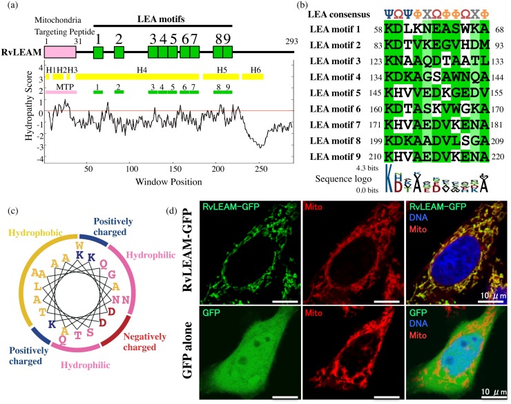
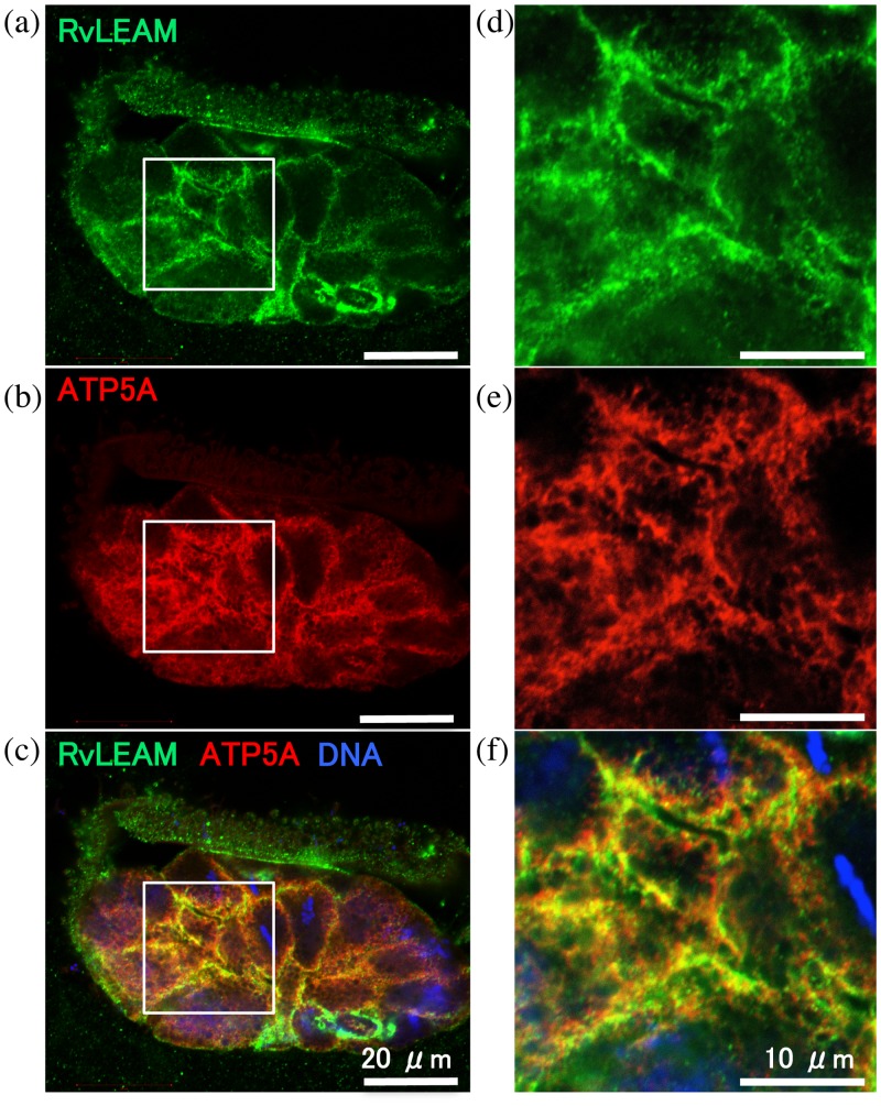
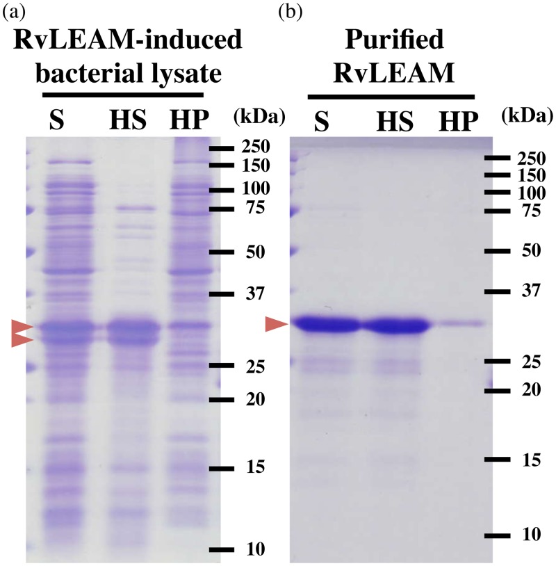
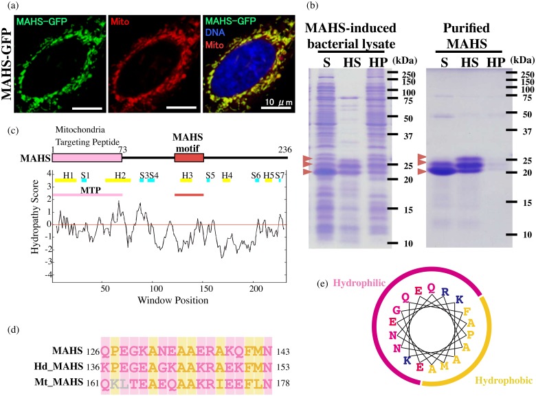
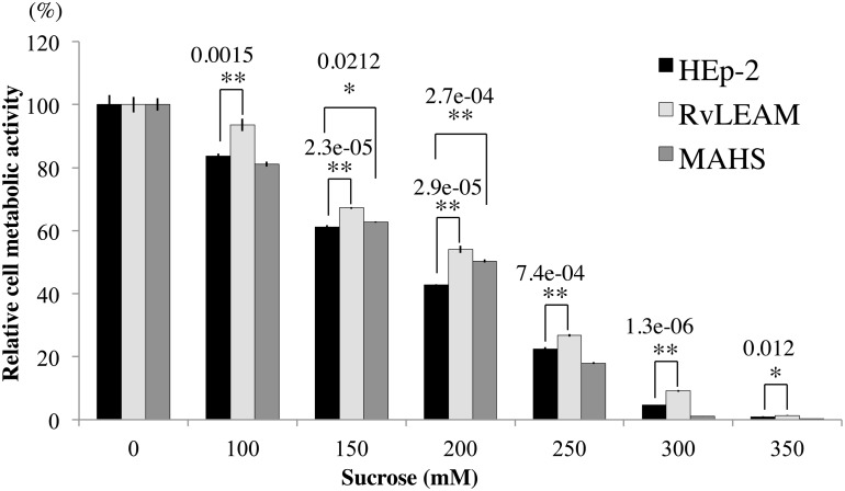
Tardigrades are able to tolerate almost complete dehydration through transition to a metabolically inactive state, called "anhydrobiosis". Late Embryogenesis Abundant (LEA) proteins are heat-soluble proteins involved in the desiccation tolerance of many anhydrobiotic organisms. Tardigrades, Ramazzottius varieornatus, however, express predominantly tardigrade-unique heat-soluble proteins: CAHS (Cytoplasmic Abundant Heat Soluble) and SAHS (Secretory Abundant Heat Soluble) proteins, which are secreted or localized in most intracellular compartments, except the mitochondria. Although mitochondrial integrity is crucial to ensure cellular survival, protective molecules for mitochondria have remained elusive. Here, we identified two novel mitochondrial heat-soluble proteins, RvLEAM and MAHS (Mitochondrial Abundant Heat Soluble), as potent mitochondrial protectants from Ramazzottius varieornatus. RvLEAM is a group3 LEA protein and immunohistochemistry confirmed its mitochondrial localization in tardigrade cells. MAHS-green fluorescent protein fusion protein localized in human mitochondria and was heat-soluble in vitro, though no sequence similarity with other known proteins was found, and one region was conserved among tardigrades. Furthermore, we demonstrated that RvLEAM protein as well as MAHS protein improved the hyperosmotic tolerance of human cells. The findings of the present study revealed that tardigrade mitochondria contain at least two types of heat-soluble proteins that might have protective roles in water-deficient environments.
Overview
The claims and statistics of this paper haven’t been checked yet.
Run the check — free, no account needed. It reads the open-access text and checks the sources it cites.
Integrity signals
Registry
Cites
Statistics
Figures
Verification
Claims the paper makes
The claims and statistics of this paper haven’t been checked yet.
Run the check — free, no account needed. It reads the open-access text and checks the sources it cites.
Claims other papers make about this one
Each is checked against this paper’s own text: does it say what the citing paper says it does?
The cited source says what the claim attributes to it.
In the citing paper
“These abundantly expressed proteins included previously identified tardigrade-unique heat-soluble proteins, CAHS and SAHS, both of which maintain solubility even after heat treatment and are proposed to be involved in the protection of biomolecules during desiccation3132.”
From Extremotolerant tardigrade genome and improved radiotolerance of human cultured cells by tardigrade-unique protein (2016) · cites this paper as [32]
Self-check
Percentages, sums and reported differences re-computed from the paper’s own numbers.
The self-check needs the paper’s full text.
Citations
Cites
A simple method for displaying the hydropathic character of a protein
Kyte, J., & Doolittle, R. F. (1982) · Journal of Molecular Biology · doi:10.1016/0022-2836(82)90515-0 · Cited by 22,927
Identification of prokaryotic and eukaryotic signal peptides and prediction of their cleavage sites
Nielsen, H., Engelbrecht, J., Brunak, S., & von HEIJNE, G. (1997) · Protein Engineering Design and Selection · doi:10.1093/protein/10.1.1 · Cited by 5,343
Fitting a mixture model by expectation maximization to discover motifs in biopolymers.
Bailey, T. L., & Elkan, C. (1994) · PubMed · Cited by 4,982
Danial, N. N., & Korsmeyer, S. J. (2004) · Cell · doi:10.1016/s0092-8674(04)00046-7 · Cited by 4,651
Predicting subcellular localization of proteins based on their N-terminal amino acid sequence.
Emanuelsson, O., Nielsen, H., Brunak, S., & von Heijne, G. (2000) · Journal of Molecular Biology · doi:10.1006/jmbi.2000.3903 · Cited by 4,419
WoLF PSORT: protein localization predictor
Horton, P., Park, K., Obayashi, T., Fujita, N., Harada, H., Adams-Collier, C. J., & Nakai, K. (2007) · Nucleic Acids Research · doi:10.1093/nar/gkm259 · Cited by 4,154
Mitochondrial membrane permeabilization in cell death
Kroemer, G., Galluzzi, L., & Brenner, C. (2007) · Physiological Reviews · doi:10.1152/physrev.00013.2006 · Cited by 3,598
Mitochondrial control of cell death
Kroemer, G., & Reed, J. C. (2000) · Nature Medicine · doi:10.1038/74994 · Cited by 3,103
InterProScan – an integration platform for the signature-recognition methods in InterPro
Zdobnov, E. M., & Apweiler, R. (2001) · Bioinformatics · doi:10.1093/bioinformatics/17.9.847 · Cited by 2,915
Computational Method to Predict Mitochondrially Imported Proteins and their Targeting Sequences
Claros, M. G., & Vincens, P. (1996) · European Journal of Biochemistry · doi:10.1111/j.1432-1033.1996.00779.x · Cited by 1,650
The Enigmatic LEA Proteins and Other Hydrophilins
Battaglia, M. E., Olvera-Carrillo, Y., Garcíarrubio, A., Campos, F., & Covarrubias, A. A. (2008) · PLANT PHYSIOLOGY · doi:10.1104/pp.108.120725 · Cited by 895
LEA proteins prevent protein aggregation due to water stress
Goyal, K., Walton, L. J., & Tunnacliffe, A. (2005) · Biochemical Journal · doi:10.1042/bj20041931 · Cited by 789
The continuing conundrum of the LEA proteins
Tunnacliffe, A., & Wise, M. J. (2007) · Die Naturwissenschaften · doi:10.1007/s00114-007-0254-y · Cited by 755
Porter: a new, accurate server for protein secondary structure prediction
Pollastri, G., & McLysaght, A. (2004) · Computer applications in the biosciences · doi:10.1093/bioinformatics/bti203 · Cited by 472
A repeating 11‐mer amino acid motif and plant desiccation
Dure, L. S. (1993) · The Plant Journal · doi:10.1046/j.1365-313x.1993.t01-19-00999.x · Cited by 432
LEA Proteins During Water Stress: Not Just for Plants Anymore
Hand, S. C., Menze, M. A., Toner, M., Boswell, L. C., & Moore, D. S. (2011) · Annual Review of Physiology · doi:10.1146/annurev-physiol-012110-142203 · Cited by 432
Tardigrades survive exposure to space in low Earth orbit
Jönsson, K. I., Rabbow, E., Schill, R. O., Harms-Ringdahl, M., & Rettberg, P. (2008) · Current Biology · doi:10.1016/j.cub.2008.06.048 · Cited by 415 · cited 1×
“Dehydrated tardigrades exhibit extreme tolerance against various environmental stresses [2,3].”
BackgroundChakrabortee, S., Boschetti, C., Walton, L. J., Sarkar, S., Rubinsztein, D. C., & Tunnacliffe, A. (2007) · Proceedings of the National Academy of Sciences · doi:10.1073/pnas.0706964104 · Cited by 315
Anhydrobiosis in nematodes: Carbohydrate and lipid metabolism during dehydration
Madin, K. A. C., & Crowe, J. H. (1975) · Journal of Experimental Zoology · doi:10.1002/jez.1401930309 · Cited by 276
Survival in extreme environments – on the current knowledge of adaptations in tardigrades
Møbjerg, N., Halberg, K. A., Jørgensen, A., Persson, D. K., Bjørn, M. E., Ramløv, H., & Kristensen, R. M. (2011) · Acta Physiologica · doi:10.1111/j.1748-1716.2011.02252.x · Cited by 261
Trehalose and anhydrobiosis in tardigrades – evidence for divergence in responses to dehydration
Hengherr, S., Heyer, A. G., Köhler, H., & Schill, R. O. (2007) · FEBS Journal · doi:10.1111/j.1742-4658.2007.06198.x · Cited by 234
Clegg, J. S. (1965) · Comparative Biochemistry and Physiology · doi:10.1016/0010-406x(65)90014-9 · Cited by 219
Dehydration-induced expression of LEA proteins in an anhydrobiotic chironomid
Kikawada, T., Nakahara, Y., Kanamori, Y., Iwata, K.-I., Watanabe, M., McGee, B., Tunnacliffe, A., & Okuda, T. (2006) · Biochemical and Biophysical Research Communications · doi:10.1016/j.bbrc.2006.07.003 · Cited by 211
Two Novel Heat-Soluble Protein Families Abundantly Expressed in an Anhydrobiotic Tardigrade
Yamaguchi, A., Tanaka, S., Yamaguchi, S., Kuwahara, H., Takamura, C., Imajoh-Ohmi, S., Horikawa, D. D., Toyoda, A., Katayama, T., Arakawa, K., et al. (2012) · PLoS ONE · doi:10.1371/journal.pone.0044209 · Cited by 176
Self-citationFunctional Divergence of Former Alleles in an Ancient Asexual Invertebrate
Pouchkina-Stantcheva, N. N., McGee, B., Boschetti, C., Tolleter, D., Chakrabortee, S., Popova, A. V., Meersman, F., Macherel, D., Hincha, D. K., & Tunnacliffe, A. (2007) · Science · doi:10.1126/science.1144363 · Cited by 152
Horikawa, D. D., Kunieda, T., Abe, W., Watanabe, M., Nakahara, Y., Yukuhiro, F., Sakashita, T., Hamada, N., Wada, S., Funayama, T., et al. (2008) · Astrobiology · doi:10.1089/ast.2007.0139 · Cited by 146
Self-citationBrowne, J. A., Dolan, K. M., Tyson, T., Goyal, K., Tunnacliffe, A., & Burnell, A. M. (2004) · Eukaryotic Cell · doi:10.1128/ec.3.4.966-975.2004 · Cited by 140
Tolerance to oxidative stress induced by desiccation in Porphyra columbina (Bangiales, Rhodophyta)
Porcia, L. C., Thomas, D. N., Flores, V., & Correa, J. A. (2010) · Journal of Experimental Botany · doi:10.1093/jxb/erq364 · Cited by 140
p23, a simple protein with complex activities
Felts, S. J., & Toft, D. O. (2003) · Cell Stress and Chaperones · doi:10.1379/1466-1268(2003)008 · Cited by 116
Late embryogenesis abundant proteins protect human hepatoma cells during acute desiccation
Li, S., Chakraborty, N., Borcar, A., Menze, M. A., Toner, M., & Hand, S. C. (2012) · Proceedings of the National Academy of Sciences · doi:10.1073/pnas.1214893109 · Cited by 112
A Putative LEA Protein, but no Trehalose, is Present in Anhydrobiotic Bdelloid Rotifers
Tunnacliffe, A., Lapinski, J., & McGee, B. (2005) · Hydrobiologia · doi:10.1007/s10750-005-4239-6 · Cited by 112
Extreme stress tolerance in tardigrades: surviving space conditions in low earth orbit
Persson, D. K., Halberg, K. A., Jørgensen, A., Ricci, C. N., Møbjerg, N., & Kristensen, R. M. (2011) · Journal of Zoological Systematics & Evolutionary Research · doi:10.1111/j.1439-0469.2010.00605.x · Cited by 106
A putative LEA protein, but no trehalose, is present in anhydrobiotic bdelloid rotifers
Tunnacliffe, A., Lapinski, J., & McGee, B. (2006) · Springer eBooks · doi:10.1007/1-4020-4408-9_32 · Cited by 104
Watanabe, M., Kikawada, T., & Okuda, T. (2003) · Journal of Experimental Biology · doi:10.1242/jeb.00418 · Cited by 99
Hatanaka, R., Hagiwara‐Komoda, Y., Furuki, T., Kanamori, Y., Fujita, M., Cornette, R., Sakurai, M., Okuda, T., & Kikawada, T. (2013) · Insect Biochemistry and Molecular Biology · doi:10.1016/j.ibmb.2013.08.004 · Cited by 85
Förster, F., Liang, C., Shkumatov, A. V., Beißer, D., Engelmann, J. C., Schnölzer, M., Frohme, M., Müller, T., Schill, R. O., & Dandekar, T. (2009) · BMC Genomics · doi:10.1186/1471-2164-10-469 · Cited by 70
de Carvalho, R. C., Catalá, M., da Silva, J. M., Branquinho, C., & Barreno, E. (2012) · Annals of Botany · doi:10.1093/aob/mcs180 · Cited by 66
Effects of Group 3 LEA protein model peptides on desiccation-induced protein aggregation
Furuki, T., Shimizu, T., Chakrabortee, S., Yamakawa, K., Hatanaka, R., Takahashi, T., Kikawada, T., Okuda, T., Mihara, H., Tunnacliffe, A., et al. (2012) · Biochimica et Biophysica Acta (BBA) - Proteins and Proteomics · doi:10.1016/j.bbapap.2012.04.013 · Cited by 62
Sharon, M. A., Kozarova, A., Clegg, J. S., Vacratsis, P. O., & Warner, A. H. (2009) · Biochemistry and Cell Biology · doi:10.1139/o09-001 · Cited by 61
Mali, B., Grohme, M. A., Förster, F., Dandekar, T., Schnölzer, M., Reuter, D., Wełnicz, W., Schill, R. O., & Frohme, M. (2010) · BMC Genomics · doi:10.1186/1471-2164-11-168 · Cited by 59
Förster, F., Beißer, D., Grohme, M. A., Liang, C., Mali, B., Siegl, A., Engelmann, J. C., Shkumatov, A. V., Schokraie, E., Müller, T., et al. (2012) · Bioinformatics and Biology Insights · doi:10.4137/bbi.s9150 · Cited by 54
Construction of Epstein–Barr Virus-Based Expression Vector Containing Mini-OriP
Tanaka, J., Miwa, Y., Miyoshi, K., Ueno, A., & Inoue, H. (1999) · Biochemical and Biophysical Research Communications · doi:10.1006/bbrc.1999.1617 · Cited by 53
Self-citationMarunde, M. R., Samarajeewa, D. A., Anderson, J., Li, S., Hand, S. C., & Menze, M. A. (2013) · Journal of Insect Physiology · doi:10.1016/j.jinsphys.2013.01.004 · Cited by 49
Group 3 LEA protein model peptides protect liposomes during desiccation
Furuki, T., & Sakurai, M. (2014) · Biochimica et Biophysica Acta (BBA) - Biomembranes · doi:10.1016/j.bbamem.2014.07.009 · Cited by 47
Trafficking of bdelloid rotifer late embryogenesis abundant proteins
Tripathi, R., Boschetti, C., McGee, B., & Tunnacliffe, A. (2012) · Journal of Experimental Biology · doi:10.1242/jeb.071647 · Cited by 30
The LEA proteins and trehalose loving couple: a step forward in anhydrobiotic engineering
Iturriaga, G. (2008) · Biochemical Journal · doi:10.1042/bj20071633 · Cited by 26
Cited by
Sorted by citations
Tardigrades Use Intrinsically Disordered Proteins to Survive Desiccation
Boothby, T. C., Tapia, H., Brozena, A. H., Piszkiewicz, S., Smith, A. E., Giovannini, I., Rebecchi, L., Pielak, G. J., Koshland, D., & Goldstein, B. (2017) · Molecular Cell · doi:10.1016/j.molcel.2017.02.018 · Cited by 418
Hashimoto, T., Horikawa, D. D., Saito, Y., Kuwahara, H., Kozuka-Hata, H., Shin-I, T., Minakuchi, Y., Ohishi, K., Motoyama, A., Aizu, T., et al. (2016) · Nature Communications · doi:10.1038/ncomms12808 · Cited by 394
Self-citationComparative genomics of the tardigrades Hypsibius dujardini and Ramazzottius varieornatus
Yoshida, Y., Koutsovoulos, G., Laetsch, D. R., Stevens, L., Kumar, S., Horikawa, D. D., Ishino, K., Komine, S., Kunieda, T., Tomita, M., et al. (2017) · PLoS Biology · doi:10.1371/journal.pbio.2002266 · Cited by 229
Self-citationHibshman, J. D., Clegg, J. S., & Goldstein, B. (2020) · Frontiers in Physiology · doi:10.3389/fphys.2020.592016 · Cited by 115
The biology of tardigrade disordered proteins in extreme stress tolerance
Hesgrove, C., & Boothby, T. C. (2020) · Cell Communication and Signaling · doi:10.1186/s12964-020-00670-2 · Cited by 113
Metadata
- Authors
- Tanaka, S., Tanaka, J., Miwa, Y., Horikawa, D. D., Katayama, T., Arakawa, K., Toyoda, A., Kubo, T., & Kunieda, T.
- Affiliations
- The University of Tokyo (JP)
- University of Tsukuba (JP)
- Keio University (JP)
- Database Center for Life Science (JP)
- National Institute of Genetics (JP)
- Corresponding author
- Kunieda, Takekazu
- ORCID
- Venue
- PLOS ONE
- Publisher
- Public Library of Science (PLoS)
- Published
- 2015-02-12
- Volume
- 10
- Issue
- 2
- Pages
- e0118272
- ISSN
- 1932-6203 (ISSN-L)
- PMC
- PMC4326354
- OpenAlex
- W1964745114
- Cited by
- 128
- References
- 46
- Citations by year
- 2026: 7 · 2025: 8 · 2024: 32 · 2023: 15 · 2022: 13 · 2021: 16 · 2020: 7 · 2019: 12 · 2018: 6 · 2017: 7 · 2016: 4 · 2015: 1
- FWCI
- 7.02
- Type
- journal article
- Language
- en
- Open access
- gold
- doi.org
- Licence
- Locations
- PLoS ONE (publishedVersion, cc-by)
- Europe PMC (PubMed Central) (publishedVersion, cc-by)
- PubMed Central (publishedVersion, cc-by)
- PubMed (publishedVersion)
- DOAJ (DOAJ: Directory of Open Access Journals) (submittedVersion, cc-by-sa)
- Figshare (submittedVersion, cc-by)
- Funders
- Ministry of Education, Culture, Sports, Science and Technology (No. 221S0002, 221S0002)
- Japan Society for the Promotion of Science (26660288, 25281016, 24657069, 13J10474, No. 221S0002, 221S0002)
- Topics
- Life Sciences › Agricultural and Biological Sciences › Ecology, Evolution, Behavior and Systematics › Tardigrade Biology and Ecology
- Physical Sciences › Environmental Science › Ecology › Physiological and biochemical adaptations
- Physical Sciences › Environmental Science › Ecology › Polar Research and Ecology
- Keywords
- tardigrades, Ramazzottius varieornatus, anhydrobiosis, late embryogenesis abundant proteins, mitochondrial protection, desiccation tolerance, mitochondrial localization, human cells
- MeSH terms
- Amino Acid Sequence, Animals, Hot Temperature, Humans, Mitochondria, Molecular Sequence Data, Osmotic Pressure, Solubility, Amino Acid Motifs, Protein Transport, Mitochondrial Proteins, Tardigrada, Osmoregulation
About these checks
Registry, retraction, Cites and statistics results are computed, not judged, with one exception: a printed reference that no index lists is read once by a model before it counts as a citation error. Claims are extracted by a model and kept only when their quote appears verbatim in the text; each cited source is then checked, with a model’s help, for whether it says what the claim relies on. Those verdicts can be wrong — read the claim’s quote and the cited source before relying on one.
Statistics and arithmetic are recomputed, not judged.
Confidence bands are shown only for fields calibrated on our annotated test set. None is calibrated yet, so no band is shown.
What counts as a claim ›How we measure confidence ›How we check this ›
Are you an author, or do you see a problem with this page? Request a review
