

The DOI matches its Crossref record; 35 of the 36 references it cites were found in a registry or an index.
Enhanced Performance of Brain Tumor Classification via Tumor Region Augmentation and Partition
Open accessAbstract
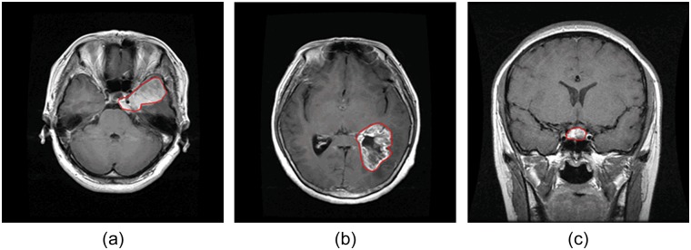
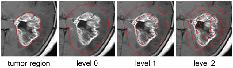
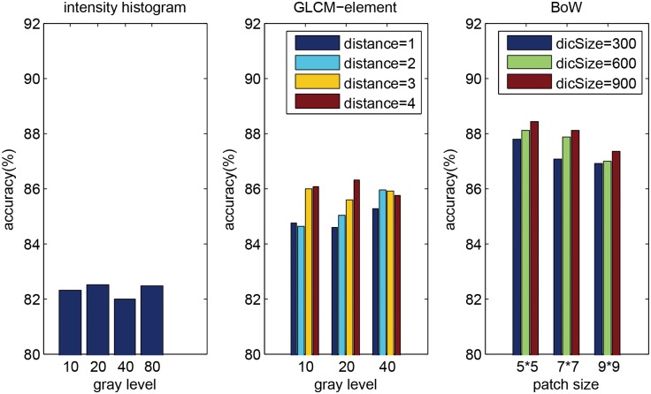
Automatic classification of tissue types of region of interest (ROI) plays an important role in computer-aided diagnosis. In the current study, we focus on the classification of three types of brain tumors (i.e., meningioma, glioma, and pituitary tumor) in T1-weighted contrast-enhanced MRI (CE-MRI) images. Spatial pyramid matching (SPM), which splits the image into increasingly fine rectangular subregions and computes histograms of local features from each subregion, exhibits excellent results for natural scene classification. However, this approach is not applicable for brain tumors, because of the great variations in tumor shape and size. In this paper, we propose a method to enhance the classification performance. First, the augmented tumor region via image dilation is used as the ROI instead of the original tumor region because tumor surrounding tissues can also offer important clues for tumor types. Second, the augmented tumor region is split into increasingly fine ring-form subregions. We evaluate the efficacy of the proposed method on a large dataset with three feature extraction methods, namely, intensity histogram, gray level co-occurrence matrix (GLCM), and bag-of-words (BoW) model. Compared with using tumor region as ROI, using augmented tumor region as ROI improves the accuracies to 82.31% from 71.39%, 84.75% from 78.18%, and 88.19% from 83.54% for intensity histogram, GLCM, and BoW model, respectively. In addition to region augmentation, ring-form partition can further improve the accuracies up to 87.54%, 89.72%, and 91.28%. These experimental results demonstrate that the proposed method is feasible and effective for the classification of brain tumors in T1-weighted CE-MRI.
Overview
The claims and statistics of this paper haven’t been checked yet.
Run the check — free, no account needed. It reads the open-access text and checks the sources it cites.
Integrity signals
Registry
Cites
Statistics
Figures
Verification
Claims the paper makes
The claims and statistics of this paper haven’t been checked yet.
Run the check — free, no account needed. It reads the open-access text and checks the sources it cites.
Claims other papers make about this one
Each is checked against this paper’s own text: does it say what the citing paper says it does?
In the citing paper
“Data collected from three different kinds of brain tumors consisted of 708 meningiomas, 1426 gliomas, and 930 pituitaries images from the General Hospital and Nanfang Hospital, Tianjin Medical University, China [72].”
From A Comprehensive Analysis of Recent Deep and Federated-Learning-Based Methodologies for Brain Tumor Diagnosis (2022) · cites this paper as [72]
In the citing paper
“This contains 3064 T1-weighted contrast-enhanced images collected from 233 patients with a resolution of 512 × 512 pixels.”
From A Comprehensive Analysis of Recent Deep and Federated-Learning-Based Methodologies for Brain Tumor Diagnosis (2022) · cites this paper as [72]
Self-check
Percentages, sums and reported differences re-computed from the paper’s own numbers.
The self-check needs the paper’s full text.
Citations
Cites
Distinctive Image Features from Scale-Invariant Keypoints
Lowe, D. G. (2004) · International Journal of Computer Vision · doi:10.1023/b:visi.0000029664.99615.94 · Cited by 55,589 · cited 1×
“(cid:129) Local features: The next step is to extract local features within the ROI, such as scale-invariant feature transform (SIFT) descriptor [20] and raw patch [21].”
Background$rm K$-SVD: An Algorithm for Designing Overcomplete Dictionaries for Sparse Representation
Aharon, M., Elad, M., & Bruckstein, A. M. (2006) · IEEE Transactions on Signal Processing · doi:10.1109/tsp.2006.881199 · Cited by 9,464
Robust Face Recognition via Sparse Representation
Wright, J. N., Yang, A., Ganesh, A., Sastry, S., & Ma, Y. (2009) · IEEE Transactions on Pattern Analysis and Machine Intelligence · doi:10.1109/tpami.2008.79 · Cited by 9,398 · cited 1×
“We also compared three classification methods (i.e. SVM, sparse representation-based classification (SRC) [31], and k nearest neighbors (kNN) classifier) using 2-region BoW representations.”
ResultBeyond Bags of Features: Spatial Pyramid Matching for Recognizing Natural Scene Categories
Lazebnik, S., Schmid, C., & Ponce, J. (2006) · 2006 IEEE Computer Society Conference on Computer Vision and Pattern Recognition - Volume 2 (CVPR'06) · doi:10.1109/cvpr.2006.68 · Cited by 7,916 · cited 4×
“Among those methods, the most notable work is SPM [15], which splits the image into hierarchical cells, computes BoW representation for each cell, and finally weights and concatenates the results.”
BackgroundMethodInfluentialVideo Google: a text retrieval approach to object matching in videos
Sivic, & Zisserman (2003) · Proceedings Ninth IEEE International Conference on Computer Vision · doi:10.1109/iccv.2003.1238663 · Cited by 6,367 · cited 1×
“The BoW method was originally used in the text retrieval domain, and it has been successfully adapted to the visual analysis domain [13,14].”
MethodRobust wide-baseline stereo from maximally stable extremal regions
Matas, J., Chum, O., Urban, M., & Pajdla, T. (2004) · Image and Vision Computing · doi:10.1016/j.imavis.2004.02.006 · Cited by 3,760 · cited 1×
“As pointed out in [29,30], it is beneficial to capture a certain amount of context.”
BackgroundA Bayesian Hierarchical Model for Learning Natural Scene Categories
Fei-Fei, L., & Perona, P. (2005) · IEEE Computer Society Conference on Computer Vision and Pattern Recognition/Proceedings - IEEE Computer Society Conference on Computer Vision and Pattern Recognition/Proceedings · doi:10.1109/cvpr.2005.16 · Cited by 3,665 · cited 1×
“The BoW method was originally used in the text retrieval domain, and it has been successfully adapted to the visual analysis domain [13,14].”
MethodLocality-constrained Linear Coding for image classification
Wang, J., Yang, J., Yu, K., Lv, F., Huang, T., & Gong, Y. (2010) · 2010 IEEE Computer Society Conference on Computer Vision and Pattern Recognition · doi:10.1109/cvpr.2010.5540018 · Cited by 3,100 · cited 2×
“Other more sophisticated methods may also be used, such as SC, soft assignment (SA) [24,25], orthogonal matching pursuit (OMP), locality-constrained linear coding [26], and local anchor embedding [27].”
MethodA comparison of affine region detectors
Mikolajczyk, K., Tuytelaars, T., Schmid, C., Zisserman, A., Matas, J., Schaffalitzky, F., Kadir, T., & Gool, L. V. (2005) · International Journal of Computer Vision · doi:10.1007/s11263-005-3848-x · Cited by 2,939
Linear spatial pyramid matching using sparse coding for image classification
Yang, J., Yu, K., Gong, Y., & Huang, T. S. (2009) · IEEE Computer Society Conference on Computer Vision and Pattern Recognition/Proceedings - IEEE Computer Society Conference on Computer Vision and Pattern Recognition/Proceedings · doi:10.1109/cvpr.2009.5206757 · Cited by 2,895
Research works printed but not linked: 1
- S Patil, 2013, A Computer Aided Diagnostic System For Classification Of Brain Tumors Using Texture Features And Probabilistic Neural Network, Int J Comput Sci Eng Inf Technol Res, 3, 61
Cited by
Sorted by citations
Brain tumor classification using deep CNN features via transfer learning
Deepak, S., & Ameer, P. M. (2019) · Computers in Biology and Medicine · doi:10.1016/j.compbiomed.2019.103345 · Cited by 1,370
Multi-grade brain tumor classification using deep CNN with extensive data augmentation
Sajjad, M., Khan, S., Muhammad, K., Wu, W., Ullah, A., & Baik, S. W. (2018) · Journal of Computational Science · doi:10.1016/j.jocs.2018.12.003 · Cited by 895
Brain tumor classification for MR images using transfer learning and fine-tuning
Swati, Z. N. K., Zhao, Q., Kabir, M., Ali, F., Ali, Z., Ahmad, S., & Lu, J. (2019) · Computerized Medical Imaging and Graphics · doi:10.1016/j.compmedimag.2019.05.001 · Cited by 895
Multi-Classification of Brain Tumor Images Using Deep Neural Network
Sultan, H. H., Salem, N. M., & Al-Atabany, W. (2019) · IEEE Access · doi:10.1109/access.2019.2919122 · Cited by 775
“First, data augmentation is used to avoid overfitting by making a geometric and color distortion on the original images [23], [31], [38].”
BackgroundMethodInfluentialClassification of Brain Tumors from MRI Images Using a Convolutional Neural Network
Atanasijević, M. B., & Barjaktarović, M. Č. (2020) · Applied Sciences · doi:10.3390/app10061999 · Cited by 753
“In the literature, there are also studies that used the same database for classification with pre-trained networks [23,35–40] or, as input, they use only tumor region or some features that are extracted from the tumor region [7,21,23,41,42].”
Background
Metadata
- Authors
- Cheng, J., Huang, W., Cao, S., Yang, R., Yang, W., Yun, Z., Wang, Z., & Feng, Q.
- Affiliations
- Southern Medical University (CN)
- Nanfang Hospital (CN)
- Corresponding author
- Feng, Qianjin
- Venue
- PLOS ONE
- Publisher
- Public Library of Science (PLoS)
- Published
- 2015-10-08
- Volume
- 10
- Issue
- 10
- Pages
- e0140381
- ISSN
- 1932-6203 (ISSN-L)
- PMC
- PMC4598126
- OpenAlex
- W2182098131
- Cited by
- 879
- References
- 36
- Citations by year
- 2026: 110 · 2025: 134 · 2024: 142 · 2023: 152 · 2022: 128 · 2021: 85 · 2020: 63 · 2019: 44 · 2018: 14 · 2017: 4 · 2016: 2 · 2015: 1
- FWCI
- 3.74
- Type
- journal article
- Language
- en
- Update notices
- correction (2015-12-02)
- Open access
- gold
- doi.org
- Licence
- Locations
- PLoS ONE (publishedVersion, cc-by)
- Europe PMC (PubMed Central) (publishedVersion, cc-by)
- PubMed Central (publishedVersion, cc-by)
- PubMed (publishedVersion)
- DOAJ (DOAJ: Directory of Open Access Journals) (submittedVersion)
- Funders
- National Natural Science Foundation of China (81101109, 31371009, 2012BAI14B02, 863 Program)
- National High-tech Research and Development Program
- Topics
- Physical Sciences › Computer Science › Computer Vision and Pattern Recognition › Brain Tumor Detection and Classification
- Health Sciences › Medicine › Radiology, Nuclear Medicine and Imaging › Radiomics and Machine Learning in Medical Imaging
- Physical Sciences › Computer Science › Artificial Intelligence › AI in cancer detection
- Keywords
- brain tumor classification, meningioma, glioma, pituitary tumors, region of interest, image dilation, spatial pyramid matching, contrast-enhanced T1-weighted imaging, computer-aided diagnosis, gray level co-occurrence matrix, bag-of-words model, intensity histogram
- MeSH terms
- Brain Neoplasms, Glioma, Humans, Image Interpretation, Computer-Assisted, Magnetic Resonance Imaging, Meningioma, Sensitivity and Specificity
About these checks
Registry, retraction, Cites and statistics results are computed, not judged, with one exception: a printed reference that no index lists is read once by a model before it counts as a citation error. Claims are extracted by a model and kept only when their quote appears verbatim in the text; each cited source is then checked, with a model’s help, for whether it says what the claim relies on. Those verdicts can be wrong — read the claim’s quote and the cited source before relying on one.
Statistics and arithmetic are recomputed, not judged.
Confidence bands are shown only for fields calibrated on our annotated test set. None is calibrated yet, so no band is shown.
What counts as a claim ›How we measure confidence ›How we check this ›
Are you an author, or do you see a problem with this page? Request a review
