

The DOI matches its Crossref record; 35 of the 36 references it cites were found in a registry or an index.
Enhanced Performance of Brain Tumor Classification via Tumor Region Augmentation and Partition
Open accessAbstract
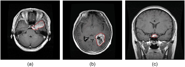
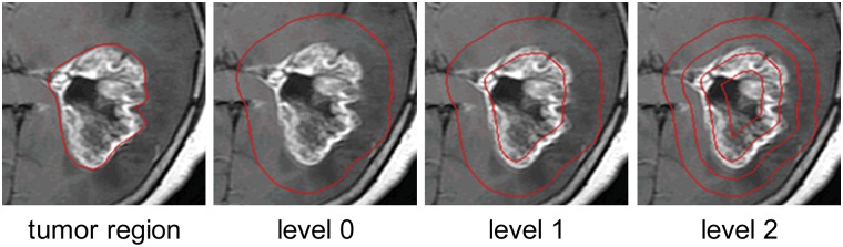
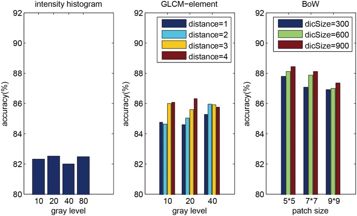
Automatic classification of tissue types of region of interest (ROI) plays an important role in computer-aided diagnosis. In the current study, we focus on the classification of three types of brain tumors (i.e., meningioma, glioma, and pituitary tumor) in T1-weighted contrast-enhanced MRI (CE-MRI) images. Spatial pyramid matching (SPM), which splits the image into increasingly fine rectangular subregions and computes histograms of local features from each subregion, exhibits excellent results for natural scene classification. However, this approach is not applicable for brain tumors, because of the great variations in tumor shape and size. In this paper, we propose a method to enhance the classification performance. First, the augmented tumor region via image dilation is used as the ROI instead of the original tumor region because tumor surrounding tissues can also offer important clues for tumor types. Second, the augmented tumor region is split into increasingly fine ring-form subregions. We evaluate the efficacy of the proposed method on a large dataset with three feature extraction methods, namely, intensity histogram, gray level co-occurrence matrix (GLCM), and bag-of-words (BoW) model. Compared with using tumor region as ROI, using augmented tumor region as ROI improves the accuracies to 82.31% from 71.39%, 84.75% from 78.18%, and 88.19% from 83.54% for intensity histogram, GLCM, and BoW model, respectively. In addition to region augmentation, ring-form partition can further improve the accuracies up to 87.54%, 89.72%, and 91.28%. These experimental results demonstrate that the proposed method is feasible and effective for the classification of brain tumors in T1-weighted CE-MRI.
Overview
The claims and statistics of this paper haven’t been checked yet.
Run the check — free, no account needed. It reads the open-access text and checks the sources it cites.
Integrity signals
Registry
Cites
Statistics
Figures
Verification
Claims the paper makes
The claims and statistics of this paper haven’t been checked yet.
Run the check — free, no account needed. It reads the open-access text and checks the sources it cites.
Claims other papers make about this one
Each is checked against this paper’s own text: does it say what the citing paper says it does?
In the citing paper
“Data collected from three different kinds of brain tumors consisted of 708 meningiomas, 1426 gliomas, and 930 pituitaries images from the General Hospital and Nanfang Hospital, Tianjin Medical University, China [72].”
From A Comprehensive Analysis of Recent Deep and Federated-Learning-Based Methodologies for Brain Tumor Diagnosis (2022) · cites this paper as [72]
In the citing paper
“This contains 3064 T1-weighted contrast-enhanced images collected from 233 patients with a resolution of 512 × 512 pixels.”
From A Comprehensive Analysis of Recent Deep and Federated-Learning-Based Methodologies for Brain Tumor Diagnosis (2022) · cites this paper as [72]
Self-check
Percentages, sums and reported differences re-computed from the paper’s own numbers.
The self-check needs the paper’s full text.
Citations
Cites
Distinctive Image Features from Scale-Invariant Keypoints
Lowe, D. G. (2004) · International Journal of Computer Vision · doi:10.1023/b:visi.0000029664.99615.94 · Cited by 55,589 · cited 1×
“(cid:129) Local features: The next step is to extract local features within the ROI, such as scale-invariant feature transform (SIFT) descriptor [20] and raw patch [21].”
Background$rm K$-SVD: An Algorithm for Designing Overcomplete Dictionaries for Sparse Representation
Aharon, M., Elad, M., & Bruckstein, A. M. (2006) · IEEE Transactions on Signal Processing · doi:10.1109/tsp.2006.881199 · Cited by 9,464
Robust Face Recognition via Sparse Representation
Wright, J. N., Yang, A., Ganesh, A., Sastry, S., & Ma, Y. (2009) · IEEE Transactions on Pattern Analysis and Machine Intelligence · doi:10.1109/tpami.2008.79 · Cited by 9,398 · cited 1×
“We also compared three classification methods (i.e. SVM, sparse representation-based classification (SRC) [31], and k nearest neighbors (kNN) classifier) using 2-region BoW representations.”
ResultBeyond Bags of Features: Spatial Pyramid Matching for Recognizing Natural Scene Categories
Lazebnik, S., Schmid, C., & Ponce, J. (2006) · 2006 IEEE Computer Society Conference on Computer Vision and Pattern Recognition - Volume 2 (CVPR'06) · doi:10.1109/cvpr.2006.68 · Cited by 7,916 · cited 4×
“Among those methods, the most notable work is SPM [15], which splits the image into hierarchical cells, computes BoW representation for each cell, and finally weights and concatenates the results.”
BackgroundMethodInfluentialVideo Google: a text retrieval approach to object matching in videos
Sivic, & Zisserman (2003) · Proceedings Ninth IEEE International Conference on Computer Vision · doi:10.1109/iccv.2003.1238663 · Cited by 6,367 · cited 1×
“The BoW method was originally used in the text retrieval domain, and it has been successfully adapted to the visual analysis domain [13,14].”
MethodRobust wide-baseline stereo from maximally stable extremal regions
Matas, J., Chum, O., Urban, M., & Pajdla, T. (2004) · Image and Vision Computing · doi:10.1016/j.imavis.2004.02.006 · Cited by 3,760 · cited 1×
“As pointed out in [29,30], it is beneficial to capture a certain amount of context.”
BackgroundA Bayesian Hierarchical Model for Learning Natural Scene Categories
Fei-Fei, L., & Perona, P. (2005) · IEEE Computer Society Conference on Computer Vision and Pattern Recognition/Proceedings - IEEE Computer Society Conference on Computer Vision and Pattern Recognition/Proceedings · doi:10.1109/cvpr.2005.16 · Cited by 3,665 · cited 1×
“The BoW method was originally used in the text retrieval domain, and it has been successfully adapted to the visual analysis domain [13,14].”
MethodLocality-constrained Linear Coding for image classification
Wang, J., Yang, J., Yu, K., Lv, F., Huang, T., & Gong, Y. (2010) · 2010 IEEE Computer Society Conference on Computer Vision and Pattern Recognition · doi:10.1109/cvpr.2010.5540018 · Cited by 3,100 · cited 2×
“Other more sophisticated methods may also be used, such as SC, soft assignment (SA) [24,25], orthogonal matching pursuit (OMP), locality-constrained linear coding [26], and local anchor embedding [27].”
MethodA comparison of affine region detectors
Mikolajczyk, K., Tuytelaars, T., Schmid, C., Zisserman, A., Matas, J., Schaffalitzky, F., Kadir, T., & Gool, L. V. (2005) · International Journal of Computer Vision · doi:10.1007/s11263-005-3848-x · Cited by 2,939
Linear spatial pyramid matching using sparse coding for image classification
Yang, J., Yu, K., Gong, Y., & Huang, T. S. (2009) · IEEE Computer Society Conference on Computer Vision and Pattern Recognition/Proceedings - IEEE Computer Society Conference on Computer Vision and Pattern Recognition/Proceedings · doi:10.1109/cvpr.2009.5206757 · Cited by 2,895
Research works printed but not linked: 1
- S Patil, 2013, A Computer Aided Diagnostic System For Classification Of Brain Tumors Using Texture Features And Probabilistic Neural Network, Int J Comput Sci Eng Inf Technol Res, 3, 61
Cited by
Sorted by citations
Brain tumor classification using deep CNN features via transfer learning
Deepak, S., & Ameer, P. M. (2019) · Computers in Biology and Medicine · doi:10.1016/j.compbiomed.2019.103345 · Cited by 1,370
Multi-grade brain tumor classification using deep CNN with extensive data augmentation
Sajjad, M., Khan, S., Muhammad, K., Wu, W., Ullah, A., & Baik, S. W. (2018) · Journal of Computational Science · doi:10.1016/j.jocs.2018.12.003 · Cited by 895
Brain tumor classification for MR images using transfer learning and fine-tuning
Swati, Z. N. K., Zhao, Q., Kabir, M., Ali, F., Ali, Z., Ahmad, S., & Lu, J. (2019) · Computerized Medical Imaging and Graphics · doi:10.1016/j.compmedimag.2019.05.001 · Cited by 895
Multi-Classification of Brain Tumor Images Using Deep Neural Network
Sultan, H. H., Salem, N. M., & Al-Atabany, W. (2019) · IEEE Access · doi:10.1109/access.2019.2919122 · Cited by 775
“First, data augmentation is used to avoid overfitting by making a geometric and color distortion on the original images [23], [31], [38].”
BackgroundMethodInfluentialClassification of Brain Tumors from MRI Images Using a Convolutional Neural Network
Atanasijević, M. B., & Barjaktarović, M. Č. (2020) · Applied Sciences · doi:10.3390/app10061999 · Cited by 753
“In the literature, there are also studies that used the same database for classification with pre-trained networks [23,35–40] or, as input, they use only tumor region or some features that are extracted from the tumor region [7,21,23,41,42].”
BackgroundBrain Tumor Classification Using Convolutional Neural Network
Abiwinanda, N., Hanif, M., Hesaputra, S. T., Handayani, A., & Mengko, T. R. (2018) · World Congress on Medical Physics and Biomedical Engineering, September 7 - 12, 2009, Munich, Germany · doi:10.1007/978-981-10-9035-6_33 · Cited by 677
A Deep Learning-Based Framework for Automatic Brain Tumors Classification Using Transfer Learning
Rehman, A., Naz, S., Razzak, I., Akram, F., & Imran, M. A. (2019) · Circuits Systems and Signal Processing · doi:10.1007/s00034-019-01246-3 · Cited by 717
Towards a guideline for evaluation metrics in medical image segmentation
Müller, D., Soto-Rey, I., & Kramer, F. (2022) · BMC Research Notes · doi:10.1186/s13104-022-06096-y · Cited by 668
“[30,31] and cell nuclei detection in pathology microscopy from Caicedo et al.”
Anaraki, A. K., Ayati, M., & Kazemi, F. (2018) · Journal of Applied Biomedicine · doi:10.1016/j.bbe.2018.10.004 · Cited by 640
Brain Tumor Type Classification via Capsule Networks
Afshar, P., Mohammadi, A., & Plataniotis, K. N. (2018) · Proceedings - International Conference on Image Processing · doi:10.1109/icip.2018.8451379 · Cited by 594
Pernas, F. J. D., Martínez‐Zarzuela, M., Antón‐Rodríguez, M., & González‐Ortega, D. (2021) · Healthcare · doi:10.3390/healthcare9020153 · Cited by 607
“methodologies [5,32] start with segmentations performed by experienced radiologists, and thus, they are semi-automatic methods.”
MethodInfluentialLoyola-Gonzalez, O. (2019) · IEEE Access · doi:10.1109/access.2019.2949286 · Cited by 559
“The brain cancer images were taken from [160] and the human brain cancer characteristics were extracted from [161].”
MethodINet: Convolutional Networks for Biomedical Image Segmentation
Weng, W., & Zhu, X. (2021) · IEEE Access · doi:10.1109/access.2021.3053408 · Cited by 569
“The BTD [43] dataset contains 3,064 T1C images from 233 patients with three kinds of brain tumor: meningioma (708 slices), glioma (1,426 slices), and pituitary tumor (930 slices).”
BackgroundBrain Tumor Detection Based on Deep Learning Approaches and Magnetic Resonance Imaging
Abdusalomov, A. B., Mukhiddinov, M., & Whangbo, T. K. (2023) · Cancers · doi:10.3390/cancers15164172 · Cited by 606
Irmak, E. (2021) · Iranian Journal of Science and Technology Transactions of Electrical Engineering · doi:10.1007/s40998-021-00426-9 · Cited by 479
Deep CNN for Brain Tumor Classification
Elhamzi, W., Elhamzi, W., Charfi, I., & Atri, M. (2021) · Neural Processing Letters · doi:10.1007/s11063-020-10398-2 · Cited by 481
Ghassemi, N., Shoeibi, A., & Rouhani, M. (2019) · Biomedical Signal Processing and Control · doi:10.1016/j.bspc.2019.101678 · Cited by 449
Ismael, S. A. A., Mohammed, A., & Hefny, H. A. (2019) · Artificial Intelligence in Medicine · doi:10.1016/j.artmed.2019.101779 · Cited by 443
Capsule Networks for Brain Tumor Classification Based on MRI Images and Coarse Tumor Boundaries
Afshar, P., Plataniotis, K. N., & Mohammadi, A. (2019) · IEEE International Conference on Acoustics Speech and Signal Processing · doi:10.1109/icassp.2019.8683759 · Cited by 450
Accurate brain tumor detection using deep convolutional neural network
Khan, M. S. I., Rahman, A., Debnath, T., Karim, M. R., Nasir, M. K., Band, S. S., Mosavi, A., & Dehzangi, A. (2022) · Computational and Structural Biotechnology Journal · doi:10.1016/j.csbj.2022.08.039 · Cited by 453
Deep Learning for Medical Anomaly Detection – A Survey
Fernando, T., Gammulle, H., Denman, S., Sridharan, S., & Fookes, C. (2021) · ACM Computing Surveys · doi:10.1145/3464423 · Cited by 449
“This framework was evaluated using the dataset of [63] which contains MRI images captured from 233 patients with different types of brain tumours: meningioma (708 samples), glioma (1426 samples), and pituitary (930 samples).”
MethodMuhammad, K., Khan, S., Ser, J. D., & de Albuquerque, V. H. C. (2020) · IEEE Transactions on Neural Networks and Learning Systems · doi:10.1109/tnnls.2020.2995800 · Cited by 411
A Hybrid Deep Learning-Based Approach for Brain Tumor Classification
Raza, A., Ayub, H., Khan, J. A., Ahmad, I., Salama, A. S., Daradkeh, Y. I., Javeed, D., Rehman, A. U., & Hamam, H. (2022) · Electronics · doi:10.3390/electronics11071146 · Cited by 357
“Additionally, the tumor region in the MRI was bordered manually by three experienced radiologists [56].”
ResultSrinivas, C., Shivaprasad, N. P. K., Zakariah, M., Alotaibi, Y. A., Shaukat, K., Partibane, B., & Halifa, A. (2022) · Journal of Healthcare Engineering · doi:10.1155/2022/3264367 · Cited by 348
Deep learning for medical image segmentation: State-of-the-art advancements and challenges
Rayed, M. E., Islam, S. M. S., Niha, S. I., Jim, J. R., Kabir, M. M., & Mridha, M. F. (2024) · Informatics in Medicine Unlocked · doi:10.1016/j.imu.2024.101504 · Cited by 410
A deep learning approach for brain tumor classification using MRI images
Aamir, M., Rahman, Z.-U., Dayo, Z. A., Abro, W. A., Uddin, M. I., Khan, I. U., Imran, A. S., Ali, Z., Ishfaq, M., Guan, Y., et al. (2022) · Computers & Electrical Engineering · doi:10.1016/j.compeleceng.2022.108105 · Cited by 338
Deep learning for brain tumor classification
Paul, J. S., Plassard, A. J., Landman, B. A., & Fabbri, D. (2017) · Proceedings of SPIE, the International Society for Optical Engineering/Proceedings of SPIE · doi:10.1117/12.2254195 · Cited by 346
Multi-Modal Brain Tumor Detection Using Deep Neural Network and Multiclass SVM
Maqsood, S., Damaševičius, R., & Maskeliūnas, R. (2022) · Medicina · doi:10.3390/medicina58081090 · Cited by 313
“[48] Linear discriminant analysis (LDA) 93.”
Brain Tumor Classification via Convolutional Neural Network and Extreme Learning Machines
Pashaei, A., Sajedi, H., & Jazayeri, N. (2018) · International Conference on Computer and Knowledge Engineering · doi:10.1109/iccke.2018.8566571 · Cited by 320
“Cheng et al [13] proposed a new method for brain tumor classification.”
BackgroundMethodResultInfluentialBrain tumor classification using modified local binary patterns (LBP) feature extraction methods
Kaplan, K., Kaya, Y., Kuncan, M., & Ertunç, H. M. (2020) · Medical Hypotheses · doi:10.1016/j.mehy.2020.109696 · Cited by 304
Mensah, P. K., Adekoya, A. F., Ayidzoe, M. A., & Baagyere, E. Y. (2019) · Journal of King Saud University - Computer and Information Sciences · doi:10.1016/j.jksuci.2019.09.014 · Cited by 303
Deep neural network correlation learning mechanism for CT brain tumor detection
Woźniak, M., Siłka, J., & Wieczorek, M. (2021) · Neural Computing and Applications · doi:10.1007/s00521-021-05841-x · Cited by 292
Özyurt, F., Sert, E., Avcı, E., & Doğantekin, E. (2019) · Measurement · doi:10.1016/j.measurement.2019.07.058 · Cited by 283
Brain Tumor Classification via Statistical Features and Back-Propagation Neural Network
Ismael, M. R., & Abdel-Qader, I. M. (2018) · IEEE International Conference on Electro Information Technology · doi:10.1109/eit.2018.8500308 · Cited by 264
“[20], they implemented an algorithm based on the three feature extraction methods, intensity histogram, gray level co-occurrence matrix (GLCM), and bag of words model and SVM model for classification.”
BackgroundMethodClassification of Brain Tumor from Magnetic Resonance Imaging Using Vision Transformers Ensembling
Tummala, S., Kadry, S., Bukhari, S. A. C., & Rauf, H. T. (2022) · Current Oncology · doi:10.3390/curroncol29100590 · Cited by 283
“More details about the dataset are available in [40,41].”
BackgroundBrain Tumor Analysis Empowered with Deep Learning: A Review, Taxonomy, and Future Challenges
Nadeem, M. W., Ghamdi, M. A. A., Hussain, M., Khan, M. A., Khan, K. M., Almotiri, S. H., & Butt, S. A. (2020) · Brain Sciences · doi:10.3390/brainsci10020118 · Cited by 262
Automated Categorization of Brain Tumor from MRI Using CNN features and SVM
Deepak, S., & Ameer, P. M. (2020) · Journal of Ambient Intelligence and Humanized Computing · doi:10.1007/s12652-020-02568-w · Cited by 274
“Among the works using hand-crafted designs, SVM on the BoW model of feature set was an effective approach (Cheng et al. 2015).”
BackgroundMethodA Transfer Learning approach for AI-based classification of brain tumors
Mehrotra, R., Ansari, M. A., Agrawal, R. K., & Anand, R. S. (2020) · Machine Learning with Applications · doi:10.1016/j.mlwa.2020.100003 · Cited by 272
Alanazi, M. F., Ali, M. U., Hussain, S. J., Zafar, A., Mohatram, M., Irfan, M., AlRuwaili, R., Alruwaili, M., Ali, N. H., & Albarrak, A. M. (2022) · Sensors · doi:10.3390/s22010372 · Cited by 259
Jiang, H., Diao, Z., Shi, T., Zhou, Y., Wang, F., Hu, W., Zhu, X., Luo, S., Tong, G., & Yao, Y. (2023) · Computers in Biology and Medicine · doi:10.1016/j.compbiomed.2023.106726 · Cited by 244
Detection of brain tumors from MRI images base on deep learning using hybrid model CNN and NADE
Hashemzehi, R., Chabok, S. J. S. M., Kheirabadi, M., & Kamel, S. R. (2020) · Journal of Applied Biomedicine · doi:10.1016/j.bbe.2020.06.001 · Cited by 254
Deep convolutional neural networks with transfer learning for automated brain image classification
Kaur, T., & Gandhi, T. K. (2020) · Machine Vision and Applications · doi:10.1007/s00138-020-01069-2 · Cited by 234
Asif, S., Yi, W., Ain, Q. U., Hou, J., Tao, Y., & Si, J. (2022) · IEEE Access · doi:10.1109/access.2022.3153306 · Cited by 221
Current AI technologies in cancer diagnostics and treatment
Tiwari, A., Mishra, S. N., & Kuo, T. (2025) · Molecular Cancer · doi:10.1186/s12943-025-02369-9 · Cited by 257
A review of deep learning for brain tumor analysis in MRI
Dorfner, F. J., Patel, J., Kalpathy–Cramer, J., Gerstner, E. R., & Bridge, C. P. (2025) · npj Precision Oncology · doi:10.1038/s41698-024-00789-2 · Cited by 265
Edge U-Net: Brain Tumor Segmentation using MRI based on Deep U-Net Model with boundary information
Allah, A. M. G., Sarhan, A. M., & Elshennawy, N. M. (2022) · Expert systems with applications · doi:10.1016/j.eswa.2022.118833 · Cited by 215
A classification of MRI brain tumor based on two stage feature level ensemble of deep CNN models
Aurna, N. F., Yousuf, M. A., Taher, K. A., Azad, A., & Moni, M. A. (2022) · Computers in Biology and Medicine · doi:10.1016/j.compbiomed.2022.105539 · Cited by 212
Automated Brain Tumor Segmentation and Classification in MRI Using YOLO-Based Deep Learning
Almufareh, M. F., Imran, M., Khan, A., Humayun, M., & Asim, M. F. (2024) · IEEE Access · doi:10.1109/access.2024.3359418 · Cited by 218
“The Brain Tumor dataset of Southern Medical University, Guangzhou, is utilized for this research acquired from [24] and [53].”
MethodMulti-class classification of brain tumor types from MR images using EfficientNets
Zulfiqar, F., Bajwa, U. I., & Mehmood, Y. (2023) · Biomedical Signal Processing and Control · doi:10.1016/j.bspc.2023.104777 · Cited by 204
Islam, T., Hafiz, M. S., Jim, J. R., Kabir, M. M., & Mridha, M. F. (2024) · Healthcare Analytics · doi:10.1016/j.health.2024.100340 · Cited by 234
A hybrid deep CNN model for brain tumor image multi-classification
Srinivasan, S., Francis, D., Mathivanan, S. K., Rajadurai, H., Shivahare, B. D., & Shah, M. A. (2024) · BMC Medical Imaging · doi:10.1186/s12880-024-01195-7 · Cited by 188
“The Cancer Genome Atlas Low-Grade Glioma data-set is the third dataset that was analyzed (TCGA-LGG) [37], and it has 242,185 MRI images of patients with low-grade gliomas (G-I and G-II) and incorporates data from 198 patients.”
MethodBrain Tumor and Glioma Grade Classification Using Gaussian Convolutional Neural Network
Rizwan, M., Shabbir, A., Javed, A. R., Shabbir, M., Baker, T., & Al‐Jumeily, D. (2022) · IEEE Access · doi:10.1109/access.2022.3153108 · Cited by 187
Celard, P., Iglesias, E. L., Sorribes-Fdez, J. M., Romero, R., Vieira, A. S., & Borrajo, L. (2023) · Neural Computing and Applications · doi:10.1007/s00521-022-07953-4 · Cited by 188
Brain tumor detection from images and comparison with transfer learning methods and 3-layer CNN
Khaliki, M. Z., & Başarslan, M. S. (2024) · Scientific Reports · doi:10.1038/s41598-024-52823-9 · Cited by 220
“In this study, the accuracy result was 91.28% 14 .”
BackgroundInfluentialDifferential Deep Convolutional Neural Network Model for Brain Tumor Classification
Kader, I. A. E., Xu, G., Zhang, S., Saminu, S., Javaid, I., & Ahmad, I. S. (2021) · Brain Sciences · doi:10.3390/brainsci11030352 · Cited by 198
Detection and Classification of Brain Tumor in MRI Images using Deep Convolutional Network
Bhanothu, Y., Kamalakannan, A., & Rajamanickam, G. (2020) · International Conference on Advanced Computing and Communication Systems (ICACCS) · doi:10.1109/icaccs48705.2020.9074375 · Cited by 189
“The MR image dataset for the proposed work was constructed from the dataset of 233 patients collected by Jun Cheng et al. [12, 13].”
MethodAn Effective Approach to Detect and Identify Brain Tumors Using Transfer Learning
Ullah, N., Khan, J. A., Khan, M. S., Khan, W., Hassan, I., Obayya, M. I., Negm, N., & Salama, A. S. (2022) · Applied Sciences · doi:10.3390/app12115645 · Cited by 189
“[31] CNN–DWA 80% 98.00 Jun Cheng [4] BoW–SVM 80% 91.28 Ismael [24] DWT–Gabor NN 70% 91.90 Pashaei [16] CNN–ELM 70% 93.68 Abiwinanda [11] Proposed CNN 70% 84.19 Sawati [45] Block-wise fine tuning 25–50–75% 94.82 Arshia Rehman [17] Transfer Learning 70% 98.69 Proposed approach Inceptionresnetv2 80%…”
BackgroundResultInfluentialAsif, S., Zhao, M., Tang, F., & Zhu, Y. (2023) · Multimedia Tools and Applications · doi:10.1007/s11042-023-14828-w · Cited by 174
“Most of the latest models use the Figshare benchmark brain tumor dataset [11] to assess performance.”
MethodHuang, Z., Du, X., Chen, L., Li, Y., Liu, M., Chou, Y., & Jin, L. (2020) · IEEE Access · doi:10.1109/access.2020.2993618 · Cited by 159
“This dataset is collected from 233 patients and consists of 708 meningioma images, 1426 glioma images, and 930 pituitary tumor images [39], [40].”
MethodBrain Tumor Classification Using ResNet-101 Based Squeeze and Excitation Deep Neural Network
Ghosal, P., Nandanwar, L., Kanchan, S., Bhadra, A. K., Chakraborty, J., & Nandi, D. (2019) · International Conference on Advanced Computational and Communication Paradigms (ICACCP) · doi:10.1109/icaccp.2019.8882973 · Cited by 165
“The proposed method was tested and evaluated on the Brain tumor database which has been collected in the year from 2005 to 2010, from Nanfang Hospital, Guangzhou, China, and General Hospital, Tianjin Medical University, China [12], [13].”
MethodInfluentialChelghoum, R., Ikhlef, A., Hameurlaine, A., & Jacquir, S. (2020) · IFIP advances in information and communication technology · doi:10.1007/978-3-030-49161-1_17 · Cited by 164
“It has already used in previous works like in [32, 33].”
MethodÇELİK, M., & İni̇k, Ö. (2023) · Expert Systems with Applications · doi:10.1016/j.eswa.2023.122159 · Cited by 166
Rammurthy, D., & Mahesh, P. K. (2020) · Journal of King Saud University - Computer and Information Sciences · doi:10.1016/j.jksuci.2020.08.006 · Cited by 152
Intelligent Ultra-Light Deep Learning Model for Multi-Class Brain Tumor Detection
Qureshi, S. A., Raza, S. E. A., Hussain, L., Malibari, A., Nour, M. K., Rehman, A. U., Al‐Wesabi, F. N., & Hilal, A. M. (2022) · Applied Sciences · doi:10.3390/app12083715 · Cited by 164
“[9] used a publicly available T1-weighted Contrast-Enhanced Magnetic Resonance Images (CE-MRI) dataset [10], consisting of brain MRI scans having: glioma-; meningioma-; and pituitary-tumors, and applied three features extraction methods: intensity histogram; bag-of-words (BoW) model; and Gray Level Co-occurrence Matri…”
BackgroundMethodA New Deep Hybrid Boosted and Ensemble Learning-Based Brain Tumor Analysis Using MRI
Zahoor, M. M., Qureshi, S. A., Bibi, S., Khan, S. H., Khan, A., Ghafoor, U., & Bhutta, M. R. (2022) · Sensors · doi:10.3390/s22072726 · Cited by 169
Classification of brain tumors from MR images using deep transfer learning
Polat, Ö., & Güngen, C. (2021) · The Journal of Supercomputing · doi:10.1007/s11227-020-03572-9 · Cited by 146
Multi-level attention network: application to brain tumor classification
Shaik, N. S., & Cherukuri, T. K. (2021) · Signal Image and Video Processing · doi:10.1007/s11760-021-02022-0 · Cited by 144
Rasheed, Z., Ma, Y.-K., Ullah, I., Ghadi, Y. Y., Khan, M. Z., Khan, M. A., Abdusalomov, A. B., Alqahtani, F. H., & Shehata, A. M. (2023) · Brain Sciences · doi:10.3390/brainsci13091320 · Cited by 140
“Brain tumors exhibit various sizes, dimensions, and densities [21].”
BackgroundHaq, A. U., Li, J., Khan, S., Alshara, M. A., Alotaibi, R. M., & Mawuli, C. B. (2022) · Scientific Reports · doi:10.1038/s41598-022-19465-1 · Cited by 137
A hybrid image enhancement based brain MRI images classification technique
Ullah, Z., Farooq, M. U., Lee, S.-H., & An, D. (2020) · Medical Hypotheses · doi:10.1016/j.mehy.2020.109922 · Cited by 143
A Novel MRI Diagnosis Method for Brain Tumor Classification Based on CNN and Bayesian Optimization
Amou, M. A., Xia, K., Kamhi, S., & Mouhafid, M. (2022) · Healthcare · doi:10.3390/healthcare10030494 · Cited by 150
RanMerFormer: Randomized vision transformer with token merging for brain tumor classification
Wang, J., Lu, S., Wang, S., & Zhang, Y. (2024) · Neurocomputing · doi:10.1016/j.neucom.2023.127216 · Cited by 135
An ensemble learning approach for brain cancer detection exploiting radiomic features
Brunese, L., Mercaldo, F., Reginelli, A., & Santone, A. (2019) · Computer Methods and Programs in Biomedicine · doi:10.1016/j.cmpb.2019.105134 · Cited by 130
Cheng, J., Huang, W., Cao, S., Yang, R., Yang, W., Yun, Z., Wang, Z., & Feng, Q. (2015) · PLoS ONE · doi:10.1371/journal.pone.0144479 · Cited by 128
Self-citationBrain tumor classification based on hybrid approach
Ayadi, W., Charfi, I., Elhamzi, W., & Atri, M. (2020) · The Visual Computer · doi:10.1007/s00371-020-02005-1 · Cited by 135
“Other systems proposed in the literature [28] exploit a region segmentation for MRI image before using it to the proposed method.”
BackgroundAhmad, B., Sun, J., You, Q., Palade, V., & Mao, Z. (2022) · Biomedicines · doi:10.3390/biomedicines10020223 · Cited by 139
A Comprehensive Survey on the Detection, Classification, and Challenges of Neurological Disorders
Lima, A. A., Mridha, M. F., Das, S. C., Kabir, M. M., Islam, M. R., & Watanobe, Y. (2022) · Biology · doi:10.3390/biology11030469 · Cited by 139
Kutlu, H., & Avcı, E. (2019) · Sensors · doi:10.3390/s19091992 · Cited by 123
“[31] focused on the classification of three types of brain tumors (i.”
BackgroundIslam, M., Reza, M. T., Kaosar, M. G., & Parvez, M. Z. (2022) · Neural Processing Letters · doi:10.1007/s11063-022-11014-1 · Cited by 120
“Data augmentation is a process that artificially generates more images while the primary input data remain the same [32].”
BackgroundReddy, C. K. K., Reddy, P. A., Janapati, H., Assiri, B., Shuaib, M., Alam, S., & Sheneamer, A. M. (2024) · Frontiers in Oncology · doi:10.3389/fonc.2024.1400341 · Cited by 121
Computer-assisted brain tumor type discrimination using magnetic resonance imaging features
Iqbal, S., Khan, M. U. G., Saba, T., & Rehman, A. U. (2017) · Biomedical Engineering Letters · doi:10.1007/s13534-017-0050-3 · Cited by 119
“[90] Custom developed 2005–2010 Nanfang Hospital and General Hospital, Tianjin Medical University”
BackgroundMethodFeature enhancement framework for brain tumor segmentation and classification
Tahir, B., Iqbal, S., Khan, M. U. G., Saba, T., Mehmood, Z., Anjum, A., & Mahmood, T. (2019) · Microscopy Research and Technique · doi:10.1002/jemt.23224 · Cited by 117
“These image-sets include Lung CT scans (Armato et al., 2015), Brain MRI (Cheng et al., 2015), and BRATS images (Menze et al., 2015).”
InfluentialJoint training of two-channel deep neural network for brain tumor classification
Bodapati, J. D., Shaik, N. S., Veeranjaneyulu, N., & Mundukur, N. B. (2020) · Signal Image and Video Processing · doi:10.1007/s11760-020-01793-2 · Cited by 115
“Inthefirst strategy, the suggested model is validated using the approach suggested (hold-out cross-validation) as in [22], where the dataset is partitionedinto70:15:15,respectively,fortrain,development and test sets and in the second strategy we adopt the fivefold cross-validation approach as in [12].”
MethodDutta, T. K., Nayak, D. R., & Zhang, Y. (2023) · Biomedical Signal Processing and Control · doi:10.1016/j.bspc.2023.105421 · Cited by 123
Asiri, A. A. M., Shaf, A., Ali, T. M., Aamir, M., Irfan, M., Alqahtani, S., Mehdar, K. M., Halawani, H. T., Alghamdi, A., Alshamrani, A. F. A., et al. (2023) · Life · doi:10.3390/life13071449 · Cited by 128
“The research used the Figshare dataset in [22] to apply an adaptive spatial division algorithm to increase tumor areas as a zone of concern before further dividing into sub-sections and extracting the intensity histogram, grey-level co-occurrence matrix value, and a bag of words (BoW)…”
MethodHolistic Brain Tumor Screening and Classification Based on DenseNet and Recurrent Neural Network
Zhou, Y., Li, Z., Zhu, H., Chen, C., Gao, M., Xu, K. M., & Xu, J. (2019) · Lecture notes in computer science · doi:10.1007/978-3-030-11723-8_21 · Cited by 110
Brain tumor categorization from imbalanced MRI dataset using weighted loss and deep feature fusion
Deepak, S., & Ameer, P. M. (2022) · Neurocomputing · doi:10.1016/j.neucom.2022.11.039 · Cited by 113
An MRI-based deep learning approach for efficient classification of brain tumors
Haq, E. U., Jianjun, H., Li, K., Haq, H. U., & Zhang, T. (2021) · Journal of Ambient Intelligence and Humanized Computing · doi:10.1007/s12652-021-03535-9 · Cited by 112
“Studies conducted by Zacharaki et al. (2009) and Cheng et al. (2015) used feature engineering to effectively perform extraction of features, which were then used for a classification task after reducing their dimensions.”
MethodComparison of fine-tuning strategies for transfer learning in medical image classification
Davila, A., Colan, J., & Hasegawa, Y. (2024) · Image and Vision Computing · doi:10.1016/j.imavis.2024.105012 · Cited by 136
Brain tumor image generation using an aggregation of GAN models with style transfer
Mukherkjee, D., Saha, P., Kaplun, D., Sinitca, A., & Sarkar, R. (2022) · Scientific Reports · doi:10.1038/s41598-022-12646-y · Cited by 106
“First, a side-by-side comparison of the images generated by these two models and AGGrGAN for the brain tumor dataset 34 is shown in Fig.”
BackgroundMethodInfluentialBayesCap: A Bayesian Approach to Brain Tumor Classification Using Capsule Networks
Afshar, P., Mohammadi, A., & Plataniotis, K. N. (2020) · IEEE Signal Processing Letters · doi:10.1109/lsp.2020.3034858 · Cited by 105
“To test the proposed BayesCap, we used a benchmark brain cancer dataset [31] that includes 3,064 brain MRI images, diagnosed with one of the three tumor types, i,e.”
MethodBrain tumor classification utilizing deep features derived from high-quality regions in MRI images
Aamir, M., Rahman, Z., Abro, W. A., Bhatti, U. A., Dayo, Z. A., & Ishfaq, M. (2023) · Biomedical Signal Processing and Control · doi:10.1016/j.bspc.2023.104988 · Cited by 105
Bačanin, N., Zivkovic, M. Z., Al-Turjman, F. M., Venkatachalam, K., Trojovský, P., Strumberger, I., & Bezdan, T. (2022) · Scientific Reports · doi:10.1038/s41598-022-09744-2 · Cited by 107
“The benchmark dataset (https:// figsh are. com/ artic les/ datas et/ brain_ tumor_ datas et/ 15124 27) is comprised of 3064 T1-weighted MRIs with Glioma, Meningioma, and Pituitary brain tumor classes, obtained from 233 subjects by Cheng et. al 65 .”
MethodNaeem, A., Anees, T., Naqvi, R. A., & Loh, W.-K. (2022) · Journal of Personalized Medicine · doi:10.3390/jpm12020275 · Cited by 103
“The first dataset was acquired from brain tumor patients referred to the General Hospital from 2005 to 2010, and the second dataset was collected from Nanfang Hospital, Tianjin Medical University, China.”
BackgroundMüezzinoğlu, T., Bayğın, N., Tuncer, I., Barua, P. D., Bayğın, M., Doğan, Ş., Tuncer, T., Palmer, E. E., Cheong, K. H., & Acharya, U. R. (2023) · Journal of Imaging Informatics in Medicine · doi:10.1007/s10278-023-00789-x · Cited by 105
“The dataset presented by Cheng et al. [27] contains 3064 images belonging to three classes: GBM (1426), Meningioma (708), and Pituitary (930) [53].”
BackgroundAlqhtani, S. M., Soomro, T. A., Ali, A., Memon, A. A., Irfan, M., Rahman, S., Jalalah, M., Almawgani, A. H. M., & Eljak, L. A. B. (2024) · IEEE Access · doi:10.1109/access.2024.3394541 · Cited by 124
“Utilizing software-based tools alongside MRI scans facilitates the segmentation, identification, and categorization of tumors, expediting treatment strategies and contributing to improved patient survival rates [18], [21], [22], [23].”
MethodCapsNet topology to classify tumours from brain images and comparative evaluation
Göçeri, E. (2019) · IET Image Processing · doi:10.1049/iet-ipr.2019.0312 · Cited by 100
A framework for efficient brain tumor classification using MRI images
Guan, Y., Aamir, M., Rahman, Z.-U., Ali, A., Abro, W. A., Dayo, Z. A., Bhutta, M. S., & Hu, Z. (2021) · Mathematical Biosciences & Engineering · doi:10.3934/mbe.2021292 · Cited by 107
Basha, J., Bačanin, N., Vukobrat, N., Zivkovic, M. Z., Venkatachalam, K., Hubálovský, Š., & Trojovský, P. (2021) · Sensors · doi:10.3390/s21196654 · Cited by 101
“Finally, Figure 5 shows three brain tumor cases from the second dataset utilized in this research [81].”
BackgroundMethodCombining the Transformer and Convolution for Effective Brain Tumor Classification Using MRI Images
Aloraini, M., Khan, A. A., Aladhadh, S., Habib, S., Alsharekh, M. F., & Islam, M. (2023) · Applied Sciences · doi:10.3390/app13063680 · Cited by 115
Metadata
- Authors
- Cheng, J., Huang, W., Cao, S., Yang, R., Yang, W., Yun, Z., Wang, Z., & Feng, Q.
- Affiliations
- Southern Medical University (CN)
- Nanfang Hospital (CN)
- Corresponding author
- Feng, Qianjin
- Venue
- PLOS ONE
- Publisher
- Public Library of Science (PLoS)
- Published
- 2015-10-08
- Volume
- 10
- Issue
- 10
- Pages
- e0140381
- ISSN
- 1932-6203 (ISSN-L)
- PMC
- PMC4598126
- OpenAlex
- W2182098131
- Cited by
- 879
- References
- 36
- Citations by year
- 2026: 110 · 2025: 134 · 2024: 142 · 2023: 152 · 2022: 128 · 2021: 85 · 2020: 63 · 2019: 44 · 2018: 14 · 2017: 4 · 2016: 2 · 2015: 1
- FWCI
- 3.74
- Type
- journal article
- Language
- en
- Update notices
- correction (2015-12-02)
- Open access
- gold
- doi.org
- Licence
- Locations
- PLoS ONE (publishedVersion, cc-by)
- Europe PMC (PubMed Central) (publishedVersion, cc-by)
- PubMed Central (publishedVersion, cc-by)
- PubMed (publishedVersion)
- DOAJ (DOAJ: Directory of Open Access Journals) (submittedVersion)
- Funders
- National Natural Science Foundation of China (81101109, 31371009, 2012BAI14B02, 863 Program)
- National High-tech Research and Development Program
- Topics
- Physical Sciences › Computer Science › Computer Vision and Pattern Recognition › Brain Tumor Detection and Classification
- Health Sciences › Medicine › Radiology, Nuclear Medicine and Imaging › Radiomics and Machine Learning in Medical Imaging
- Physical Sciences › Computer Science › Artificial Intelligence › AI in cancer detection
- Keywords
- brain tumor classification, meningioma, glioma, pituitary tumors, region of interest, image dilation, spatial pyramid matching, contrast-enhanced T1-weighted imaging, computer-aided diagnosis, gray level co-occurrence matrix, bag-of-words model, intensity histogram
- MeSH terms
- Brain Neoplasms, Glioma, Humans, Image Interpretation, Computer-Assisted, Magnetic Resonance Imaging, Meningioma, Sensitivity and Specificity
About these checks
Registry, retraction, Cites and statistics results are computed, not judged, with one exception: a printed reference that no index lists is read once by a model before it counts as a citation error. Claims are extracted by a model and kept only when their quote appears verbatim in the text; each cited source is then checked, with a model’s help, for whether it says what the claim relies on. Those verdicts can be wrong — read the claim’s quote and the cited source before relying on one.
Statistics and arithmetic are recomputed, not judged.
Confidence bands are shown only for fields calibrated on our annotated test set. None is calibrated yet, so no band is shown.
What counts as a claim ›How we measure confidence ›How we check this ›
Are you an author, or do you see a problem with this page? Request a review
