

The DOI matches its Crossref record; 19 of the 19 references it cites were found in a registry or an index.
Deep neural networks show an equivalent and often superior performance to dermatologists in onychomycosis diagnosis: automatic construction of onychomycosis datasets by region-based convolutional deep neural network.
Open accessAbstract
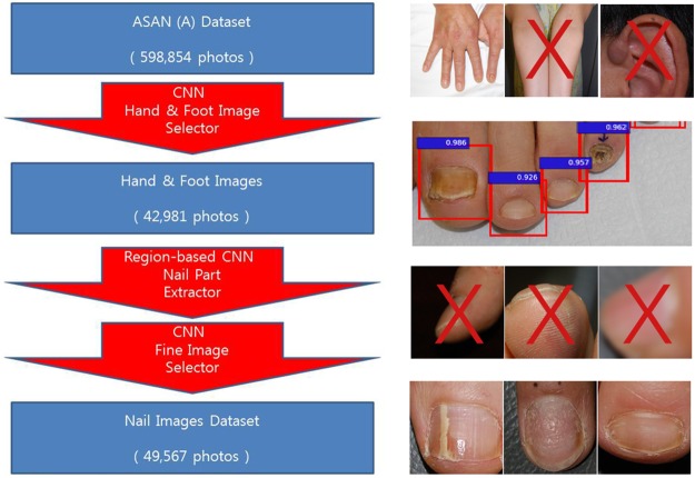
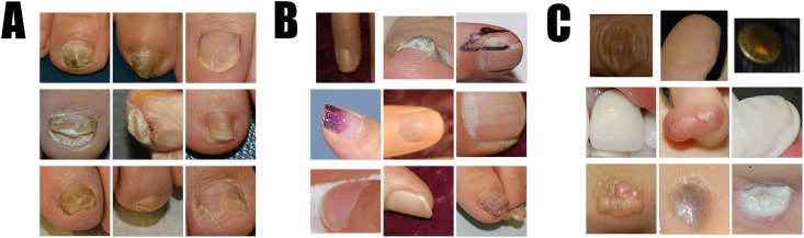
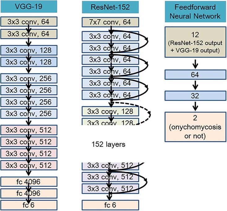
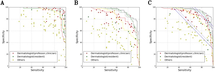
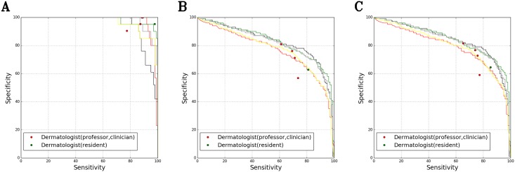
Although there have been reports of the successful diagnosis of skin disorders using deep learning, unrealistically large clinical image datasets are required for artificial intelligence (AI) training. We created datasets of standardized nail images using a region-based convolutional neural network (R-CNN) trained to distinguish the nail from the background. We used R-CNN to generate training datasets of 49,567 images, which we then used to fine-tune the ResNet-152 and VGG-19 models. The validation datasets comprised 100 and 194 images from Inje University (B1 and B2 datasets, respectively), 125 images from Hallym University (C dataset), and 939 images from Seoul National University (D dataset). The AI (ensemble model; ResNet-152 + VGG-19 + feedforward neural networks) results showed test sensitivity/specificity/ area under the curve values of (96.0 / 94.7 / 0.98), (82.7 / 96.7 / 0.95), (92.3 / 79.3 / 0.93), (87.7 / 69.3 / 0.82) for the B1, B2, C, and D datasets. With a combination of the B1 and C datasets, the AI Youden index was significantly (p = 0.01) higher than that of 42 dermatologists doing the same assessment manually. For B1+C and B2+ D dataset combinations, almost none of the dermatologists performed as well as the AI. By training with a dataset comprising 49,567 images, we achieved a diagnostic accuracy for onychomycosis using deep learning that was superior to that of most of the dermatologists who participated in this study.
Overview
The claims and statistics of this paper haven’t been checked yet.
Run the check — free, no account needed. It reads the open-access text and checks the sources it cites.
Integrity signals
Registry
Cites
Statistics
Figures
Verification
Claims the paper makes
The claims and statistics of this paper haven’t been checked yet.
Run the check — free, no account needed. It reads the open-access text and checks the sources it cites.
Claims other papers make about this one
Each is checked against this paper’s own text: does it say what the citing paper says it does?
In the citing paper
“Several algorithms incorporating artificial intelligence (AI) have been developed to help interpret both clinical11,12,13,14,15 and dermoscopic16,17,18,19,20,21,22,23 images for a variety of skin conditions”
Self-check
Percentages, sums and reported differences re-computed from the paper’s own numbers.
The self-check needs the paper’s full text.
Citations
Cites
Deep Residual Learning for Image Recognition
He, K., Zhang, X., Ren, S., & Sun, J. (2016) · 2016 IEEE Conference on Computer Vision and Pattern Recognition (CVPR) · doi:10.1109/cvpr.2016.90 · Cited by 235,474
Very Deep Convolutional Networks for Large-Scale Image Recognition
Simonyan, K., & Zisserman, A. (2014) · arXiv · doi:10.48550/arxiv.1409.1556 · Cited by 114,148
Faster R-CNN: Towards Real-Time Object Detection with Region Proposal Networks
Ren, S., He, K., Girshick, R., & Sun, J. (2017) · IEEE Transactions on Pattern Analysis and Machine Intelligence · doi:10.1109/tpami.2016.2577031 · Cited by 73,472
Faster R-CNN: Towards Real-Time Object Detection with Region Proposal Networks
Ren, S., He, K., Girshick, R., & Sun, J. (2015) · arXiv · doi:10.48550/arxiv.1506.01497 · Cited by 70,812
ImageNet Large Scale Visual Recognition Challenge
Russakovsky, O., Deng, J., Su, H., Krause, J., Satheesh, S., Ma, S., Huang, Z., Karpathy, A., Khosla, A., Bernstein, M., et al. (2015) · International Journal of Computer Vision · doi:10.1007/s11263-015-0816-y · Cited by 39,380
Rich Feature Hierarchies for Accurate Object Detection and Semantic Segmentation
Girshick, R., Donahue, J., Darrell, T., & Malik, J. (2014) · 2014 IEEE Conference on Computer Vision and Pattern Recognition · doi:10.1109/cvpr.2014.81 · Cited by 32,684
Delving Deep into Rectifiers: Surpassing Human-Level Performance on ImageNet Classification
He, K., Zhang, X., Ren, S., & Sun, J. (2015) · 2015 IEEE International Conference on Computer Vision (ICCV) · doi:10.1109/iccv.2015.123 · Cited by 19,741
Dermatologist-level classification of skin cancer with deep neural networks
Esteva, A., Kuprel, B., Novoa, R. A., Ko, J., Swetter, S. M., Blau, H. M., & Thrun, S. (2017) · Nature · doi:10.1038/nature21056 · Cited by 14,362
Caffe: Convolutional Architecture for Fast Feature Embedding
Jia, Y., Shelhamer, E., Donahue, J., Karayev, S., Long, J., Girshick, R., Guadarrama, S., & Darrell, T. (2014) · Proceedings of the 22nd ACM international conference on Multimedia · doi:10.1145/2647868.2654889 · Cited by 11,077
Gulshan, V., Peng, L., Coram, M., Stumpe, M. C., Wu, D., Narayanaswamy, A., Venugopalan, S., Widner, K., Madams, T., Cuadros, J., et al. (2016) · JAMA · doi:10.1001/jama.2016.17216 · Cited by 7,697
Research works printed but not linked: 1
- He K, Zhang X, Ren S, Sun J, editors. Deep residual learning for image recognition. Proceedings of the IEEE conference on computer vision and pattern recognition; 2016. · DOI · Paper page
Cited by
Sorted by citations
Tschandl, P., Rosendahl, C., & Kittler, H. (2018) · Scientific Data · doi:10.1038/sdata.2018.161 · Cited by 3,628
Liu, X., Faes, L., Kale, A. U., Wagner, S. K., Fu, D. J., Bruynseels, A., Mahendiran, T., Moraes, G., Shamdas, M., Kern, C., et al. (2019) · The Lancet Digital Health · doi:10.1016/s2589-7500(19)30123-2 · Cited by 1,883
Nagendran, M., Chen, Y., Lovejoy, C. A., Gordon, A. C., Komorowski, M., Harvey, H., Topol, E. J., Ioannidis, J. P. A., Collins, G. S., & Maruthappu, M. (2020) · BMJ · doi:10.1136/bmj.m689 · Cited by 1,152
Transfer learning for medical image classification: a literature review
Kim, H. E., Cosa‐Linan, A., Santhanam, N., Jannesari, M., Maros, M. E., & Ganslandt, T. (2022) · BMC Medical Imaging · doi:10.1186/s12880-022-00793-7 · Cited by 1,117
A deep learning system for differential diagnosis of skin diseases
Liu, Y., Jain, A., Eng, C., Way, D. H., Lee, K., Bui, P., Kanada, K., de Oliveira Marinho, G., Gallegos, J., Gabriele, S., et al. (2020) · Nature Medicine · doi:10.1038/s41591-020-0842-3 · Cited by 853
Han, S. S., Kim, M. S., Lim, W., Park, G., Park, I., & Chang, S. E. (2018) · Journal of Investigative Dermatology · doi:10.1016/j.jid.2018.01.028 · Cited by 740
Artificial intelligence in retina
Schmidt-Erfurth, U., Sadeghipour, A., Gerendas, B. S., Waldstein, S. M., & Bogunović, H. (2018) · Progress in Retinal and Eye Research · doi:10.1016/j.preteyeres.2018.07.004 · Cited by 779
Goyal, M., Knackstedt, T., Yan, S., & Hassanpour, S. (2020) · Computers in Biology and Medicine · doi:10.1016/j.compbiomed.2020.104065 · Cited by 476
Daneshjou, R., Smith, M. P., Sun, M. D., Rotemberg, V., & Zou, J. (2021) · JAMA Dermatology · doi:10.1001/jamadermatol.2021.3129 · Cited by 403
Artificial Intelligence Versus Clinicians in Disease Diagnosis: Systematic Review
Shen, J., Zhang, C. J. P., Jiang, B., Chen, J., Song, J., Liu, Z., He, Z., Wong, S. Y., Fang, P.-H., & Ming, W.-K. (2019) · JMIR Medical Informatics · doi:10.2196/10010 · Cited by 373
What is AI? Applications of artificial intelligence to dermatology
Du‐Harpur, X., Watt, F. M., Luscombe, N. M., & Lynch, M. D. (2020) · British Journal of Dermatology · doi:10.1111/bjd.18880 · Cited by 355
Machine Learning in Dermatology: Current Applications, Opportunities, and Limitations
Chan, S. Y., Reddy, V. D., Myers, B. A., Thibodeaux, Q. G., Brownstone, N. D., & Liao, W. J. (2020) · Dermatology and Therapy · doi:10.1007/s13555-020-00372-0 · Cited by 285
Artificial Intelligence in Dermatology Image Analysis: Current Developments and Future Trends
Li, Z., Koban, K. C., Schenck, T. L., Giunta, R. E., Li, Q., & Sun, Y. (2022) · Journal of Clinical Medicine · doi:10.3390/jcm11226826 · Cited by 291
Artificial Intelligence in Dermatology: A Primer
Young, A. T., Xiong, M., Pfau, J., Keiser, M. J., & Wei, M. L. (2020) · Journal of Investigative Dermatology · doi:10.1016/j.jid.2020.02.026 · Cited by 249
Sollini, M., Antunovic, L., Chiti, A., & Kirienko, M. (2019) · European Journal of Nuclear Medicine and Molecular Imaging · doi:10.1007/s00259-019-04372-x · Cited by 227
Han, S. S., Park, I., Eun Chang, S., Lim, W., Kim, M. S., Park, G. H., Chae, J. B., Huh, C. H., & Na, J.-I. (2020) · Journal of Investigative Dermatology · doi:10.1016/j.jid.2020.01.019 · Cited by 248
Hirani, R., Noruzi, K. A., Khuram, H., Hussaini, A. S., Aifuwa, E., Ely, K., Lewis, J. M., Gabr, A. E., Smiley, A., Tiwari, R. K., et al. (2024) · Life · doi:10.3390/life14050557 · Cited by 280
Artificial Intelligence in Skin Cancer Diagnostics: The Patients' Perspective
Jutzi, T. B., Krieghoff-Henning, E. I., Holland-Letz, T., Utikal, J. S., Hauschild, A., Schadendorf, D., Sondermann, W., Fröhling, S., Hekler, A., Schmitt, M., et al. (2020) · Frontiers in Medicine · doi:10.3389/fmed.2020.00233 · Cited by 196
Artificial Intelligence in Dermatology—Where We Are and the Way to the Future: A Review
Hogarty, D. T., Su, J. C., Phan, K. H., Attia, M., Hossny, M., Nahavandi, S., Lenane, P., Moloney, F. J., & Yazdabadi, A. (2019) · American Journal of Clinical Dermatology · doi:10.1007/s40257-019-00462-6 · Cited by 195
Algorithmic Management: Bright and Dark Sides, Practical Implications, and Research Opportunities
Benlian, A., Wiener, M., Cram, W. A., Krasnova, H., Maedche, A., Möhlmann, M., Recker, J., & Remus, U. (2022) · Business & Information Systems Engineering · doi:10.1007/s12599-022-00764-w · Cited by 180
Skin disease diagnosis with deep learning: a review
Li, H., Pan, Y., Zhao, J., & Zhang, L. (2021) · Neurocomputing · doi:10.1016/j.neucom.2021.08.096 · Cited by 178
Pre-trained multimodal large language model enhances dermatological diagnosis using SkinGPT-4
Zhou, J., He, X., Sun, L., Xu, J., Chen, X., Chu, Y., Zhou, L., Liao, X., Zhang, B., Afvari, S., et al. (2024) · Nature Communications · doi:10.1038/s41467-024-50043-3 · Cited by 189
Jain, A., Way, D., Gupta, V., Gao, Y., de Oliveira Marinho, G., Hartford, J., Sayres, R., Kanada, K., Eng, C., Nagpal, K., et al. (2021) · JAMA Network Open · doi:10.1001/jamanetworkopen.2021.7249 · Cited by 164
Machine Learning and Its Application in Skin Cancer
Das, K., Cockerell, C. J., Patil, A. D., Pietkiewicz, P., Giulini, M., Grabbe, S., & Goldust, M. (2021) · International Journal of Environmental Research and Public Health · doi:10.3390/ijerph182413409 · Cited by 175
Keratinocytic Skin Cancer Detection on the Face Using Region-Based Convolutional Neural Network
Han, S. S., Moon, I. J., Lim, W., Suh, I. S., Lee, S. Y., Na, J., Kim, S. H., & Chang, S. E. (2019) · JAMA Dermatology · doi:10.1001/jamadermatol.2019.3807 · Cited by 150
Deep learning in dermatology: a systematic review of current approaches, outcomes, and limitations
Jeong, H. K., Park, C., Henao, R., & Kheterpal, M. (2023) · JID Innovations · doi:10.1016/j.xjidi.2022.100150 · Cited by 134
Use of artificial intelligence in dermatology
De, A., Sarda, A., Gupta, S., & DAS, S. K. (2020) · Indian Journal of Dermatology · doi:10.4103/ijd.ijd_418_20 · Cited by 119
Malik, H., Farooq, M. S., Khelifi, A., Abid, A., Nasir Qureshi, J., & Hussain, M. (2020) · IEEE Access · doi:10.1109/access.2020.3004766 · Cited by 104
Deep learning in electron microscopy
Ede, J. M. (2020) · Machine Learning Science and Technology · doi:10.1088/2632-2153/abd614 · Cited by 118
Systematic review of machine learning for diagnosis and prognosis in dermatology
Thomsen, K. G., Iversen, L., Titlestad, T. L., & Winther, O. (2019) · Journal of Dermatological Treatment · doi:10.1080/09546634.2019.1682500 · Cited by 108
Luca, A. R., Ursuleanu, T. F., Gheorghe, L., Grigorovici, R., Iancu, S., Hlusneac, M., & Grigorovici, A. (2022) · Informatics in Medicine Unlocked · doi:10.1016/j.imu.2022.100911 · Cited by 110
Ultrasound image analysis using deep learning algorithm for the diagnosis of thyroid nodules
Song, J., Chai, Y. J., Masuoka, H., Park, S., Kim, S., Choi, J. Y., Kong, H., Lee, K. E., Lee, J., Kwak, N., et al. (2019) · Medicine · doi:10.1097/md.0000000000015133 · Cited by 93
Wang, X., Xuan, H., Evers, B., Shrestha, S. K., Pless, R. B., & Poland, J. (2019) · GigaScience · doi:10.1093/gigascience/giz120 · Cited by 97
Updated Perspectives on the Diagnosis and Management of Onychomycosis
Falotico, J. M., & Lipner, S. R. (2022) · Clinical Cosmetic and Investigational Dermatology · doi:10.2147/ccid.s362635 · Cited by 97
Han, S. S., Kim, Y. J., Moon, I. J., Jung, J. M., Lee, M. Y., Lee, W. J., Won, C. H., Lee, M. W., Kim, S. H., Navarrete‐Dechent, C., et al. (2022) · Journal of Investigative Dermatology · doi:10.1016/j.jid.2022.02.003 · Cited by 84
Basic of machine learning and deep learning in imaging for medical physicists
Manco, L., Maffei, N., Strolin, S., Vichi, S., Bottazzi, L., & Strigari, L. (2021) · Physica Medica · doi:10.1016/j.ejmp.2021.03.026 · Cited by 75
Early automated detection system for skin cancer diagnosis using artificial intelligent techniques
Mahmoud, N. M., & Soliman, A. M. (2024) · Scientific Reports · doi:10.1038/s41598-024-59783-0 · Cited by 72
Deep Learning for Diagnostic Binary Classification of Multiple-Lesion Skin Diseases
Thomsen, K. G., Christensen, A. L., Iversen, L., Lomholt, H. B., & Winther, O. (2020) · Frontiers in Medicine · doi:10.3389/fmed.2020.574329 · Cited by 72
Deep learning for dermatologists: Part II. Current applications
Puri, P., Comfere, N. I., Drage, L. A., Shamim, H., Bezalel, S. A., Pittelkow, M. R., Davis, M. D. P., Wang, M., Mangold, A. R., Tollefson, M. M., et al. (2020) · Journal of the American Academy of Dermatology · doi:10.1016/j.jaad.2020.05.053 · Cited by 70
Machine learning applications in the evaluation and management of psoriasis: a systematic review
Yu, K., Syed, M. N., Bernardis, E., & Gelfand, J. M. (2020) · Journal of Psoriasis and Psoriatic Arthritis · doi:10.1177/2475530320950267 · Cited by 65
Improving Skin Color Diversity in Cancer Detection: Deep Learning Approach
Rezk, E., Eltorki, M., & El-Dakhakhni, W. W. (2022) · JMIR Dermatology · doi:10.2196/39143 · Cited by 75
Yoo, T. K., Choi, J. Y., Kim, H. K., Ryu, I. H., & Kim, J. K. (2021) · Computer Methods and Programs in Biomedicine · doi:10.1016/j.cmpb.2021.106086 · Cited by 64
Cho, S. I., Sun, S., Mun, J., Kim, C., Kim, S. Y., Cho, S., Youn, S. W., Kim, H., & Chung, J. H. (2019) · British Journal of Dermatology · doi:10.1111/bjd.18459 · Cited by 63
Diagnosis of Onychomycosis: From Conventional Techniques and Dermoscopy to Artificial Intelligence
Lim, S. S., Ohn, J., & Mun, J. (2021) · Frontiers in Medicine · doi:10.3389/fmed.2021.637216 · Cited by 59
Multi-view Convolutional Recurrent Neural Networks for Lung Cancer Nodule Identification
Abid, M. M. N., Zia, T., Ghafoor, M., & Windridge, D. (2021) · Neurocomputing · doi:10.1016/j.neucom.2020.06.144 · Cited by 57
Koo, T., Kim, M.-H., & Jue, M. (2021) · PLoS ONE · doi:10.1371/journal.pone.0256290 · Cited by 55
A survey of deep learning models in medical therapeutic areas
Nogales, A., García-Tejedor, Á. J., Martín, D. M., Vara, J. S., & Rodríguez, C. A. (2021) · Artificial Intelligence in Medicine · doi:10.1016/j.artmed.2021.102020 · Cited by 59
Background selection schema on deep learning-based classification of dermatological disease
Zhou, J., Wu, Z., Jiang, Z., Huang, K., Guo, K., & Zhao, S. (2022) · Computers in Biology and Medicine · doi:10.1016/j.compbiomed.2022.105966 · Cited by 53
Yang, Y., Ge, Y., Guo, L., Wu, Q., Peng, L., Zhang, E., Xie, J., Li, Y., & Lin, T. (2020) · Skin Research and Technology · doi:10.1111/srt.12911 · Cited by 53
Artificial intelligence in dermatology and healthcare: An overview
Pai, V. V., & Pai, R. B. (2021) · Indian Journal of Dermatology Venereology and Leprology · doi:10.25259/ijdvl_518_19 · Cited by 57
Skin Disease Classification System Based on Machine Learning Technique: A Survey
Mohammed, S. S., & Al-Tuwaijari, J. M. (2021) · IOP Conference Series Materials Science and Engineering · doi:10.1088/1757-899x/1076/1/012045 · Cited by 59
Yanagisawa, Y., Shido, K., Kojima, K., & Yamasaki, K. (2023) · Journal of Dermatological Science · doi:10.1016/j.jdermsci.2023.01.005 · Cited by 58
Decroos, F., Springenberg, S., Lang, T., Päpper, M., Zapf, A., Metze, D., Steinkraus, V, & Böer-Auer, A. (2021) · Acta Dermato Venereologica · doi:10.2340/00015555-3893 · Cited by 51
Ground truthing from multi-rater labeling with three-way decision and possibility theory
Campagner, A., Ciucci, D., Svensson, C., Figge, M. T., & Cabitza, F. (2020) · Information Sciences · doi:10.1016/j.ins.2020.09.049 · Cited by 53
Artificial Intelligence in Emergency Medicine: Benefits, Risks, and Recommendations
Vearrier, L., Derse, A. R., Basford, J. B., Larkin, G. L., & Moskop, J. C. (2022) · The Journal of Emergency Medicine · doi:10.1016/j.jemermed.2022.01.001 · Cited by 49
Fatima, F., Imran, M., Ullah, A., Arif, M., & Noor, R. (2022) · Computers in Biology and Medicine · doi:10.1016/j.compbiomed.2022.105424 · Cited by 50
Kim, Y. J., Han, S. S., Yang, H. J., & Chang, S. E. (2020) · PLoS ONE · doi:10.1371/journal.pone.0234334 · Cited by 47
Hossain, S. I., de Hervé, J. D. G., Hassan, M. S., Martineau, D., Petrosyan, E., Corbin, V., Beytout, J., Lebert, I., Durand, J., Carravieri, I., et al. (2022) · Computer Methods and Programs in Biomedicine · doi:10.1016/j.cmpb.2022.106624 · Cited by 52
Review of Machine Learning in Predicting Dermatological Outcomes
Du, A., Emam, S., & Gniadecki, R. (2020) · Frontiers in Medicine · doi:10.3389/fmed.2020.00266 · Cited by 44
Xu, H., Tang, T., Zhang, B., & Liu, Y. (2022) · Progress in Nuclear Energy · doi:10.1016/j.pnucene.2022.104191 · Cited by 44
Research Techniques Made Simple: Deep Learning for the Classification of Dermatological Images
Cullell-Dalmau, M., Otero-Viñas, M., & Manzo, C. (2020) · Journal of Investigative Dermatology · doi:10.1016/j.jid.2019.12.029 · Cited by 42
Diagnosing Onychomycosis: What’s New?
Gupta, A. K., Hall, D. C., Cooper, E. A., & Ghannoum, M. A. (2022) · Journal of Fungi · doi:10.3390/jof8050464 · Cited by 45
Artificial intelligence in dermatology: A practical introduction to a paradigm shift
Eapen, B. R. (2020) · Indian Dermatology Online Journal · doi:10.4103/idoj.idoj_388_20 · Cited by 45
Improving electron micrograph signal-to-noise with an atrous convolutional encoder-decoder
Ede, J. M., & Beanland, R. (2019) · Ultramicroscopy · doi:10.1016/j.ultramic.2019.03.017 · Cited by 38
Toward automated severe pharyngitis detection with smartphone camera using deep learning networks
Yoo, T. K., Choi, J. Y., Jang, Y., Oh, E., & Ryu, I. H. (2020) · Computers in Biology and Medicine · doi:10.1016/j.compbiomed.2020.103980 · Cited by 43
Artificial Intelligence in Diagnostic Dermatology: Challenges and the Way Forward
Sengupta, D. (2023) · Indian Dermatology Online Journal · doi:10.4103/idoj.idoj_462_23 · Cited by 40
Application of artificial intelligence in modern medicine
Xu, N., Yang, D., Arikawa, K., & Bai, C. (2023) · Clinical eHealth · doi:10.1016/j.ceh.2023.09.001 · Cited by 45
Automated severity scoring of atopic dermatitis patients by a deep neural network
Bang, C. H., Yoon, J. W., Ryu, J. Y., Chun, J. H., Han, J. H., Lee, Y. B., Lee, J. Y., Park, Y. M., Lee, S. J., & Lee, J. H. (2021) · Scientific Reports · doi:10.1038/s41598-021-85489-8 · Cited by 38
Clinical spectrum of cutaneous melanoma morphology
Klebanov, N., Gunasekera, N. S., Lin, W. M., Hawryluk, E. B., Miller, D. M., Reddy, B. Y., Christman, M. P., Beaulieu, D., Rajadurai, S., Duncan, L. M., et al. (2018) · Journal of the American Academy of Dermatology · doi:10.1016/j.jaad.2018.08.028 · Cited by 35
Deep learning‐based diagnosis models for onychomycosis in dermoscopy
Zhu, X., Zheng, B., Cai, W., Zhang, J., Lu, S., Li, X., Xi, L., & Kong, Y. (2022) · Mycoses · doi:10.1111/myc.13427 · Cited by 35
Sinha, M., Paul, S., Ghosh, M., Mohanty, S. N., & Pattanayak, R. M. (2024) · Scientific Reports · doi:10.1038/s41598-024-58438-4 · Cited by 34
Skin Type Diversity in Skin Lesion Datasets: A Review
Alipour, N., Burke, T., & Courtney, J. (2024) · Current Dermatology Reports · doi:10.1007/s13671-024-00440-0 · Cited by 46
Lesion-Based Convolutional Neural Network in Diagnosis of Early Gastric Cancer
Yoon, H. J., & Kim, J. (2020) · Clinical Endoscopy · doi:10.5946/ce.2020.046 · Cited by 32
Zaar, O., Larson, A., Polesie, S., Saleh, K., Tarstedt, M., Olives, A., Suárez, A. L., Gillstedt, M., & Neittaanmäki, N. (2020) · Acta Dermato Venereologica · doi:10.2340/00015555-3624 · Cited by 30
The biomedical applications of artificial intelligence: an overview of decades of research
Naskar, S., Sharma, S., Kuotsu, K., Halder, S., Pal, G., Saha, S., Mondal, S., Biswas, U. K., Jana, M., & Bhattacharjee, S. (2025) · Journal of drug targeting · doi:10.1080/1061186x.2024.2448711 · Cited by 35
The Role of DICOM in Artificial Intelligence for Skin Disease
Caffery, L. J., Rotemberg, V. M., Weber, J., Soyer, H. P., Malvehy, J., & Clunie, D. (2021) · Frontiers in Medicine · doi:10.3389/fmed.2020.619787 · Cited by 32
Comparing Artificial Intelligence Platforms for Histopathologic Cancer Diagnosis.
Borkowski, A., Wilson, C. P., Borkowski, S. A., Thomas, L. B., Deland, L. A., Grewe, S., & Mastorides, S. (2019) · PubMed Central · Cited by 30
Wang, S., Zhang, X.-Y., Liu, J., Tao, C., Zhu, C., Shu, C., Xu, T., & Jin, H. (2020) · Chinese Medical Journal · doi:10.1097/cm9.0000000000001023 · Cited by 31
Sendín‐Martín, M., Lara-Caro, M., Harris, U., Moronta, M., Rossi, A., Lee, E. H., Chen, C. J., Nehal, K. S., Conejo‐Mir, J., Rodríguez, J., et al. (2021) · Journal of Investigative Dermatology · doi:10.1016/j.jid.2021.09.029 · Cited by 30
Gupta, A. K., Wang, T., Cooper, E. A., Summerbell, R. C., Piguet, V., Tosti, A., & Piraccini, B. M. (2023) · Journal of the European Academy of Dermatology and Venereology · doi:10.1111/jdv.19644 · Cited by 29
Web-based study on Chinese dermatologists’ attitudes towards artificial intelligence
Shen, C., Li, C., Xu, F., Wang, Z., Shen, X., Gao, J., Ko, R., Jing, Y., Tang, X., Yu, R., et al. (2020) · Annals of Translational Medicine · doi:10.21037/atm.2019.12.102 · Cited by 31
Taylor, M., Liu, X., Denniston, A. K., Esteva, A., Ko, J., Daneshjou, R., & Chan, A. (2021) · Journal of Investigative Dermatology · doi:10.1016/j.jid.2021.02.744 · Cited by 28
Artificial intelligence in psoriasis: Where we are and where we are going
Liu, Z., Wang, X., Ma, Y., Lin, Y., & Wang, G. (2023) · Experimental Dermatology · doi:10.1111/exd.14938 · Cited by 33
PENERAPAN METODE RESIDUAL NETWORK (RESNET) DALAM KLASIFIKASI PENYAKIT PADA DAUN GANDUM
Ridhovan, A., & Suharso, A. (2022) · JIPI (Jurnal Ilmiah Penelitian dan Pembelajaran Informatika) · doi:10.29100/jipi.v7i1.2410 · Cited by 26
Kim, J., Oh, I., Lee, Y. N., Lee, J. H., Lee, Y. I., Kim, J., & Lee, J. H. (2023) · Scientific Reports · doi:10.1038/s41598-023-40395-z · Cited by 26
Zhu, A.-Q., Wang, Q., Shi, Y.-L., Ren, W.-W., Cao, X., Ren, T.-T., Wang, J., Zhang, Y.-Q., Sun, Y.-K., Chen, X.-W., et al. (2024) · EClinicalMedicine · doi:10.1016/j.eclinm.2023.102391 · Cited by 25
Singla, N., Kundu, R., & Dey, P. (2023) · Cytopathology · doi:10.1111/cyt.13336 · Cited by 25
Yılmaz, A., Göktay, F., Varol, R., Gençoğlan, G., & Üvet, H. (2022) · Mycoses · doi:10.1111/myc.13498 · Cited by 34
Interpretation of the Outputs of a Deep Learning Model Trained with a Skin Cancer Dataset
Han, S. S., Lim, W., Kim, M. S., Park, I., Park, G., & Chang, S. E. (2018) · Journal of Investigative Dermatology · doi:10.1016/j.jid.2018.05.014 · Cited by 24
Alzamili, A. H., & Ruhaiyem, N. I. R. (2025) · Journal of Intelligent Systems · doi:10.1515/jisys-2024-0381 · Cited by 24
Out-of-Distribution Detection for Skin Lesion Images with Deep Isolation Forest
Li, X., Lu, Y., Desrosiers, C., & Liu, X. (2020) · Lecture notes in computer science · doi:10.1007/978-3-030-59861-7_10 · Cited by 23
Bozkurt, A., Köse, K., Coll‐Font, J., Alessi‐Fox, C., Brooks, D. H., Dy, J., & Rajadhyaksha, M. M. (2021) · Scientific Reports · doi:10.1038/s41598-021-90328-x · Cited by 28
Graham, S. S. (2022) · doi:10.1093/oso/9780197644461.001.0001 · Cited by 25
Artificial Intelligence in Diagnosis and Management of Nail Disorders: A Narrative Review
Gaurav, V., Grover, C., Tyagi, M., & Saurabh, S. (2024) · Indian Dermatology Online Journal · doi:10.4103/idoj.idoj_460_24 · Cited by 28
Deep Neural Forest for Out-of-Distribution Detection of Skin Lesion Images
Li, X., Desrosiers, C., & Liu, X. (2022) · IEEE Journal of Biomedical and Health Informatics · doi:10.1109/jbhi.2022.3171582 · Cited by 23
Using DeepLabCut as a Real-Time and Markerless Tool for Cardiac Physiology Assessment in Zebrafish
Suryanto, M. E., Saputra, F., Kurnia, K. A., Vasquez, R. D., Roldan, M. J. M., Chen, K. H. ‐., Huang, J., & Hsiao, C. (2022) · Biology · doi:10.3390/biology11081243 · Cited by 21
Pre-trained Multimodal Large Language Model Enhances Dermatological Diagnosis using SkinGPT-4Preprint
Zhou, J., He, X., Sun, L., Xu, J., Chen, X., Chu, Y., Zhou, L., Liao, X., Zhang, B., & Gao, X. (2023) · medRxiv · doi:10.1101/2023.06.10.23291127 · Cited by 20
Willem, T., Krammer, S., Böhm, A. ‐., French, L. E., Hartmann, D., Lasser, T., & Buyx, A. (2022) · Journal of the European Academy of Dermatology and Venereology · doi:10.1111/jdv.18192 · Cited by 20
Tao, Y., Hu, H. M., Li, J., Li, M., Zheng, Q., Zhang, G., & Ni, M. (2022) · Arthroplasty · doi:10.1186/s42836-022-00145-4 · Cited by 19
New Frontiers in Explainable AI: Understanding the GI to Interpret the GO
Cabitza, F., Campagner, A., & Ciucci, D. (2019) · Lecture notes in computer science · doi:10.1007/978-3-030-29726-8_3 · Cited by 19
Metadata
- Authors
- Han, S. S., Park, G. H., Lim, W., Kim, M. S., Na, J. I., Park, I., & Chang, S. E.
- Affiliations
- Hallym University (KR)
- Inje University (KR)
- Seoul National University (KR)
- Chonnam National University (KR)
- Asan Medical Center (KR)
- Corresponding author
- Chang, Sung Eun
- ORCID
- Venue
- PLoS ONE
- Publisher
- Public Library of Science (PLoS)
- Published
- 2018-01-19
- Volume
- 13
- Issue
- 1
- Pages
- e0191493
- ISSN
- 1932-6203 (ISSN-L)
- PMC
- PMC5774804
- OpenAlex
- W2785200097
- Cited by
- 305
- References
- 18
- Citations by year
- 2026: 24 · 2025: 32 · 2024: 40 · 2023: 33 · 2022: 58 · 2021: 37 · 2020: 49 · 2019: 24 · 2018: 7
- FWCI
- 22.75
- Type
- journal article
- Language
- en
- Open access
- gold
- doi.org
- Licence
- Locations
- PLoS ONE (publishedVersion, cc-by)
- Europe PMC (PubMed Central) (publishedVersion, cc-by)
- PubMed Central (publishedVersion, cc-by)
- PubMed (publishedVersion)
- DOAJ (DOAJ: Directory of Open Access Journals) (submittedVersion, cc-by-sa)
- Topics
- Health Sciences › Medicine › Dermatology › Nail Diseases and Treatments
- Physical Sciences › Computer Science › Computer Vision and Pattern Recognition › Advanced Neural Network Applications
- Health Sciences › Medicine › Health Informatics › Artificial Intelligence in Healthcare and Education
- Keywords
- onychomycosis, automated diagnosis, dermatologist comparison, R-CNN, ResNet-152, VGG19, ensemble learning, dermatological image analysis
- MeSH terms
- Machine Learning, Dermatologists, Adult, Aged, Algorithms, Artificial Intelligence, Diagnosis, Computer-Assisted, Female, Foot Dermatoses, Hand Dermatoses, Humans, Image Interpretation, Computer-Assisted, Male, Middle Aged, Onychomycosis, Databases, Factual, Neural Networks, Computer, Area Under Curve, Young Adult
About these checks
Registry, retraction, Cites and statistics results are computed, not judged, with one exception: a printed reference that no index lists is read once by a model before it counts as a citation error. Claims are extracted by a model and kept only when their quote appears verbatim in the text; each cited source is then checked, with a model’s help, for whether it says what the claim relies on. Those verdicts can be wrong — read the claim’s quote and the cited source before relying on one.
Statistics and arithmetic are recomputed, not judged.
Confidence bands are shown only for fields calibrated on our annotated test set. None is calibrated yet, so no band is shown.
What counts as a claim ›How we measure confidence ›How we check this ›
Are you an author, or do you see a problem with this page? Request a review
