

The DOI matches its Crossref record; 47 of the 47 references it cites were found in a registry or an index.
In vitro immunotherapy potency assays using real-time cell analysis
Open accessAbstract
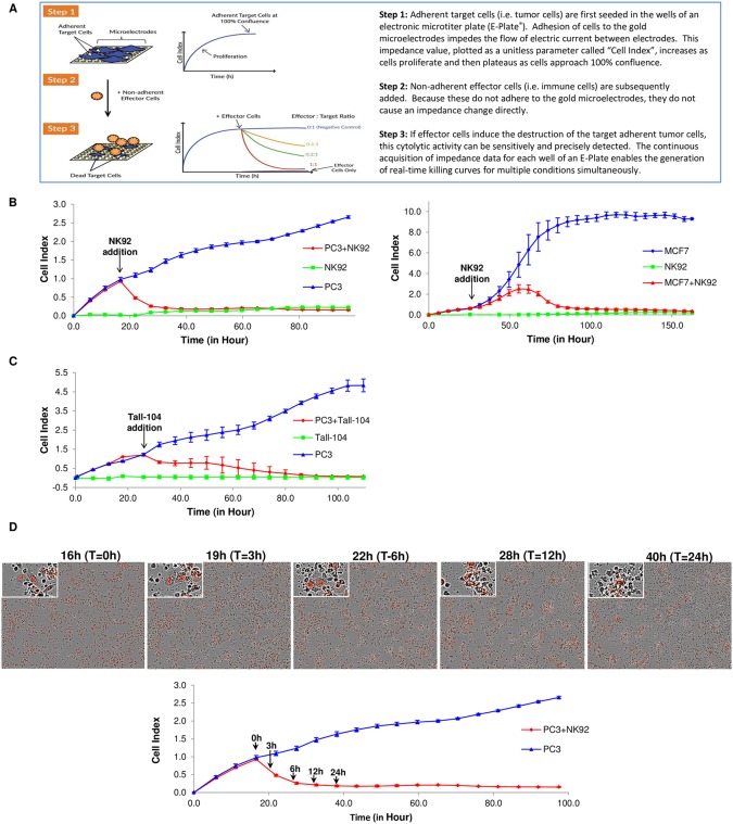
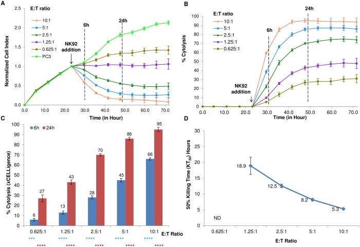
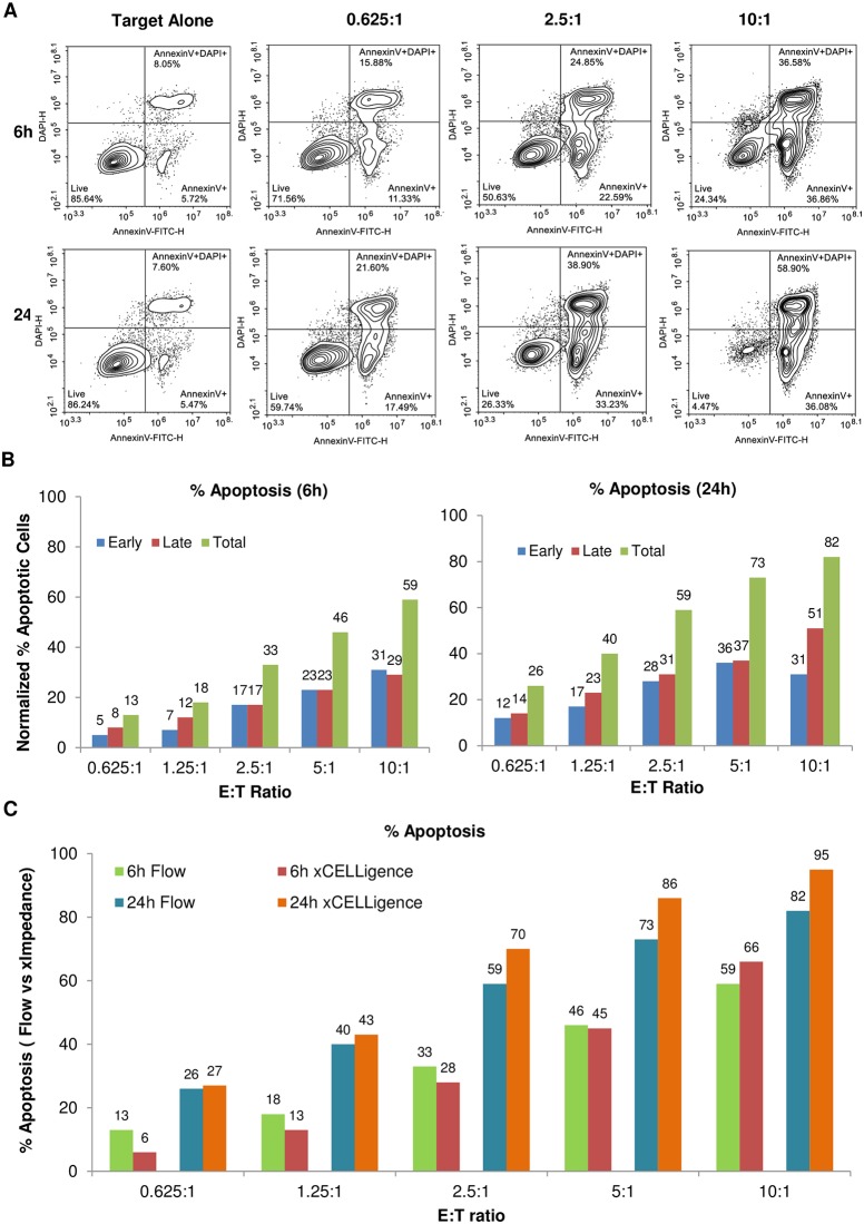
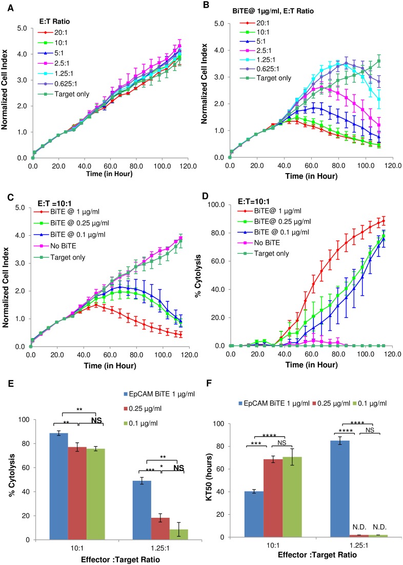
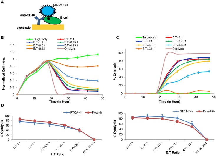
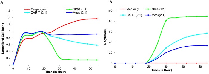
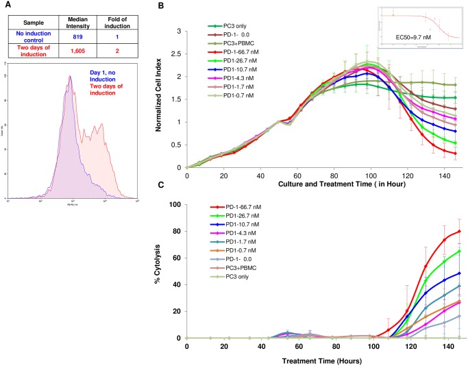
A growing understanding of the molecular interactions between immune effector cells and target tumor cells, coupled with refined gene therapy approaches, are giving rise to novel cancer immunotherapeutics with remarkable efficacy in the clinic against both solid and liquid tumors. While immunotherapy holds tremendous promise for treatment of certain cancers, significant challenges remain in the clinical translation to many other types of cancers and also in minimizing adverse effects. Therefore, there is an urgent need for functional potency assays, in vitro and in vivo, that could model the complex interaction of immune cells with tumor cells and can be used to rapidly test the efficacy of different immunotherapy approaches, whether it is small molecule, biologics, cell therapies or combinations thereof. Herein we report the development of an xCELLigence real-time cytolytic in vitro potency assay that uses cellular impedance to continuously monitor the viability of target tumor cells while they are being subjected to different types of treatments. Specialized microtiter plates containing integrated gold microelectrodes enable the number, size, and surface attachment strength of adherent target tumor cells to be selectively monitored within a heterogeneous mixture that includes effector cells, antibodies, small molecules, etc. Through surface-tethering approach, the killing of liquid cancers can also be monitored. Using NK92 effector cells as example, results from RTCA potency assay are very well correlated with end point data from image-based assays as well as flow cytometry. Several effector cells, i.e., PBMC, NK, CAR-T were tested and validated as well as biological molecules such as Bi-specific T cell Engagers (BiTEs) targeting the EpCAM protein expressed on tumor cells and blocking antibodies against the immune checkpoint inhibitor PD-1. Using the specifically designed xCELLigence immunotherapy software, quantitative parameters such as KT50 (the amount of time it takes to kill 50% of the target tumor cells) and % cytolysis are calculated and used for comparing the relative efficacy of different reagents. In summary, our results demonstrate the xCELLigence platform to be well suited for potency assays, providing quantitative assessment with high reproducibility and a greatly simplified work flow.
Overview
The claims and statistics of this paper haven’t been checked yet.
Run the check — free, no account needed. It reads the open-access text and checks the sources it cites.
Integrity signals
Registry
Cites
Statistics
Figures
Verification
Claims the paper makes
The claims and statistics of this paper haven’t been checked yet.
Run the check — free, no account needed. It reads the open-access text and checks the sources it cites.
Claims other papers make about this one
Each is checked against this paper’s own text: does it say what the citing paper says it does?
The check wasn’t confident either way, so it gives no verdict — never disputed.
In the citing paper
“A similar clinical trial phase structure exists in the European Union, although some GMP regulations are stricter in Europe (132,133).”
From Scalable Manufacturing of CAR T Cells for Cancer Immunotherapy (2021) · cites this paper as [132]
Experimental — accuracy not yet measured
Self-check
Percentages, sums and reported differences re-computed from the paper’s own numbers.
The self-check needs the paper’s full text.
Citations
Cites
PD-1 and PD-L1 Immune Checkpoint Blockade to Treat Breast Cancer
Hartkopf, A. D., Taran, F., Wallwiener, M., Walter, C. B., Krämer, B., Grischke, E., & Brucker, S. Y. (2016) · Breast Care · doi:10.1159/000453569 · Cited by 30,989
Mutational landscape determines sensitivity to PD-1 blockade in non–small cell lung cancer
Rizvi, N. A., Hellmann, M. D., Snyder, A., Kvistborg, P., Makarov, V., Havel, J. J., Lee, W., Yuan, J., Wong, P., Ho, T. S., et al. (2015) · Science · doi:10.1126/science.aaa1348 · Cited by 7,802
Zhang, J.-H., Chung, T. D. Y., & Oldenburg, K. R. (1999) · SLAS Discovery · doi:10.1177/108705719900400206 · Cited by 6,847
Self-citationCancer immunotherapy: the beginning of the end of cancer?
Farkona, S., Diamandis, E. P., & Blasutig, I. M. (2016) · BMC Medicine · doi:10.1186/s12916-016-0623-5 · Cited by 1,231
Human cancer immunotherapy with antibodies to the PD-1 and PD-L1 pathway
Ohaegbulam, K. C., Assal, A., Lázár‐Molnár, E., Yao, Y., & Zang, X. (2014) · Trends in Molecular Medicine · doi:10.1016/j.molmed.2014.10.009 · Cited by 861
Tandem CAR T cells targeting HER2 and IL13Rα2 mitigate tumor antigen escape
Hegde, M. G., Mukherjee, M., Grada, Z., Pignata, A., Landi, D. B., Navai, S. A., Wakefield, A., Fousek, K., Bielamowicz, K., Chow, K. K. H., et al. (2016) · Journal of Clinical Investigation · doi:10.1172/jci83416 · Cited by 694
The Next Immune-Checkpoint Inhibitors: PD-1/PD-L1 Blockade in Melanoma
Mahoney, K. M., Freeman, G. J., & McDermott, D. F. (2015) · Clinical Therapeutics · doi:10.1016/j.clinthera.2015.02.018 · Cited by 589
The xCELLigence System for Real-Time and Label-Free Monitoring of Cell Viability
Ning, K., Wang, X., Xu, X., & Abassi, Y. A. (2011) · Methods in molecular biology · doi:10.1007/978-1-61779-108-6_6 · Cited by 441
Self-citationApplication of Real-Time Cell Electronic Sensing (RT-CES) Technology to Cell-Based Assays
Solly, K. J., Wang, X., Xu, X. Y., Strulovici, B., & Zheng, W. (2004) · Assay and Drug Development Technologies · doi:10.1089/adt.2004.2.363 · Cited by 383
Conditional reprogramming and long-term expansion of normal and tumor cells from human biospecimens
Liu, X., Krawczyk, E., Suprynowicz, F. A., Palechor-Ceron, N., Yuan, H., Dakic, A., Simić, V., Zheng, Y., Sripadhan, P., Chen, C., et al. (2017) · Nature Protocols · doi:10.1038/nprot.2016.174 · Cited by 305
Cited by
Sorted by citations
Scalable Manufacturing of CAR T Cells for Cancer Immunotherapy
Abou-el-Enein, M., Elsallab, M., Feldman, S. A., Fesnak, A. D., Heslop, H. E., Marks, P., Till, B. G., Bauer, G., & Savoldo, B. (2021) · Blood Cancer Discovery · doi:10.1158/2643-3230.bcd-21-0084 · Cited by 321
“An automated QC testing setup that produces real-time measurements of cell binding and killing allowing for continuous product quality monitoring during the manufacturing process may prove advantageous (132).”
MethodThe Tim-3-Galectin-9 Pathway and Its Regulatory Mechanisms in Human Breast Cancer
Yasinska, I. M., Sakhnevych, S. S., Pavlova, L., Selnø, A. T. H., Abeleira, A. M. T., Benlaouer, O., Silva, I. G., Mosimann, M., Varani, L., Bardelli, M., et al. (2019) · Frontiers in Immunology · doi:10.3389/fimmu.2019.01594 · Cited by 178
“Intriguingly, in line with our observations that galectin-9 levels in PC3 prostate cancer cells are low compared to other cancer cells, these cells were recently reported to be rapidly killed by TALL-104 cells used in our work (32).”
BackgroundComparative analysis of assays to measure CAR T cell-mediated cytotoxicity
Kiesgen, S., Messinger, J. C., Chintala, N. K., Tano, Z., & Adusumilli, P. S. (2021) · Nature Protocols · doi:10.1038/s41596-020-00467-0 · Cited by 138
Nakazawa, T., Natsume, A., Nishimura, F., Morimoto, T., Matsuda, R., Nakamura, M., Yamada, S., Nakagawa, I., Motoyama, Y., Park, Y.-S., et al. (2020) · Cells · doi:10.3390/cells9040998 · Cited by 102
Invariant natural killer T cells coordinate removal of senescent cells
Arora, S., Thompson, P. J., Wang, Y., Bhattacharyya, A., Apostolopoulou, H., Hatano, R., Naikawadi, R. P., Shah, A., Wolters, P. J., Koliwad, S., et al. (2021) · Med · doi:10.1016/j.medj.2021.04.014 · Cited by 77
Takahashi, N., Hoshi, H., Higa, A., Hiyama, G., Tamura, H., Ogawa, M., Takagi, K., Goda, K., Okabe, N., Muto, S., et al. (2019) · Cells · doi:10.3390/cells8050481 · Cited by 75
“developed the real-time, impedance-monitoring system to assess immune-checkpoint inhibitors using prostate cancer PC3 cells, in combination with an anti- programmed cell death-1 (PD-1) antibody and peripheral blood mononuclear cells (PBMCs) [27].”
BackgroundRoute of 41BB/41BBL Costimulation Determines Effector Function of B7-H3-CAR.CD28ζ T Cells
Nguyen, P., Okeke, E., Clay, M. R., Haydar, D., Justice, J., O'Reilly, C., Pruett‐Miller, S. M., Papizan, J. B., Moore, J., Zhou, S., et al. (2020) · Molecular Therapy — Oncolytics · doi:10.1016/j.omto.2020.06.018 · Cited by 66
“Percent cytotoxicity was calculated using the RTCA Software Pro immunotherapy module (Agilent).(52) For repeat killing assays, 72 h after T cell plating, media and T cells were gently removed to avoid disrupting adherent LM7 cells and plated on 30,000 fresh LM7 cells adhered to a new 96-well E-Plate.”
MethodPreclinical Evaluation of CAR T Cell Function: In Vitro and In Vivo Models
Si, X., Xiao, L., Brown, C. E., & Wang, D. (2022) · International Journal of Molecular Sciences · doi:10.3390/ijms23063154 · Cited by 70
“Further, with the technical advances of real-time imaging [71], CAR T cell cytotoxicity can be monitored overtime at varying E:T ratios, allowing for mathematical modeling and more precise evaluation of killing kinetics [72].”
BackgroundNaghizadeh, A., Tsao, W.-C., Cho, J. H., Xu, H., Mohamed, M., Li, D., Xiong, W., Metaxas, D., Ramos, C. A., & Liu, D. (2022) · PLoS Computational Biology · doi:10.1371/journal.pcbi.1009883 · Cited by 60
“A cell impedance system by xCELLigence RealTime Cell Analysis (RTCA) is also common in industry [60].”
MethodOptimization and validation of CAR transduction into human primary NK cells using CRISPR and AAV
Kararoudi, M. N., Likhite, S. B., Elmas, E., Yamamoto, K., Schwartz, M., Sorathia, K., Pereira, M. D. S. F., Sezgin, Y., Devine, R. D., Lyberger, J. M., et al. (2022) · Cell Reports Methods · doi:10.1016/j.crmeth.2022.100236 · Cited by 59
Li, W., Zhu, X., Xu, Y., Chen, J., Zhang, H., Yang, Z., Qi, Y., Hong, J., Li, Y., Wang, G., et al. (2022) · Frontiers in Immunology · doi:10.3389/fimmu.2022.1052717 · Cited by 49
“provide a real-time and informative view of CAR-T killing capacity continuously (33).”
BackgroundLymphocyte expansion in bioreactors: upgrading adoptive cell therapy
García‐Aponte, O. F., Herwig, C., & Kozma, B. (2021) · Journal of Biological Engineering · doi:10.1186/s13036-021-00264-7 · Cited by 48
Designing Clinical Trials for Combination Immunotherapy: A Framework for Glioblastoma
Singh, K., Batich, K. A., Wen, P. Y., Tan, A. C., Bagley, S. J., Lim, M. K., Platten, M., Colman, H., Ashley, D. M., Chang, S. M., et al. (2021) · Clinical Cancer Research · doi:10.1158/1078-0432.ccr-21-2681 · Cited by 42
“Neoadjuvant and surgical window of opportunity studies (where tumor tissue can be collected after combinatorial immunotherapy) may offer the most sensitive PD and PK analysis, but other surrogate markers of effect such as KT50 are also useful.”
MethodInfluentialModelling the tumor immune microenvironment for precision immunotherapy
Mackenzie, N. J., Nicholls, C., Templeton, A. R., Perera, O. M., Jeffery, P. L., Zimmermann, K., Kulasinghe, A., Kenna, T., Vela, I., Williams, E. D., et al. (2022) · Clinical & Translational Immunology · doi:10.1002/cti2.1400 · Cited by 45
“Cerignoli et al.78 developed a realtime potency assay employing sensitive impedance measurements to directly investigate PC-3 prostate cancer cell cytotoxicity changes when co-cultured with healthy donor PBMCs. Demonstrating the utility of this model to quantitively assess immunotherapeutic approaches, anti-PD-1 treat…”
BackgroundInfluentialThe application of patient-derived organoid in the research of lung cancer
Li, Y., Gao, X., Ni, C., Zhao, B. T., & Cheng, X. (2023) · Cellular Oncology · doi:10.1007/s13402-023-00771-3 · Cited by 41
Gargett, T., Truong, N. T., Ebert, L. M., Yu, W., & Brown, M. P. (2019) · Cytotherapy · doi:10.1016/j.jcyt.2019.03.003 · Cited by 39
Wang, L., Smith, B. A., Balanis, N. G., Tsai, B. L., Nguyen, K. N., Cheng, M. W., Obusan, M. B., Esedebe, F. N., Patel, S. J., Zhang, H., et al. (2019) · Proceedings of the National Academy of Sciences · doi:10.1073/pnas.1915770117 · Cited by 38
“Cancer cell lines are useful tools for preclinical evaluation of CSPs as potential immunotherapy targets (48).”
MethodTalbot, L. J., Chabot, A., Funk, A., Nguyen, P., Wagner, J., Ross, A. B., Tillman, H. S., Davidoff, A. M., Gottschalk, S. M., & DeRenzo, C. C. (2021) · Frontiers in Immunology · doi:10.3389/fimmu.2021.691741 · Cited by 39
“Percent cytotoxicity was calculated using the RTCA Software Pro immunotherapy module (Agilent) (20).”
MethodCancer Immunotherapies and Humanized Mouse Drug Testing Platforms
Chen, Q., Wang, J., Liu, W. N., & Zhao, Y. (2019) · Translational Oncology · doi:10.1016/j.tranon.2019.04.020 · Cited by 36
“[8] Cerignoli F, Abassi YA, Lamarche BJ, Guenther G, Santa Ana D, Guimet D, Zhang W, Zhang J, and Xi B (2018).”
Aehnlich, P., Simões, A. M. C., Skadborg, S. K., Olofsson, G. H., & Straten, P. T. (2020) · Frontiers in Immunology · doi:10.3389/fimmu.2020.01868 · Cited by 34
“Data were analyzed with the immunotherapy module of the xCELLigence RTCA Software Pro (ACEA Biosciences) as reported previously (33).”
MethodSITC cancer immunotherapy resource document: a compass in the land of biomarker discovery
Hu‐Lieskovan, S., Bhaumik, S., Dhodapkar, K. M., Grivel, J., Gupta, S. V., Hanks, B. A., Janetzki, S., Kleen, T. O., Koguchi, Y., Lund, A. W., et al. (2020) · Journal for ImmunoTherapy of Cancer · doi:10.1136/jitc-2020-000705 · Cited by 35
Overcoming biological barriers to improve solid tumor immunotherapy
Ukidve, A., Cu, K., Kumbhojkar, N., Lahann, J., & Mitragotri, S. (2021) · Drug Delivery and Translational Research · doi:10.1007/s13346-021-00923-8 · Cited by 33
Sasaki, M., Tanaka, M., Kojima, Y., Nishie, H., Shimura, T., Kubota, E., & Kataoka, H. (2023) · Molecular Therapy — Oncolytics · doi:10.1016/j.omto.2022.12.009 · Cited by 34
“To overcome this limitation, PBMCs were treated with SEB to trigger polyclonal T cell activation.(64) Based on these results, we considered the synergism of TS-PDT and anti-PD-1 antibody in vivo using an immunocompetent mouse tumor model.”
MethodDynamic Profiling of Antitumor Activity of CAR T Cells Using Micropatterned Tumor Arrays
Wang, X., Scarfò, I., Schmidts, A., Toner, M., Maus, M. V., & Irimia, D. (2019) · Advanced Science · doi:10.1002/advs.201901829 · Cited by 29
Clinical Adoption of Advanced Therapies: Challenges and Opportunities
Sharpe, M. E., Barry, J., & Kefalas, P. (2020) · Journal of Pharmaceutical Sciences · doi:10.1016/j.xphs.2020.08.027 · Cited by 30
Kelp, G., Li, J., Lu, J., DiNapoli, N., Delgado, R., Liu, C., Fan, D., Dutta‐Gupta, S., & Shvets, G. B. (2020) · Lab on a Chip · doi:10.1039/c9lc01054h · Cited by 28
Huang, Y., Li, D., Zhang, P., Liu, M., Liang, X., Yang, X., Jiang, L., Zhang, L., Zhou, W., Su, J., et al. (2019) · The FASEB Journal · doi:10.1096/fj.201901809r · Cited by 29
Production and quality testing of multipotent mesenchymal stromal cell therapeutics for clinical use
Bieback, K., Kuçi, S., & Schäfer, R. (2019) · Transfusion · doi:10.1111/trf.15252 · Cited by 24
Real-time cell analysis (RTCA) to measure killer cell activity against adherent tumor cells in vitro
Oberg, H., Peters, C., Kabelitz, D., & Wesch, D. (2019) · Methods in enzymology on CD-ROM/Methods in enzymology · doi:10.1016/bs.mie.2019.07.020 · Cited by 25
Benefits of functional assays in personalized cancer medicine: more than just a proof-of-concept
Puch, C. B. M. D., Vanderstraete, M., Giraud, S., Lautrette, C., Christou, N., & Mathonnet, M. (2021) · Theranostics · doi:10.7150/thno.55954 · Cited by 25
“Easier to implement, biophysical parameters such as real-time cellular impedance can be converted into cytolysis measurement to study the in vitro activity of most immunotherapy drug classes and immune effector cells [162].”
BackgroundADCC: the rock band led by therapeutic antibodies, tumor and immune cells
Vincken, R., Armendáriz-Martínez, U., & Ruiz-Sáenz, A. (2025) · Frontiers in Immunology · doi:10.3389/fimmu.2025.1548292 · Cited by 29
“While it is most suited for adherent target cells (36, 59, 86), it can be adjusted to include suspension target cells by tethering the cells to interface (87).”
Evaluation of CAR-T cell cytotoxicity: Real-time impedance-based analysis
Lisby, A., Carlson, R. D., Baybutt, T. R., Weindorfer, M., & Snook, A. E. (2022) · Methods in cell biology · doi:10.1016/bs.mcb.2021.08.002 · Cited by 24
Immunophenotyping: Analytical approaches and role in preclinical development of nanomedicines
Newton, H. S., & Dobrovolskaia, M. A. (2022) · Advanced Drug Delivery Reviews · doi:10.1016/j.addr.2022.114281 · Cited by 24
Medina-Enríquez, M. M., Félix, A. J., Ciudad, C. J., & Noé, V. (2018) · PLoS ONE · doi:10.1371/journal.pone.0206818 · Cited by 20
Torchia, M. L. G., Gilbreth, R. N., Merlino, A. A., Sult, E., Monks, N. R., Chesebrough, J. W., Tammali, R., Chu, N., Tong, J., Meekin, J. H., et al. (2022) · Cytotherapy · doi:10.1016/j.jcyt.2022.03.008 · Cited by 21
Anti-myeloma activity and molecular logic operation by Natural Killer cells in microfluidic droplets
Sarkar, S., McKenney, S., Sabhachandani, P., Adler, J. H., Hu, X., Stroopinksy, D., Rosenblatt, J. M., Avigan, D., & Konry, T. (2018) · Sensors and Actuators B Chemical · doi:10.1016/j.snb.2018.11.068 · Cited by 20
Ghaderinia, M., Khayamian, M. A., Abadijoo, H., Shalileh, S., Faramarzpour, M., Zandi, A., Simaee, H., Abbasvandi, F., Esmailinejad, M. R., Tafti, S. R., et al. (2021) · Biosensors and Bioelectronics · doi:10.1016/j.bios.2021.113194 · Cited by 19
Takahashi, N., Higa, A., Hiyama, G., Tamura, H., Hoshi, H., Dobashi, Y., Katahira, K., Ishihara, H., Takagi, K., Goda, K., et al. (2021) · Oncology Letters · doi:10.3892/ol.2021.12667 · Cited by 17
“Although many efficient and simple in vitro assay systems are available for deter‐ mining clinically efficacious immunotherapy potency, such as the chromium 51 release assay, cytokine release assay, flow cytometry‐based detection of crucial biomarkers, lactate dehydrogenase release assay, these are limited in throughp…”
BackgroundCharacterization of Renal Cell Carcinoma Heterotypic 3D Co-Cultures with Immune Cell Subsets
Rausch, M., Blanc, L., Silva, O. D. S., Dormond, O., Griffioen, A. W., & Nowak-Sliwinska, P. M. (2021) · Cancers · doi:10.3390/cancers13112551 · Cited by 17
“Those that are available are usually expensive and complex [27,28], and include heterogenic 3D cell cultures [29–31] or chip systems [32–34].”
BackgroundChimeric antigen receptor NK cells for breast cancer immunotherapy
Wu, N., Yang, N., Zhang, S., Wu, H., Fang, X., Lin, W., Zhang, Y., Qi, X., & Gong, Y. (2025) · Cancer Treatment Reviews · doi:10.1016/j.ctrv.2025.102943 · Cited by 19
Bernardo, L., Corallo, L., Caterini, J., Su, J., Gisonni-Lex, L., & Gajewska, B. (2021) · PLoS ONE · doi:10.1371/journal.pone.0248491 · Cited by 17
‘Off-the-Shelf’ Immunotherapy: Manufacture of CD8+ T Cells Derived from Hematopoietic Stem Cells
Boyd, N. R., Cartledge, K., Cao, H., Evtimov, V. J., Pupovac, A., Trounson, A. O., & Boyd, R. L. (2021) · Cells · doi:10.3390/cells10102631 · Cited by 16
“Conversely, a reduction in CI is indicative of target cell death [31].”
BackgroundOchoa, S. G., Téllez‐Bañuelos, M. C., Méndez-Clemente, A. S., Bravo‐Cuéllar, A., Hernández‐Flores, G., Palafox-Mariscal, L. A., Haramati, J., Pedraza‐Brindis, E. J., Sánchez-Reyes, K., & Ortiz‐Lazareno, P. C. (2022) · Journal of Immunology Research · doi:10.1155/2022/1810804 · Cited by 16
“3.4 (Agilent Technologies Inc., Santa Clara, CA, USA) [28].”
InfluentialKararoudi, M. N., Likhite, S. B., Elmas, E., Yamamoto, K., Schwartz, M., Sorathia, K., Pereira, M. D. S. F., Devin, R. D., Lyberger, J. M., Behbehani, G. K., et al. (2021) · bioRxiv (Cold Spring Harbor Laboratory) · doi:10.1101/2021.03.17.435886 · Cited by 15
Li, Z., Liu, N., Shah, Z., Jin, L., Tian, L., Sun, X., Zhang, J., Li, Z., Caligiuri, M. A., & Yu, J. (2025) · Journal for ImmunoTherapy of Cancer · doi:10.1136/jitc-2025-011890 · Cited by 16
Self-citationVyver, A. J. V. D., Eigenmann, M. J., Ovacik, M., Pohl, C., Herter, S., Weinzierl, T., Fauti, T., Klein, C., Lehr, T., Bacac, M., et al. (2021) · The AAPS Journal · doi:10.1208/s12248-021-00637-2 · Cited by 13
“Here, we highlight the utility of real-time imag - ing systems such as incuCyte or real-time cellular imped - ance like xCELLigence ( 34 ) that generate richer datasets and are less labor-and time-intensive than analogous work - flows that use flow cytometry ( 35 ) in early drug discovery.”
Logun, M. T., Colonna, M. B., Mueller, K. P., Ventarapragada, D., Rodier, R., Tondepu, C., Piscopo, N. J., Das, A., Chvatal, S. A., Hayes, H. B., et al. (2023) · Cytotherapy · doi:10.1016/j.jcyt.2023.01.008 · Cited by 14
“While impedance is extremely sensitive to adherent cells, immune effector cells are non-adherent and do not directly affect impedance signal [23].”
BackgroundIdentification of a Promiscuous Epitope Peptide Derived From HSP70
Matsui, H., Hazama, S., Tamada, K., Udaka, K., Irie, A., Nishimura, Y., Miyakawa, T., Doi, S., Nakajima, M., Kanekiyo, S., et al. (2019) · Journal of Immunotherapy · doi:10.1097/cji.0000000000000274 · Cited by 13
Ma, A., Motyka, B., Gutfreund, K. S., Shi, Y. E., & George, R. (2019) · Human Vaccines & Immunotherapeutics · doi:10.1080/21645515.2019.1689080 · Cited by 13
Optimization and Validation of CAR Transduction into Human Primary NK Cells Using CRISPR and AAVPreprint
Kararoudi, M. N., Likhite, S. B., Elmas, E., Yamamoto, K., Schwartz, M., Sorathia, K., Pereira, M. D. S. F., Devin, R. D., Lyberger, J. M., Behbehani, G. K., et al. (2021) · SSRN Electronic Journal · doi:10.2139/ssrn.3869896 · Cited by 13
Charting the Cannabis plant chemical space with computational metabolomics
Myoli, A., Choene, M. S., Kappo, A. P., Madala, N. E., van der Hooft, J. J. J., & Tugizimana, F. (2024) · Metabolomics · doi:10.1007/s11306-024-02125-y · Cited by 13
“However, since this study was based on crude index analysis to show real-time cytotoxic activities of the extracts over time (Kho et al., 2015; Cerignoli et al., 2018).”
BackgroundGanju, V., Marx, G. M., Pattison, S. T., Amaro-Mugridge, N. B., Zhao, J., Williams, B. R. G., MacDiarmid, J. A., & Brahmbhatt, H. N. (2023) · Clinical Cancer Research · doi:10.1158/1078-0432.ccr-23-1821 · Cited by 12
Lentivirus Manufacturing Process for Primary T‐Cell Biofactory Production
Radhakrishnan, H., Javitz, H. S., & Bhatnagar, P. (2020) · Advanced Biosystems · doi:10.1002/adbi.201900288 · Cited by 12
Cesaro, G., Milia, M., Baruzzo, G., Finco, G., Morandini, F., Lazzarini, A., Alotto, P., de Miranda, N. F. C. C., Trajanoski, Z., Finotello, F., et al. (2022) · Bioinformatics Advances · doi:10.1093/bioadv/vbac092 · Cited by 11
Xi, B., Berahovich, R., Zhou, H. F., Xu, S., Wei, Y., Guan, J., Harto, H., Guan, J., Wu, L., Ana, D. S., et al. (2019) · Journal of Visualized Experiments · doi:10.3791/59033 · Cited by 11
Self-citationAbadijoo, H., Khayamian, M. A., Faramarzpour, M., Ghaderinia, M., Simaee, H., Shalileh, S., Yazdanparast, S. M., Ghabraie, B., Makarem, J., Sarrami‐Forooshani, R., et al. (2022) · Frontiers in Bioengineering and Biotechnology · doi:10.3389/fbioe.2022.850571 · Cited by 11
“Due to the HLA mismatch between the MDA-MB-231 cells and the donor WBCs, it is supposed that the cytotoxic T cells (CTLs), as well as natural killer cells (NK cells), are activated and invade the cells (Figure 3L) (Martínez-Lostao et al., 2015; Shenouda et al., 2017; Cerignoli et al., 2018).”
BackgroundNguyen, A., Ramesh, A., Fish, A., & Kulkarni, A. A. (2024) · Advanced Functional Materials · doi:10.1002/adfm.202400393 · Cited by 10
Lee, H. W., O'Reilly, C., Beckett, A., Currier, D. G., Chen, T., & DeRenzo, C. C. (2024) · Journal of Experimental & Clinical Cancer Research · doi:10.1186/s13046-024-03022-x · Cited by 11
“Percent cytotoxicity was calculated using RTCA Software Pro immuno-therapy module (Agilent) [23].”
MethodA Live Cell Protein Complementation Assay for ORFeome-Wide Probing of Human HOX Interactomes
Jia, Y., Reboulet, J., Gillet, B., Hughes, S., Forcet, C., Tribollet, V., Hajj Sleiman, N., Kundlacz, C., Vanacker, J.-M., Bleicher, F., et al. (2023) · Cells · doi:10.3390/cells12010200 · Cited by 10
Distinct functions of CAR-T cells possessing a dectin-1 intracellular signaling domain
Liang, X., Huang, Y., Li, D., Yang, X., Jiang, L., Zhou, W., Su, J., Chen, N., & Wang, W. (2021) · Gene Therapy · doi:10.1038/s41434-021-00257-7 · Cited by 10
“xCELLigence RTCA SP) as previously described [23].”
Song, X., He, N., Xing, Y., Jin, X., Li, Y., Liu, S., Gao, Z.-Y., Guo, C., Wang, J., Huang, Y., et al. (2021) · Frontiers in Genetics · doi:10.3389/fgene.2021.761926 · Cited by 9
Choi, S. Y., Kim, M., Kang, S. J., Choi, Y. W., Maeng, S., Kim, S.-H., & Chang, I. H. (2023) · BioChip Journal · doi:10.1007/s13206-023-00125-z · Cited by 8
“Current potency assays involve the direct measurement of the cytotoxic effects of effector cells or the indirect assessment of cytokine release [6].”
Haeseleer, F., Eichholz, K., Tareen, S. U., Iwamoto, N., Roederer, M., Kirchhoff, F., Park, H., Okoye, A. A., & Corey, L. (2020) · AIDS Research and Human Retroviruses · doi:10.1089/aid.2020.0163 · Cited by 8
“Impedancebased assays are another option to quantify the cytolytic activity of immune cells in real time but require target cells to be attached to the electrodes and are thus less suitable to assess killing of nonadherent target cells such as primary CD4 T cells.(23) The preparation of target cells is a critical fact…”
Release Assays and Potency Assays for CAR T-Cell Interventions
Dias, J., Cadiñanos-Garai, A., & Roddie, C. (2023) · Advances in experimental medicine and biology · doi:10.1007/978-3-031-30040-0_8 · Cited by 10
Vigil, T. N., Zhang-Hulsey, D., Santos, J. L., & Hussmann, G. P. (2021) · Analytical Biochemistry · doi:10.1016/j.ab.2021.114259 · Cited by 7
Kanemaru, H., Mizukami, Y., Kaneko, A., Kajihara, I., & Fukushima, S. (2022) · STAR Protocols · doi:10.1016/j.xpro.2022.101128 · Cited by 7
“If you use nonadherent cells as targets, such as leukemia-derived cell lines, consult previously reported approaches to tether target cells to the plate using a specific antibody, such as an anti-CD40 antibody for Raji cells (Cerignoli et al., 2018).”
BackgroundLRRC15-CAR T Cells for the Treatment of Osteosarcoma
O'Reilly, C., Tuladhar, S., Nguyen, P. T., Sheppard, H., Koo, S. C., Chockley, P. J., Houke, H., Santiago, T., Tian, L., Jeschke, J., et al. (2026) · Clinical Cancer Research · doi:10.1158/1078-0432.ccr-24-2335 · Cited by 6
Hou, R., Zhang, Z., Li, H., He, W., Wang, X., Zhao, X., Li, S., Guan, Z., Liu, D., Zheng, J.-N., et al. (2025) · Molecular Therapy — Methods & Clinical Development · doi:10.1016/j.omtm.2025.101537 · Cited by 5
“42,43 46 In Luc-based cytotoxicity assays, target cell viability is inferred from Luc activity; however, under certain assay conditions, Luc enzymatic activity may persist post lysis, leading to discrepancies between luminescence signals and actual cell death levels.”
In vitro assays to evaluate CAR-T cell cytotoxicity
Cao, M., Carlson, R. D., Staudt, R. E., & Snook, A. E. (2023) · Methods in cell biology · doi:10.1016/bs.mcb.2023.05.009 · Cited by 4
Co-SLD suppressed the growth of oral squamous cell carcinoma via disrupting mitochondrial function
Li, S., Li, G., Zhang, T., Li, J., Zhao, Q., Zhang, B., Wang, R., Zhou, R., Si, J., Gan, L., et al. (2019) · Artificial Cells Nanomedicine and Biotechnology · doi:10.1080/21691401.2019.1608218 · Cited by 4
“KT50 (the amount of time it takes to kill 50% of the target tumour cells) was selected as a quantitative parameter for evaluating the efficient killing kinetic of Co-SLD [68].”
BackgroundMethodMoetlhoa, B., Naicker, L., Hayeshi, R. K., Grobler, A., Mokoena, N. B., & Mawadza, C. (2020) · Access Microbiology · doi:10.1099/acmi.0.000191 · Cited by 4
Evaluating CD8+ T Cell Responses In Vitro
Lemonnier, F. A. (2019) · Methods in molecular biology · doi:10.1007/978-1-4939-9450-2_15 · Cited by 3
Deletion of Glycogen Synthase Kinase 3 Beta Reprograms NK Cell Metabolism
Pereira, M. D. S. F., Sorathia, K., Sezgin, Y., Thakkar, A., Maguire, C. T., Collins, P. L., Mundy-Bosse, B. L., Lee, D. A., & Kararoudi, M. N. (2023) · Cancers · doi:10.3390/cancers15030705 · Cited by 3
“Briefly, the xCELLigence SP instrument (Agilent) was utilized to record cellular impedance following the previously described [21,22].”
MethodA New Approach to In Vivo Transformation of Killer T Cells
Rodriguez, N., Hofmann, C., Yang, O. O., & Gelbart, W. M. (2025) · Journal of Molecular Biology · doi:10.1016/j.jmb.2025.169369 · Cited by 4
Chan, L. L. (2020) · Methods in pharmacology and toxicology · doi:10.1007/978-1-0716-0171-6_2 · Cited by 3
Potency Assays: The ‘Bugaboo’ of Stem Cell Therapy
Torrents, S., Grau-Vorster, M., & Vives, J. (2023) · Advances in experimental medicine and biology · doi:10.1007/978-3-031-30040-0_3 · Cited by 2
Talib, F. N. A. A., Marzuki, M., & Hoe, S. L. L. (2023) · Heliyon · doi:10.1016/j.heliyon.2023.e17480 · Cited by 2
Tang, Y., Liao, X., & Cao, Y. (2022) · Journal of Pharmaceutical Sciences · doi:10.1016/j.xphs.2023.12.008 · Cited by 2
Vozár, J., Hudáková, N., Nosálová, N., Huniadi, M., Marcinčáková, D., Horňák, S., Horňáková, Ľ., Majerová, P., & Cizkova, D. (2025) · Frontiers in Veterinary Science · doi:10.3389/fvets.2024.1517349 · Cited by 2
“On the other hand, RTCA monitors real-time changes in cell adhesion, proliferation, and morphology (36).”
Stirling, E. R., & Soto-Pantoja, D. R. (2022) · Methods in molecular biology · doi:10.1007/978-1-0716-1896-7_8 · Cited by 2
NK Cell Effector Functions and Bystander Tumor Cell Killing in Immunovirotherapy
Floerchinger, A., & Engeland, C. E. (2022) · Methods in molecular biology · doi:10.1007/978-1-0716-2441-8_12 · Cited by 2
Xi, B., Ye, P., Golubovskaya, V., & Abassi, Y. A. (2020) · Methods in pharmacology and toxicology · doi:10.1007/978-1-0716-0171-6_3 · Cited by 2
Self-citationXi, B., Berahovich, R., Zhou, H., Xu, S., Wei, Y., Guan, J., Harto, H., Guan, J., Wu, L., Ana, D. S., et al. (2019) · Journal of Visualized Experiments · doi:10.3791/59033-v · Cited by 2
Self-citationRather, A. A., Halikar, A. M., Sachdeva, R., Tiwari, S. K., & Santhoshkumar, T. R. (2026) · Biosensors and Bioelectronics · doi:10.1016/j.bios.2026.118471 · Cited by 1
Li, J., Li, D., Huang, Y., Zhou, W., Li, J., Wang, F., Zhang, Y., Yan, F., Gao, H., Gu, X., et al. (2026) · Biomedicine & Pharmacotherapy · doi:10.1016/j.biopha.2026.119110 · Cited by 1
Nahum, S., Doshi, D., Kosnik, C., Jeffries, E., Nishida, K. S., Caplan, V., & von der Leyen, H. E. (2026) · Regenerative Therapy · doi:10.1016/j.reth.2026.101112 · Cited by 1
“…analysis for mycoplasma detection, replication-competent virus (RCV) testing, vector copy number (VCN) quantification [100 – 102], and transgene confirmation, image-based functional assays for potency [103] and impedance-based functional assays for real-time cell activity monitoring [104].”
Engineering Organ-on-a-Chip Systems for Cancer Immunotherapy: Strategies and Assay Integration
Wang, J., & Wang, Z. (2026) · Bioengineering · doi:10.3390/bioengineering13050492 · Cited by 1
“The resulting killing curves correlated closely with endpoint measurements from image-based assays and flow cytometry (Figure 6a) [81].”
InfluentialLiu, W.-W., Xie, X.-S., & Yang, X.-J. (2026) · Journal of Translational Medicine · doi:10.1186/s12967-026-08278-9 · Cited by 1
“Tumor cell killing is commonly quantified using real-time impedance assays (e.g., xCELLigence), lactate dehydrogenase (LDH) release assays, or ATP-based viability assays [47, 48].”
Two-dimensional dynamic evaluation of natural killer cell-mediated lysis of adherent target cells
Frazao, A., Rethacker, L., & Caignard, A. (2019) · Methods in enzymology on CD-ROM/Methods in enzymology · doi:10.1016/bs.mie.2019.07.033 · Cited by 1
Lee, H. W., O'Reilly, C., Beckett, A., Currier, D. G., Chen, T., & DeRenzo, C. C. (2023) · bioRxiv (Cold Spring Harbor Laboratory) · doi:10.1101/2023.05.26.542130 · Cited by 1
Maruthai, M. Y. S. (2019) · UTM Institutional Repository
Data from Scalable Manufacturing of CAR T Cells for Cancer Immunotherapy
Abou‐El‐Enein, M., Elsallab, M., Feldman, S. A., Fesnak, A. D., Heslop, H. E., Marks, P. W., Till, B. G., Bauer, G., & Savoldo, B. (2023) · doi:10.1158/2643-3230.c.6550449
Shi, W., Luo, Y., Burrows, J. M., Black, D. M., Civitarese, A., Perlaza-Jiménez, L., Zhang, P., Manning, M., Tuano, N., Rentería, M. E., et al. (2024) · bioRxiv (Cold Spring Harbor Laboratory) · doi:10.1101/2024.05.10.593465
“…MCF-7, SKBR3, MDA-MB-231 and the immortalised normal breast cell line B80-T5 24 were cultured in 96 well E-Plates (ACEA Biosciences) for 24 hours followed by co-culturing with HLA-A2-HER2-restricted T cells on the xCELLigence RTCA instrument (ACEA Biosciences) until the cytolysis plateaus 25,26 .”
MethodHa, J. Y., Song, T. W., Jebamani, P., Lee, S., & Jung, S. T. (2025) · Journal of Biological Engineering · doi:10.1186/s13036-025-00536-6
“T cell-mediated tumor cell lysis was assessed by monitoring the cell index and % cytolysis using the xCELLigence real-time cell analysis (RTCA) instrument (Agilent, Santa Clara, CA, USA) [32].”
Kakuno, S., Shibata, W., Kawamoto, K., Imoto, W., Yamada, K., Niki, M., Kaneko, Y., Umeyama, T., Miyazaki, Y., & Kakeya, H. (2025) · Medical Mycology Journal · doi:10.3314/mmj.25-00003
Lee, H. W., O'Reilly, C., Beckett, A. N., Currier, D. G., Chen, T., & DeRenzo, C. C. (2024) · Figshare · doi:10.6084/m9.figshare.25522554
Tschaidse, T., Trefny, M. P., Carlini, E., Andreu-Sanz, D., Michaelides, S., Nguyen, T., & Kobold, S. (2025) · Journal of Visualized Experiments · doi:10.3791/69623
From Bench to Bedside: Innovations and Challenges in CAR-T Cell Therapy Manufacturing and Regulation
Talukdar, J., Baishya, D., Karmakar, S., Ghosh, A., Seth, T., Kumar, U., & Ghatak, A. (2026) · Springer eBooks · doi:10.1007/978-981-95-3628-3_8
Dynamics of natural killer cell function upon recurrent stimulation
One, J., Narayan, J., Cichocki, F. M., Hu, W., & Azarin, S. M. (2026) · Biotechnology Progress · doi:10.1002/btpr.70100
“This could be due to the use of a high effector to target cell ratio (10:1), which could mask subtle differences in killing efficacy, 33 particularly for cell lines that exhibit faster rates of killing by NK cells.”
Murarolli, J., Capuz, G. B. C. B., Arrojo, M. L., Mizukami, A., Caruso, S. R., Orellana, M. D., & Panepucci, R. A. (2026) · bioRxiv (Cold Spring Harbor Laboratory) · doi:10.64898/2026.01.30.702833
Metadata
- Authors
- Cerignoli, F., Abassi, Y. A., Lamarche, B. J., Guenther, G., Santa Ana, D., Guimet, D., Zhang, W., Zhang, J., & Xi, B.
- Affiliations
- ACEA Biosciences (United States) (US)
- Corresponding author
- Xi, Biao
- Venue
- PLOS ONE
- Publisher
- Public Library of Science (PLoS)
- Published
- 2018-03-02
- Volume
- 13
- Issue
- 3
- Pages
- e0193498
- ISSN
- 1932-6203 (ISSN-L)
- PMC
- PMC5834184
- OpenAlex
- W2791857742
- Cited by
- 105
- References
- 47
- Citations by year
- 2026: 13 · 2025: 9 · 2024: 6 · 2023: 13 · 2022: 15 · 2021: 19 · 2020: 11 · 2019: 17 · 2018: 2
- FWCI
- 5.27
- Type
- journal article
- Language
- en
- Open access
- gold
- doi.org
- Licence
- Locations
- PLoS ONE (publishedVersion, cc-by)
- PubMed Central (publishedVersion, cc-by)
- PubMed (publishedVersion)
- DOAJ (DOAJ: Directory of Open Access Journals) (submittedVersion, cc-by-sa)
- Figshare (submittedVersion, cc-by)
- Topics
- Life Sciences › Immunology and Microbiology › Immunology › Immune Cell Function and Interaction
- Health Sciences › Medicine › Oncology › Cancer Immunotherapy and Biomarkers
- Life Sciences › Immunology and Microbiology › Immunology › Immunotherapy and Immune Responses
- Keywords
- cancer immunotherapy, potency assay, real-time cell monitoring, xCELLigence, tumor cell lysis, chimeric antigen receptor T cells, natural killer cells, bispecific T-cell engagers, EpCAM, gold microelectrodes, liquid tumors
- MeSH terms
- Epithelial Cell Adhesion Molecule, Antibodies, Biological Assay, Cytological Techniques, Humans, Immunotherapy, Leukocytes, Mononuclear, Apoptosis, Cell Line, Tumor, Programmed Cell Death 1 Receptor, MCF-7 Cells
About these checks
Registry, retraction, Cites and statistics results are computed, not judged, with one exception: a printed reference that no index lists is read once by a model before it counts as a citation error. Claims are extracted by a model and kept only when their quote appears verbatim in the text; each cited source is then checked, with a model’s help, for whether it says what the claim relies on. Those verdicts can be wrong — read the claim’s quote and the cited source before relying on one.
Statistics and arithmetic are recomputed, not judged.
Confidence bands are shown only for fields calibrated on our annotated test set. None is calibrated yet, so no band is shown.
What counts as a claim ›How we measure confidence ›How we check this ›
Are you an author, or do you see a problem with this page? Request a review
