

The DOI matches its Crossref record; 40 of the 40 references it cites were found in a registry or an index.
Delivering genes across the blood-brain barrier: LY6A, a novel cellular receptor for AAV-PHP.B capsids
Open accessPublished version of a preprint
Abstract
The engineered AAV-PHP.B family of adeno-associated virus efficiently delivers genes throughout the mouse central nervous system. To guide their application across disease models, and to inspire the development of translational gene therapy vectors for targeting neurological diseases in humans, we sought to elucidate the host factors responsible for the CNS tropism of the AAV-PHP.B vectors. Leveraging CNS tropism differences across 13 mouse strains, we systematically determined a set of genetic variants that segregate with the permissivity phenotype, and rapidly identified LY6A as an essential receptor for the AAV-PHP.B vectors. Interfering with LY6A by CRISPR/Cas9-mediated Ly6a disruption or with blocking antibodies reduced transduction of mouse brain endothelial cells by AAV-PHP.eB, while ectopic expression of Ly6a increased AAV-PHP.eB transduction of HEK293T and CHO cells by 30-fold or more. Importantly, we demonstrate that this newly discovered mode of AAV binding and transduction can occur independently of other known AAV receptors. These findings illuminate the previously reported species- and strain-specific tropism characteristics of the AAV-PHP.B vectors and inform ongoing efforts to develop next-generation AAV vehicles for human CNS gene therapy.
Overview
The claims and statistics of this paper haven’t been checked yet.
Run the check — free, no account needed. It reads the open-access text and checks the sources it cites.
Integrity signals
Registry
Cites
Statistics
Figures
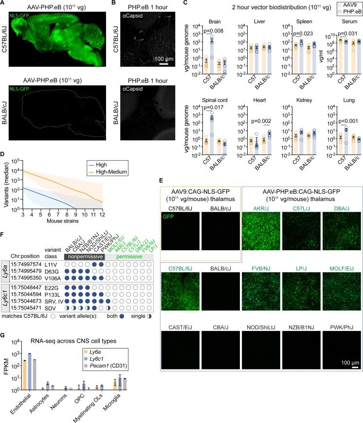
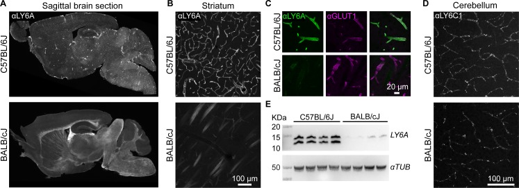
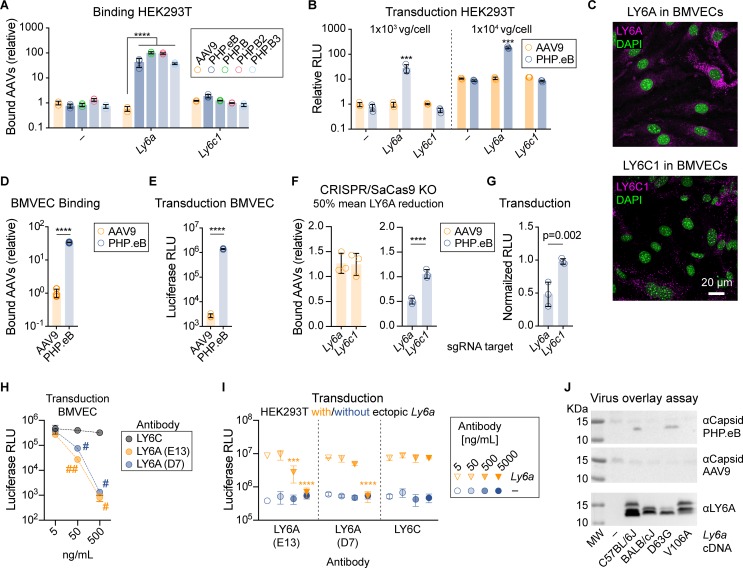
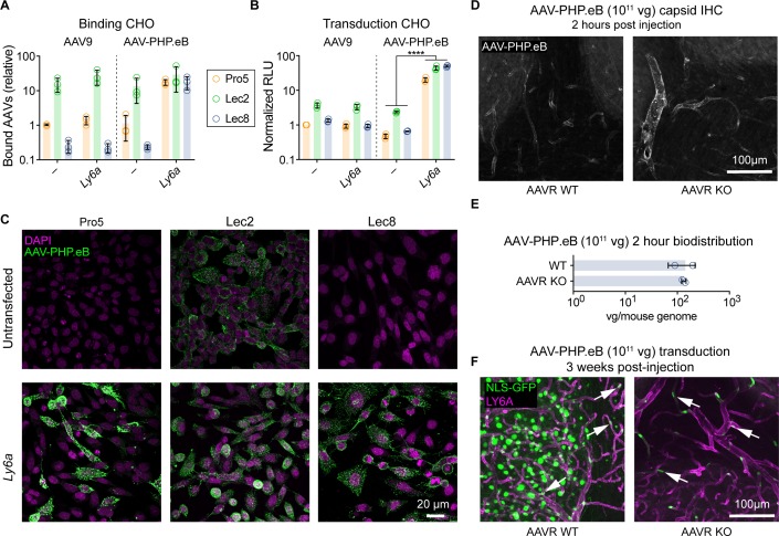
Verification
Claims the paper makes
The claims and statistics of this paper haven’t been checked yet.
Run the check — free, no account needed. It reads the open-access text and checks the sources it cites.
Claims other papers make about this one
Each is checked against this paper’s own text: does it say what the citing paper says it does?
The source backs part of the claim, not all of it — or backs it for a different population, condition or comparison.
In the citing paper
“Later, it was reported that lymphocyte antigen 6 complex, locus A (LY6A), a GPI-anchored membrane protein, responsible for specific engagement to the 7-mer insertion of PHP.B and PHP.eB and transcytosis at BBB [118, 119].”
From Designing and optimizing AAV-mediated gene therapy for neurodegenerative diseases: from bench to bedside (2024) · cites this paper as [118]
Experimental — accuracy not yet measuredThe cited source says what the claim attributes to it.
In the citing paper
“However, LY6A homolog is highly and specifically presented in brain endothelial cells of C57BL/6 J mice but not in BALB/cJ mice and primates.”
From Designing and optimizing AAV-mediated gene therapy for neurodegenerative diseases: from bench to bedside (2024) · cites this paper as [118]
Self-check
Percentages, sums and reported differences re-computed from the paper’s own numbers.
The self-check needs the paper’s full text.
Citations
Cites
Zhang, Y., Chen, K., Sloan, S. A., Bennett, M. L., Scholze, A. R., O'Keeffe, S., Phatnani, H. P., Guarnieri, P., Caneda, C., Ruderisch, N., et al. (2014) · The Journal of Neuroscience · doi:10.1523/jneurosci.1860-14.2014 · Cited by 4,875
Self-citationOptimized sgRNA design to maximize activity and minimize off-target effects of CRISPR-Cas9
Doench, J. G., Fusi, N., Sullender, M., Hegde, M., Vaimberg, E. W., Donovan, K. F., Smith, I., Tothova, Z., Wilen, C., Orchard, R., et al. (2016) · Nature Biotechnology · doi:10.1038/nbt.3437 · Cited by 4,637
In vivo genome editing using Staphylococcus aureus Cas9
Ran, F. A., Cong, L., Yan, W. X., Scott, D. A., Gootenberg, J. S., Kriz, A. J., Zetsche, B., Shalem, O., Wu, X., Makarova, K. S., et al. (2015) · Nature · doi:10.1038/nature14299 · Cited by 2,687
Single-Dose Gene-Replacement Therapy for Spinal Muscular Atrophy
Mendell, J. R., Al-Zaidy, S., Shell, R., Arnold, W. D., Rodino-Klapac, L. R., Prior, T. W., Lowes, L., Alfano, L., Berry, K., Church, K., et al. (2017) · New England Journal of Medicine · doi:10.1056/nejmoa1706198 · Cited by 2,381
Russell, S., Bennett, J., Wellman, J. A., Chung, D. C., Yu, Z.-F., Tillman, A., Wittes, J., Pappas, J., Elci, O., McCague, S., et al. (2017) · The Lancet · doi:10.1016/s0140-6736(17)31868-8 · Cited by 1,952
Mouse genomic variation and its effect on phenotypes and gene regulation.
Keane, T. M., Goodstadt, L., Danecek, P., White, M. A., Wong, K., Yalcin, B., Heger, A., Agam, A., Slater, G., Goodson, M., et al. (2011) · Nature · doi:10.1038/nature10413 · Cited by 1,580
Chan, K. Y., Jang, M. J., Yoo, B. B., Greenbaum, A., Ravi, N., Wu, W.-L., Sánchez-Guardado, L., Lois, C., Mazmanian, S. K., Deverman, B. E., et al. (2017) · Nature Neuroscience · doi:10.1038/nn.4593 · Cited by 1,578
Self-citationIdentification of host proteins required for HIV infection through a functional genomic screen
Brass, A. L., Dykxhoorn, D. M., Benita, Y., Yan, N., Engelman, A., Xavier, R. J., Lieberman, J., & Elledge, S. J. (2008) · Science · doi:10.1126/science.1152725 · Cited by 1,449 · cited 2×
“The Ly6 gene family also influences susceptibility to infection by HIV126,27, Flaviviridae (yellow fever virus, dengue, and West Nile virus28, Influenza A29, and Marek’s disease virus in chickens30.”
BackgroundA Designer AAV Variant Permits Efficient Retrograde Access to Projection Neurons
Tervo, D. G. R., Hwang, B., Viswanathan, S., Gaj, T., Lavzin, M., Ritola, K., Lindo, S. E., Michael, S. C., Кулешова, Е. П., Ojala, D. S., et al. (2016) · Neuron · doi:10.1016/j.neuron.2016.09.021 · Cited by 1,220
Cre-dependent selection yields AAV variants for widespread gene transfer to the adult brain
Deverman, B. E., Pravdo, P. L., Simpson, B. P., Kumar, S. R., Chan, K. Y., Banerjee, A., Wu, W.-L., Yang, B., Huber, N., Pasca, S. P., et al. (2016) · Nature Biotechnology · doi:10.1038/nbt.3440 · Cited by 1,061
Self-citation
Research works printed but not linked: 2
- B. Ravina, 2017, Intraputaminal AADC gene therapy for advanced Parkinson's disease: interim results of a phase 1b Trial [abstract], Human Gene Therapy, 28, A6
- S Pillay, 2017, AAV serotypes have distinctive interactions with domains of the cellular receptor AAVR, J Virol
Cited by
Sorted by citations
Strategies for delivering therapeutics across the blood–brain barrier
Terstappen, G. C., Meyer, A. H., Bell, R. D., & Zhang, W. (2021) · Nature Reviews Drug Discovery · doi:10.1038/s41573-021-00139-y · Cited by 1,138
Levy, J. M., Yeh, W.-H., Pendse, N. D., Davis, J. R., Hennessey, E., Butcher, R. M., Koblan, L. W., Comander, J., Liu, Q., & Liu, D. R. (2020) · Nature Biomedical Engineering · doi:10.1038/s41551-019-0501-5 · Cited by 476
AAV vectors: The Rubik’s cube of human gene therapy
Pupo, A., Fernández, A., Low, S. H., François, A., Suárez-Amarán, L., & Samulski, R. J. (2022) · Molecular Therapy · doi:10.1016/j.ymthe.2022.09.015 · Cited by 382
Efficient prime editing in mouse brain, liver and heart with dual AAVs
Davis, J. R., Banskota, S., Levy, J. M., Newby, G. A., Wang, X., Anzalone, A. V., Nelson, A. T., Chen, P. J., Hennes, A. D., An, M., et al. (2023) · Nature Biotechnology · doi:10.1038/s41587-023-01758-z · Cited by 248
Mathiesen, S. N., Lock, J. L., Schoderboeck, L., Abraham, W. C., & Hughes, S. M. (2020) · Molecular Therapy — Methods & Clinical Development · doi:10.1016/j.omtm.2020.10.011 · Cited by 219
Metadata
- Authors
- Huang, Q., Chan, K. Y., Tobey, I. G., Chan, Y. A., Poterba, T., Boutros, C. L., Balazs, A. B., Daneman, R., Bloom, J. M., Seed, C., & Deverman, B. E.
- Affiliations
- Broad Institute (US)
- Ragon Institute of MGH, MIT and Harvard (US)
- University of California San Diego (US)
- Corresponding author
- Deverman, Benjamin E.
- ORCID
- Venue
- PLOS ONE
- Publisher
- Public Library of Science (PLoS)
- Published
- 2019-11-14
- Volume
- 14
- Issue
- 11
- Pages
- e0225206
- ISSN
- 1932-6203 (ISSN-L)
- PMC
- PMC6855452
- OpenAlex
- W2986362275
- Cited by
- 234
- References
- 40
- Citations by year
- 2026: 28 · 2025: 33 · 2024: 39 · 2023: 30 · 2022: 34 · 2021: 38 · 2020: 28 · 2019: 4
- FWCI
- 15.67
- Type
- journal article
- Language
- en
- Open access
- gold
- doi.org
- Licence
- Locations
- PLoS ONE (publishedVersion, cc-by)
- bioRxiv (Cold Spring Harbor Laboratory) (acceptedVersion, cc-by-nc-nd)
- PubMed Central (publishedVersion, cc-by)
- PubMed (publishedVersion)
- DOAJ (DOAJ: Directory of Open Access Journals) (submittedVersion, cc-by-sa)
- Figshare (submittedVersion, cc-by)
- Funders
- Stanley Center for Psychiatric Research, Broad Institute
- National Institute of Neurological Disorders and Stroke (UG3NS111689)
- Topics
- Life Sciences › Biochemistry, Genetics and Molecular Biology › Genetics › Virus-based gene therapy research
- Life Sciences › Neuroscience › Neurology › Barrier Structure and Function Studies
- Life Sciences › Biochemistry, Genetics and Molecular Biology › Molecular Biology › RNA Interference and Gene Delivery
- Keywords
- blood-brain barrier, gene therapy vectors, receptor identification, mouse strain differences, brain endothelial cells, neurological disorders
- MeSH terms
- Dependovirus, Animals, Antigens, Ly, Blood-Brain Barrier, Brain, Cell Line, Genetic Vectors, Humans, Membrane Proteins, Neurons, Transduction, Genetic, Genetic Variation, Gene Transfer Techniques, Tropism, Transgenes, Mice
About these checks
Registry, retraction, Cites and statistics results are computed, not judged, with one exception: a printed reference that no index lists is read once by a model before it counts as a citation error. Claims are extracted by a model and kept only when their quote appears verbatim in the text; each cited source is then checked, with a model’s help, for whether it says what the claim relies on. Those verdicts can be wrong — read the claim’s quote and the cited source before relying on one.
Statistics and arithmetic are recomputed, not judged.
Confidence bands are shown only for fields calibrated on our annotated test set. None is calibrated yet, so no band is shown.
What counts as a claim ›How we measure confidence ›How we check this ›
Are you an author, or do you see a problem with this page? Request a review
