Cited by 1RNA Research and SplicingRNA regulation and diseaseRNA and protein synthesis mechanisms

The DOI matches its Crossref record; 5 of the 5 references it cites were found in a registry or an index.

Conserved domains and structural motifs that differentiate closely related Rex1 and Rex3 DEDDh exoribonucleases are required for their function in yeast

Open access
Daniels, Peter W., Kelly, Sophie, Tebbs, Iwan J., Mitchell, Phil·2025·PLOS One

Published version of a preprint

Abstract

The DEDD family of exonucleases has expanded through evolution whilst retaining a conserved catalytic domain. One subgroup with closely related catalytic DEDD domain sequences includes the yeast enzymes Rex1 (RNA exonuclease 1) and Rex3, the metazoan REXO1 (RNA exonuclease 1 homologue) and Rexo5 proteins, and the plant protein Sdn5 (small RNA degrading nuclease). Comparison of protein structure models and sequence analyses revealed that this group can be differentiated into two distinct clades consisting of Rex1, Rexo5 and Sdn5 on the one hand, and Rex3 and REXO1 on the other. The catalytic domain of Rex1-related proteins is inserted within a conserved, discontinuous alkaline phosphatase (AlkP) domain. The AlkP domain of yeast Rex1 contains three surface loops that are modelled to be directed towards the DEDD domain, one of which forms an extended helical arch that is found in homologues across fungi and plants. We show that this arch and an adjacent loop are required for Rex1-mediated processing of 5S rRNA and tRNA in Saccharomyces cerevisiae . Rex3-related proteins, including REXO1, lack the AlkP domain but contain a KIX domain (CREB kinase-inducible domain (KID) interacting domain) and a cysteine- and histidine-rich domain (CHORD) adjacent to a C-terminal DEDD domain. Deletion of the N-terminal region within yeast Rex3 spanning the KIX domain blocked its function in RNase MRP processing. In contrast to Rex1, Rex3 proteins are found in metazoans and fungi but not in plants or algae. This work identifies evolutionarily conserved structural hallmarks within Rex1 and Rex3 proteins and demonstrates that specific features are required for Rex1- and Rex3-mediated RNA processing pathways in vivo.

Overview

The claims and statistics of this paper haven’t been checked yet.

Run the check — free, no account needed. It reads the open-access text and checks the sources it cites.

A few free checks a day · papers already verified open instantly · results are public

Integrity signals

Registry

Crossref · 10.1371/journal.pone.0321120

Cites

5 of 5 references verified0 citation errors · 0 not checkable

Statistics

Figures

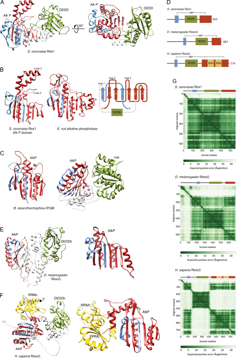
Fig 1 Rex1 and Rexo5 orthologues have a discontinuous alkaline phosphatase fold. Ribbon structure diagrams of Rex1- and AlkP-related proteins. The central DEDDh and cap domains of Rex1-related proteins and iPGM, respectively, are coloured green; N- and C-terminal portions of AlkP domains are coloured blue and red, respectively. The RRM domains inserted within human Rexo5 are coloured gold. (A) Ribbon structure (residues 53-553) of the yeast Rex1 AF model (AF-P53331). (B) Comparison of the yeast Rex1 AlkP domain with AlkP from E. coli (PDB ID 1ALK) [27]. The perspective shown is towards the surface of the AlkP domain ß-sheet. The Rex1 topology diagram shows the ß-sheet numbering, the insertion points of the DEDDh domain within the AlkP domain and the location of prominent loops. Regions before strand 1, between strands 2 and 3, and connecting strands 3 and 4 within the structure of AlkP, are omitted for clarity. (C) AlkP domain and dual domain structure of iPGM from B. stearothermophilus (PBD ID 1EJJ) [24]. The left-hand panel shows the surface of the ß-sheet, as in panel B. The right-hand panel shows the AlkP and cap domains. (D) Schematic, drawn to scale, of the domain structure of Rex1 and Rexo5 proteins. (E) Ribbon diagram (residues 145-681) of the AF model of fly Rexo5 (AF-Q9VRX7) and the AlkP domain alone, viewed with the perspective shown in panels B and C. (F) AF model of the human Rexo5 protein (AF-Q96IC2, residues 49-774) and the AlkP/RRM module, viewed from the same perspective as shown in panels B, C and E. The inferred 5’-3’ polarity of RNA bound to RRM1 in the human protein is indicated. (G) Predicted aligned error maps and domain schematics of yeast Rex1 and the fly and human Rexo5 proteins.
Fig 1 Rex1 and Rexo5 orthologues have a discontinuous alkaline phosphatase fold. Ribbon structure diagrams of Rex1- and AlkP-related proteins. The central DEDDh and cap domains of Rex1-related proteins and iPGM, respectively, are coloured green; N- and C-terminal portions of AlkP domains are coloured blue and red, respectively. The RRM domains inserted within human Rexo5 are coloured gold. (A) Ribbon structure (residues 53-553) of the yeast Rex1 AF model (AF-P53331). (B) Comparison of the yeast Rex1 AlkP domain with AlkP from E. coli (PDB ID 1ALK) [27]. The perspective shown is towards the surface of the AlkP domain ß-sheet. The Rex1 topology diagram shows the ß-sheet numbering, the insertion points of the DEDDh domain within the AlkP domain and the location of prominent loops. Regions before strand 1, between strands 2 and 3, and connecting strands 3 and 4 within the structure of AlkP, are omitted for clarity. (C) AlkP domain and dual domain structure of iPGM from B. stearothermophilus (PBD ID 1EJJ) [24]. The left-hand panel shows the surface of the ß-sheet, as in panel B. The right-hand panel shows the AlkP and cap domains. (D) Schematic, drawn to scale, of the domain structure of Rex1 and Rexo5 proteins. (E) Ribbon diagram (residues 145-681) of the AF model of fly Rexo5 (AF-Q9VRX7) and the AlkP domain alone, viewed with the perspective shown in panels B and C. (F) AF model of the human Rexo5 protein (AF-Q96IC2, residues 49-774) and the AlkP/RRM module, viewed from the same perspective as shown in panels B, C and E. The inferred 5’-3’ polarity of RNA bound to RRM1 in the human protein is indicated. (G) Predicted aligned error maps and domain schematics of yeast Rex1 and the fly and human Rexo5 proteins.CC BY · PubMed Central
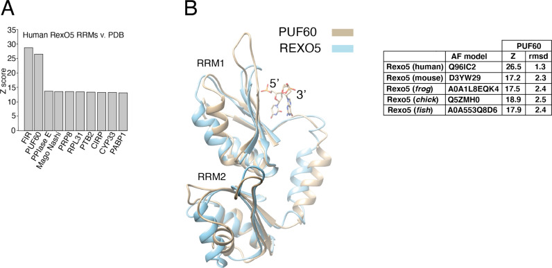
Fig 2 The RRM domains of vertebrate Rexo5 proteins are related to PUF60/FIR. (A) A plot of Dali Z scores for the twin RRMs within the human Rexo5 protein AF model and the most closely related protein structures within the PDB. (B) Structural overlay of the RRM domains in the AF model of human Rexo5 (AF-Q96IC2) and the resolved structure of PUF60 with bound dinucleotide (PDB ID 5KW1) [33]. Z-scores and rmsd values for PUF60 structure overlays with AF models of vertebrate REXO5 proteins are shown.
Fig 2 The RRM domains of vertebrate Rexo5 proteins are related to PUF60/FIR. (A) A plot of Dali Z scores for the twin RRMs within the human Rexo5 protein AF model and the most closely related protein structures within the PDB. (B) Structural overlay of the RRM domains in the AF model of human Rexo5 (AF-Q96IC2) and the resolved structure of PUF60 with bound dinucleotide (PDB ID 5KW1) [33]. Z-scores and rmsd values for PUF60 structure overlays with AF models of vertebrate REXO5 proteins are shown.CC BY · PubMed Central
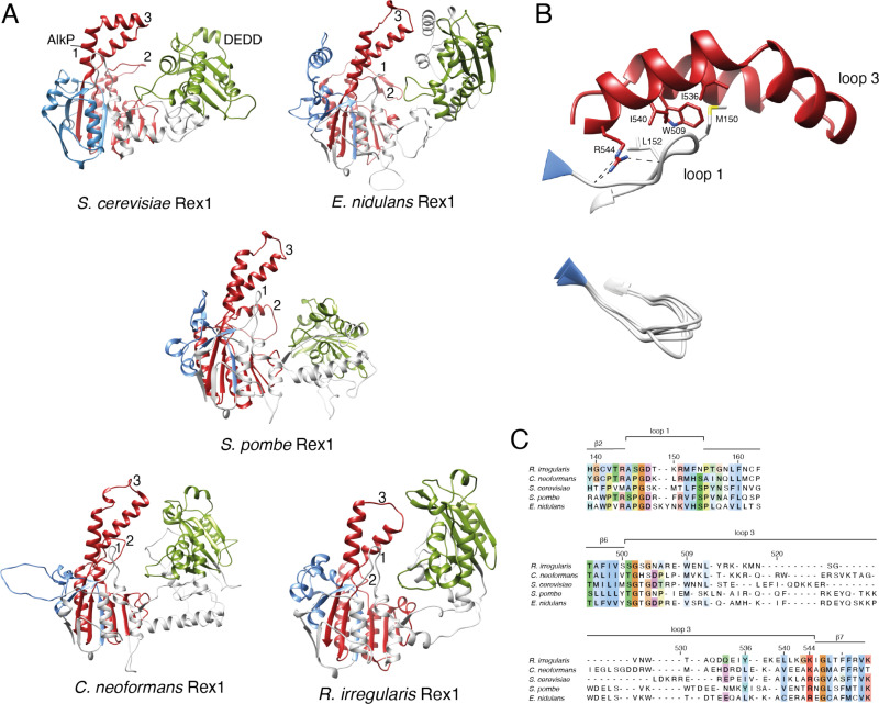
Fig 3 Surface Loops on the AlkP domain of Rex1 are conserved across fungi. (A) Comparison of the AF model of Rex1 from S. cerevisiae (AF-P53331) (Fig 1A) with homologous proteins from E. nidulans (AF-Q5AVW4), S. pombe (AF-O94443), C. neoformans (AF-A0A225XBF6) and R. irregularis (AF-A0A2I1EHK1). The AlkP and DEDD domains are coloured as in Fig 1. Unstructured N- and C-terminal regions of the protein models are omitted for clarity. The positions of loops 1, 2 and 3 are indicated. (B) Expanded view of the interaction between loop 1 and loop 3 (residues 144-155 and 504-544, respectively) in Rex1 from S. cerevisiae. The positions of key residues are indicated. Dashed lines indicate hydrogen bonding between loops 1 and 3. A structural overlay of loop 1 sequences from the five fungal proteins is included. (C) AF model structure-based sequence alignment of loops 1 and 3. Residues are coloured by conservation.
Fig 3 Surface Loops on the AlkP domain of Rex1 are conserved across fungi. (A) Comparison of the AF model of Rex1 from S. cerevisiae (AF-P53331) (Fig 1A) with homologous proteins from E. nidulans (AF-Q5AVW4), S. pombe (AF-O94443), C. neoformans (AF-A0A225XBF6) and R. irregularis (AF-A0A2I1EHK1). The AlkP and DEDD domains are coloured as in Fig 1. Unstructured N- and C-terminal regions of the protein models are omitted for clarity. The positions of loops 1, 2 and 3 are indicated. (B) Expanded view of the interaction between loop 1 and loop 3 (residues 144-155 and 504-544, respectively) in Rex1 from S. cerevisiae. The positions of key residues are indicated. Dashed lines indicate hydrogen bonding between loops 1 and 3. A structural overlay of loop 1 sequences from the five fungal proteins is included. (C) AF model structure-based sequence alignment of loops 1 and 3. Residues are coloured by conservation.CC BY · PubMed Central
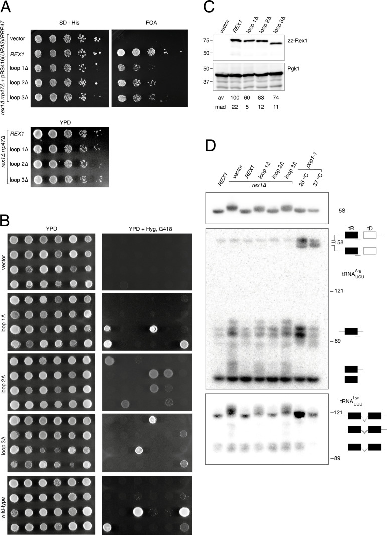
Fig 4 Loops within the AlkP domain of Rex1 are required for RNA processing in vivo. (A) Spot growth assays of rex1∆ rrp47∆ double mutants. Upper panels, growth of a rex1∆ rrp47∆ strain harbouring a plasmid expressing the RRP47 and URA3 genes after transformation with a HIS3 marker plasmid encoding rex1 alleles. Growth on selective medium lacking histidine (SD - His) or containing FOA to purge the URA3 marker plasmid was recorded after 3 days. Lower panel, growth of FOA-resistant isolates on YPD medium. Plates were photographed after 2 days incubation. Assays were performed on three independent isolates. (B) Spot growth assays of randomly selected meiotic progeny from a rex1∆::KANMX4/REX1 rrp47∆::HYGMX4/RRP47 diploid strain harbouring plasmids expressing the wild-type REX1 gene, the loop deletion mutants or the control vector. The panels show growth of haploid progeny on nonselective YPD medium and on medium containing hygromycin and G418. Plates were photographed after incubation for 1-2 days. (C) Western blot analyses of rex1 mutant protein expression levels. Wild-type and mutant rex1 proteins were expressed in a rex1∆ single mutant grown at 30°C. Electrophoretic mobilities of molecular weight markers (size in kDa) are indicated on the left-hand side. of each blot. Rex1 expression levels were normalised to Pgk1. Values for the mean average (av) and mean absolute deviation (mad) of three independent replicates are given. (D) Northern blot hybridisation analyses of total cellular RNA from a wild-type strain, rex1 mutants and the pop1-1 temperature-sensitive mutant. The northern blot was hybridised reiteratively with oligonucleotides complementary to the acceptor stem/3’ trailer sequence boundary of unprocessed tRNAArgUCU, the tRNALysUUU intron and 5S rRNA. Detected tRNA species are indicated schematically on the right. Lines under the schematics indicate the positions of sequences complementary to the oligonucleotide probes used for each panel. The electrophoretic mobilities of 5S rRNA (121 nucleotides), 5.8SS (158 nucleotides) and U24 (89 nucleotides) are indicated as size markers.
Fig 4 Loops within the AlkP domain of Rex1 are required for RNA processing in vivo. (A) Spot growth assays of rex1∆ rrp47∆ double mutants. Upper panels, growth of a rex1∆ rrp47∆ strain harbouring a plasmid expressing the RRP47 and URA3 genes after transformation with a HIS3 marker plasmid encoding rex1 alleles. Growth on selective medium lacking histidine (SD - His) or containing FOA to purge the URA3 marker plasmid was recorded after 3 days. Lower panel, growth of FOA-resistant isolates on YPD medium. Plates were photographed after 2 days incubation. Assays were performed on three independent isolates. (B) Spot growth assays of randomly selected meiotic progeny from a rex1∆::KANMX4/REX1 rrp47∆::HYGMX4/RRP47 diploid strain harbouring plasmids expressing the wild-type REX1 gene, the loop deletion mutants or the control vector. The panels show growth of haploid progeny on nonselective YPD medium and on medium containing hygromycin and G418. Plates were photographed after incubation for 1-2 days. (C) Western blot analyses of rex1 mutant protein expression levels. Wild-type and mutant rex1 proteins were expressed in a rex1∆ single mutant grown at 30°C. Electrophoretic mobilities of molecular weight markers (size in kDa) are indicated on the left-hand side. of each blot. Rex1 expression levels were normalised to Pgk1. Values for the mean average (av) and mean absolute deviation (mad) of three independent replicates are given. (D) Northern blot hybridisation analyses of total cellular RNA from a wild-type strain, rex1 mutants and the pop1-1 temperature-sensitive mutant. The northern blot was hybridised reiteratively with oligonucleotides complementary to the acceptor stem/3’ trailer sequence boundary of unprocessed tRNAArgUCU, the tRNALysUUU intron and 5S rRNA. Detected tRNA species are indicated schematically on the right. Lines under the schematics indicate the positions of sequences complementary to the oligonucleotide probes used for each panel. The electrophoretic mobilities of 5S rRNA (121 nucleotides), 5.8SS (158 nucleotides) and U24 (89 nucleotides) are indicated as size markers.CC BY · PubMed Central
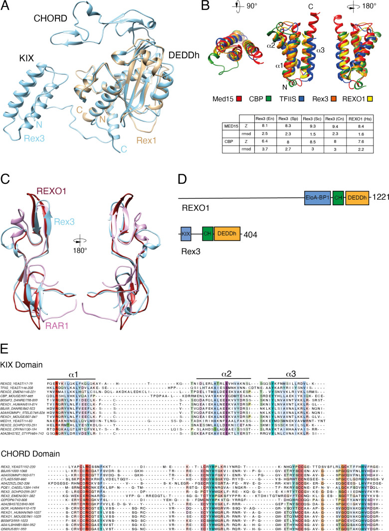
Fig 5 Rex3 related proteins contain KIX and CHORD domains. (A) Structure overlay of AF models for full-length Rex3 (blue) and the DEDDh domain of Rex1 (brown) from S. cerevisiae. Protein domains and the N’- and C’-termini are labelled. (B) Overlay ribbon diagrams of the triple helical domains within yeast Rex3 and human REXO1 proteins and the KIX domains of yeast Med15, mouse CBP and yeast TFIIS. Z-scores and rmsd values for the correlation between Rex3/REXO1 protein structural models and the structures of the KIX domains of Med15 and CBP are given. (C) Structural overlays of the AF models for the CHORD domain of yeast Rex3 (blue) and human REXO1 (red) with the crystal structure of the CHORD domain of RAR1 (purple) [40]. (D) Domain structure of human REXO1 and yeast Rex3 proteins. (E) Structure-based multiple sequence alignments of the KIX and CHORD domains of Rex3 and REOX1 proteins. Residues are coloured by conservation. The KIX domain alignment includes TFIIS, Med15 and CBP.
Fig 5 Rex3 related proteins contain KIX and CHORD domains. (A) Structure overlay of AF models for full-length Rex3 (blue) and the DEDDh domain of Rex1 (brown) from S. cerevisiae. Protein domains and the N’- and C’-termini are labelled. (B) Overlay ribbon diagrams of the triple helical domains within yeast Rex3 and human REXO1 proteins and the KIX domains of yeast Med15, mouse CBP and yeast TFIIS. Z-scores and rmsd values for the correlation between Rex3/REXO1 protein structural models and the structures of the KIX domains of Med15 and CBP are given. (C) Structural overlays of the AF models for the CHORD domain of yeast Rex3 (blue) and human REXO1 (red) with the crystal structure of the CHORD domain of RAR1 (purple) [40]. (D) Domain structure of human REXO1 and yeast Rex3 proteins. (E) Structure-based multiple sequence alignments of the KIX and CHORD domains of Rex3 and REOX1 proteins. Residues are coloured by conservation. The KIX domain alignment includes TFIIS, Med15 and CBP.CC BY · PubMed Central
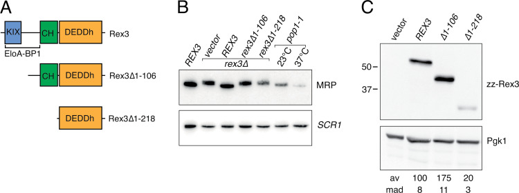
Fig 6 The N-terminal region of Rex3 is required for processing of RNase MRP RNA. (A) Schematic of the domain architecture of yeast Rex3 and the deletion mutants analysed in this study. (B) Northern blot analyses of RNA from a REX3 wild-type strain, isogenic rex3∆ mutant strains harbouring the cloning vector or expressing either full length, wild-type Rex3 or the deletion mutants, and the pop1-1 mutant during growth at 23°C and 37°C. Total cellular RNA was resolved through 6% polyacrylamide gels, transferred to Hybond N+ membranes and hybridised with a probe specific to the RNase MRP RNA. An SCR1 probe was used as a loading control. (C) Western analyses of whole cell lysates from strains expressing full length Rex3 or the N-terminal deletion mutants. Rex3 expression levels were normalised to Pgk1 and are expressed as a percentage of the wild-type protein. Mean average (av) and mean absolute deviation (mad) values of three independent replicates are given.
Fig 6 The N-terminal region of Rex3 is required for processing of RNase MRP RNA. (A) Schematic of the domain architecture of yeast Rex3 and the deletion mutants analysed in this study. (B) Northern blot analyses of RNA from a REX3 wild-type strain, isogenic rex3∆ mutant strains harbouring the cloning vector or expressing either full length, wild-type Rex3 or the deletion mutants, and the pop1-1 mutant during growth at 23°C and 37°C. Total cellular RNA was resolved through 6% polyacrylamide gels, transferred to Hybond N+ membranes and hybridised with a probe specific to the RNase MRP RNA. An SCR1 probe was used as a loading control. (C) Western analyses of whole cell lysates from strains expressing full length Rex3 or the N-terminal deletion mutants. Rex3 expression levels were normalised to Pgk1 and are expressed as a percentage of the wild-type protein. Mean average (av) and mean absolute deviation (mad) values of three independent replicates are given.CC BY · PubMed Central
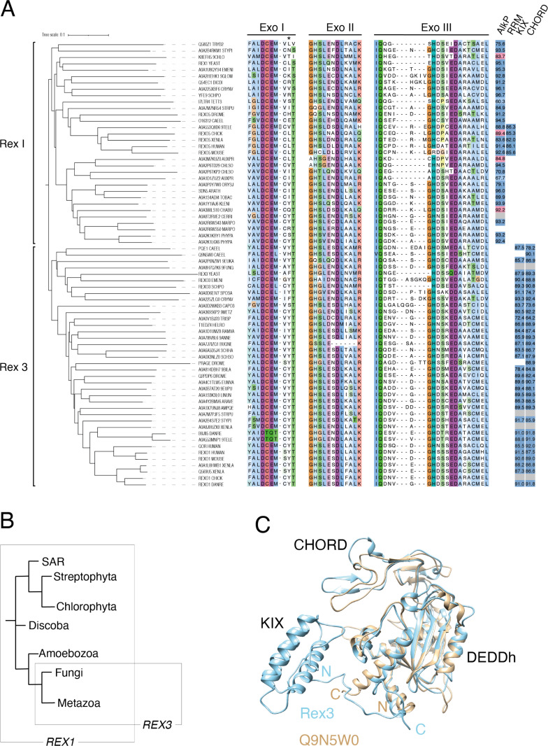
Fig 7 Phylogenetic analyses of Rex1 and Rex3 proteins. (A) Phylogenetic tree and multiple sequence alignment of Rex1 and Rex3 proteins. Exo I, II and III motif sequences are shown. The asterisk indicates a conserved Y/F residue in Rex3 proteins. The presence of AlkP, RRM, KIX and CHORD domains within AF structures are indicated to the right (dark blue indicates high confidence pLDDT scores above 70; light blue indicates low confidence pLDDT scores below 70; pink indicates a partial model of high confidence; grey indicates the absence of a model). Domain pLDDT values are given (see also S6 Table). Proteins are identified by their common names or InterPro codes. Organism codes are as follows: TRYB2, T. brucei; STYPI, S. pistillata; 9CHLO, Bathycoccus prasinos; YEAST, S. cerevisiae; EMENI, A. nidulans; 9GLOM, R. irregularis; DICDI, D. discoideum; CRYNV, C. neoformans; SCHPO, S. pombe; TETTS, T. thermophila; STRPU, S. purpuratus; DROME, D. melanogaster; CAEEL, C. elegans; 9TELE, D. translucida; XENLA, X. laevis; AUXPR, A. protothecoides; CHLSO, C. sorokiniana; ORYSJ, O. sativa, subsp. japonica; ARATH, A. thaliana; TOBAC, N. tabacum; KLENI, K. nitens; CHABU, C. braunii; CERRI, C. richardii; MARPO, M. polymorpha; PHYPA, P. patens; EUKA, Planoprotostelium fungivorum; 9FUNG, Umbelopsis vinacea; SPOSA, Sporidiobolus salmonicolor; CAPO3, Capsaspora owczarzaki; 9METZ, Trichoplax sp. H2; TRISP, Trichinella spiralis; HELRO, Helobdella robusta; RAMVA, Ramazzottius varieornatus; 9ANNE, Dimorphilus gyrociliatus; BUGNE, Bugula neritina; SCHHA, Schistosoma haematobium; SCHSO, Schistocephalus solidus; 9BILA, A. steineri; EUMVA, Eumeta variegata; 9EUPU, Arion vulgaris; LINUN, Lingula unguis; ARAVE, Araneus ventricosus; AMPQE, Amphimedon queenslandica; DANRE, D. rerio. (B) Simplified cladogram showing the phylogeny of REX1 and REX3 genes. (C) Structural overlay of AF models for Rex3 from S. cerevisiae and Q9N5W0 from C. elegans. Domains and termini are labelled.
Fig 7 Phylogenetic analyses of Rex1 and Rex3 proteins. (A) Phylogenetic tree and multiple sequence alignment of Rex1 and Rex3 proteins. Exo I, II and III motif sequences are shown. The asterisk indicates a conserved Y/F residue in Rex3 proteins. The presence of AlkP, RRM, KIX and CHORD domains within AF structures are indicated to the right (dark blue indicates high confidence pLDDT scores above 70; light blue indicates low confidence pLDDT scores below 70; pink indicates a partial model of high confidence; grey indicates the absence of a model). Domain pLDDT values are given (see also S6 Table). Proteins are identified by their common names or InterPro codes. Organism codes are as follows: TRYB2, T. brucei; STYPI, S. pistillata; 9CHLO, Bathycoccus prasinos; YEAST, S. cerevisiae; EMENI, A. nidulans; 9GLOM, R. irregularis; DICDI, D. discoideum; CRYNV, C. neoformans; SCHPO, S. pombe; TETTS, T. thermophila; STRPU, S. purpuratus; DROME, D. melanogaster; CAEEL, C. elegans; 9TELE, D. translucida; XENLA, X. laevis; AUXPR, A. protothecoides; CHLSO, C. sorokiniana; ORYSJ, O. sativa, subsp. japonica; ARATH, A. thaliana; TOBAC, N. tabacum; KLENI, K. nitens; CHABU, C. braunii; CERRI, C. richardii; MARPO, M. polymorpha; PHYPA, P. patens; EUKA, Planoprotostelium fungivorum; 9FUNG, Umbelopsis vinacea; SPOSA, Sporidiobolus salmonicolor; CAPO3, Capsaspora owczarzaki; 9METZ, Trichoplax sp. H2; TRISP, Trichinella spiralis; HELRO, Helobdella robusta; RAMVA, Ramazzottius varieornatus; 9ANNE, Dimorphilus gyrociliatus; BUGNE, Bugula neritina; SCHHA, Schistosoma haematobium; SCHSO, Schistocephalus solidus; 9BILA, A. steineri; EUMVA, Eumeta variegata; 9EUPU, Arion vulgaris; LINUN, Lingula unguis; ARAVE, Araneus ventricosus; AMPQE, Amphimedon queenslandica; DANRE, D. rerio. (B) Simplified cladogram showing the phylogeny of REX1 and REX3 genes. (C) Structural overlay of AF models for Rex3 from S. cerevisiae and Q9N5W0 from C. elegans. Domains and termini are labelled.CC BY · PubMed Central

Verification

Claims the paper makes

The claims and statistics of this paper haven’t been checked yet.

Run the check — free, no account needed. It reads the open-access text and checks the sources it cites.

A few free checks a day · papers already verified open instantly · results are public

Self-check

Percentages, sums and reported differences re-computed from the paper’s own numbers.

The self-check needs the paper’s full text.

Citations

Cites

Research works printed but not linked: 67

  1. Y Zuo, 2001, Exoribonuclease superfamilies: structural analysis and phylogenetic distribution, Nucleic Acids Research, 29, 1017, 10.1093/nar/29.5.1017 · DOI
  2. MJ Moser, 1997, The proofreading domain of Escherichia coli DNA polymerase I and other DNA and/or RNA exonuclease domains, Nucleic Acids Res, 25, 5110, 10.1093/nar/25.24.5110 · DOI
  3. A van Hoof, 2000, Three conserved members of the RNase D family have unique and overlapping functions in the processing of 5S, 5.8S, U4, U5, RNase MRP and RNase P RNAs in yeast, EMBO J, 19, 1357, 10.1093/emboj/19.6.1357 · DOI
  4. PW Piper, 1983, Altered maturation of sequences at the 3’ terminus of 5S gene transcripts in a Saccharomyces cerevisiae mutant that lacks a RNA processing endonuclease, EMBO J, 2, 353, 10.1002/j.1460-2075.1983.tb01430.x · DOI
  5. A Kempers-Veenstra, 1986, 3’-end formation of transcripts from the yeast rRNA operon, EMBO J, 5, 2703, 10.1002/j.1460-2075.1986.tb04554.x · DOI
  6. LA Copela, 2008, Competition between the Rex1 exonuclease and the La protein affects both Trf4p-mediated RNA quality control and pre-tRNA maturation, RNA, 14, 1214, 10.1261/rna.1050408 · DOI
  7. SG Ozanick, 2009, Rex1p deficiency leads to accumulation of precursor initiator tRNAMet and polyadenylation of substrate RNAs in Saccharomyces cerevisiae, Nucleic Acids Res, 37, 298, 10.1093/nar/gkn925 · DOI
  8. E Leung, 2014, Integrity of SRP RNA is ensured by La and the nuclear RNA quality control machinery, Nucleic Acids Res, 42, 10698, 10.1093/nar/gku761 · DOI
  9. D Foretek, 2016, Control of Saccharomyces cerevisiae pre-tRNA processing by environmental conditions, RNA, 22, 339, 10.1261/rna.054973.115 · DOI
  10. D Hodko, 2016, The Rtr1p CTD phosphatase autoregulates its mRNA through a degradation pathway involving the REX exonucleases, RNA, 22, 559, 10.1261/rna.055723.115 · DOI
  11. W Garland, 2013, Rrp47 functions in RNA surveillance and stable RNA processing when divorced from the exoribonuclease and exosome-binding domains of Rrp6, RNA, 19, 1659, 10.1261/rna.039388.113 · DOI
  12. W Peng, 2003, A panoramic view of yeast noncoding RNA processing, Cell, 113, 919, 10.1016/S0092-8674(03)00466-5 · DOI
  13. P Mitchell, 2003, Rrp47p is an exosome-associated protein required for the 3’ processing of stable RNAs, Mol Cell Biol, 23, 6982, 10.1128/MCB.23.19.6982-6992.2003 · DOI
  14. S Gerstberger, 2017, The Conserved RNA Exonuclease Rexo5 Is Required for 3’ End Maturation of 28S rRNA, 5S rRNA, and snoRNAs, Cell Rep, 21, 758, 10.1016/j.celrep.2017.09.067 · DOI
  15. S Silva, 2017, Characterization of the mammalian RNA exonuclease 5/NEF-sp as a testis-specific nuclear 3’ → 5’ exoribonuclease, RNA, 23, 1385, 10.1261/rna.060723.117 · DOI
  16. YJ Lee, 2024, REXO5 promotes genomic integrity through regulating R-loop using its exonuclease activity, Leukemia, 38, 2150, 10.1038/s41375-024-02362-z · DOI
  17. K Tamura, 2003, Identification of EloA-BP1, a novel Elongin A binding protein with an exonuclease homology domain, Biochem Biophys Res Commun, 309, 189, 10.1016/S0006-291X(03)01556-0 · DOI
  18. PW Daniels, 2022, Contribution of domain structure to the function of the yeast DEDD family exoribonuclease and RNase T functional homolog, Rex1, RNA, 28, 493, 10.1261/rna.078939.121 · DOI
  19. K Shirasu, 1999, A novel class of eukaryotic zinc-binding proteins is required for disease resistance signaling in barley and development in C. elegans, Cell, 99, 355, 10.1016/S0092-8674(00)81522-6 · DOI
  20. CT Heise, 2007, Biochemical characterization of RAR1 cysteine- and histidine-rich domains (CHORDs): a novel class of zinc-dependent protein-protein interaction modules, Biochemistry, 46, 1612, 10.1021/bi062174k · DOI
  21. JK Thakur, 2014, Molecular recognition by the KIX domain and its role in gene regulation, Nucleic Acids Res, 42, 2112, 10.1093/nar/gkt1147 · DOI
  22. MJ Jedrzejas, 2000, Structure and mechanism of action of a novel phosphoglycerate mutase from Bacillus stearothermophilus, EMBO J, 19, 1419, 10.1093/emboj/19.7.1419 · DOI
  23. TD Panosian, 2011, Bacillus cereus phosphopentomutase is an alkaline phosphatase family member that exhibits an altered entry point into the catalytic cycle, J Biol Chem, 286, 8043, 10.1074/jbc.M110.201350 · DOI
  24. MY Galperin, 2001, Conserved core structure and active site residues in alkaline phosphatase superfamily enzymes, Proteins, 45, 318, 10.1002/prot.1152 · DOI
  25. EE Kim, 1991, Reaction mechanism of alkaline phosphatase based on crystal structures. Two-metal ion catalysis, J Mol Biol, 218, 449, 10.1016/0022-2836(91)90724-K · DOI
  26. M Steinegger, 2018, Clustering huge protein sequence sets in linear time, Nat Commun, 9, 2542, 10.1038/s41467-018-04964-5 · DOI · Paper page
  27. C Van Buskirk, 2002, Half pint regulates alternative splice site selection in Drosophila, Dev Cell, 2, 343, 10.1016/S1534-5807(02)00128-4 · DOI
  28. A Joshi, 2014, Solution and crystal structures of a C-terminal fragment of the neuronal isoform of the polypyrimidine tract binding protein (nPTB), PeerJ, 2, 10.7717/peerj.305 · DOI
  29. N Safaee, 2012, Interdomain allostery promotes assembly of the poly(A) mRNA complex with PABP and eIF4G, Mol Cell, 48, 375, 10.1016/j.molcel.2012.09.001 · DOI
  30. H-HT Hsiao, 2020, Unraveling the mechanism of recognition of the 3’ splice site of the adenovirus major late promoter intron by the alternative splicing factor PUF60, PLoS One, 15
  31. GV Crichlow, 2008, Dimerization of FIR upon FUSE DNA binding suggests a mechanism of c-myc inhibition, EMBO J, 27, 277, 10.1038/sj.emboj.7601936 · DOI
  32. GM Daubner, 2013, RRM-RNA recognition: NMR or crystallography…and new findings, Curr Opin Struct Biol, 23, 100, 10.1016/j.sbi.2012.11.006 · DOI
  33. PW Piper, 1989, Processing of transcripts of a dimeric tRNA gene in yeast uses the nuclease responsible for maturation of the 3’ termini upon 5 S and 37 S precursor rRNAs, FEBS Lett, 250, 311, 10.1016/0014-5793(89)80745-8 · DOI
  34. D Engelke, 1985, Nucleolytic processing of a tRNAArg-tRNAAsp dimeric precursor by a homologous component from Saccharomyces cerevisiae, J Biol Chem, 260, 1271, 10.1016/S0021-9258(20)71239-1 · DOI
  35. Z Lygerou, 1994, The POP1 gene encodes a protein component common to the RNase MRP and RNase P ribonucleoproteins, Genes Dev, 8, 1423, 10.1101/gad.8.12.1423 · DOI
  36. PP Chan, 2016, GtRNAdb 2.0: an expanded database of transfer RNA genes identified in complete and draft genomes, Nucleic Acids Res, 44
  37. M Zhang, 2010, Structural basis for assembly of Hsp90-Sgt1-CHORD protein complexes: implications for chaperoning of NLR innate immunity receptors, Mol Cell, 39, 269, 10.1016/j.molcel.2010.05.010 · DOI
  38. SA Kassube, 2013, Structural mimicry in transcription regulation of human RNA polymerase II by the DNA helicase RECQL5, Nat Struct Mol Biol, 20, 892, 10.1038/nsmb.2596 · DOI
  39. R Gopalakrishnan, 2021, The histone chaperone Spt6 is required for normal recruitment of the capping enzyme Abd1 to transcribed regions, J Biol Chem, 297, 101205, 10.1016/j.jbc.2021.101205 · DOI
  40. D Blears, 2024, Redundant pathways for removal of defective RNA polymerase II complexes at a promoter-proximal pause checkpoint, Mol Cell
  41. A Takahashi, 2003, HSP90 interacts with RAR1 and SGT1 and is essential for RPS2-mediated disease resistance in Arabidopsis, Proc Natl Acad Sci U S A, 100, 11777, 10.1073/pnas.2033934100 · DOI
  42. MB Ardehali, Elongin A associates with actively transcribed genes and modulates enhancer RNA levels with limited impact on transcription elongation rate in vivo, J Biol Chem
  43. ME Schmitt, 1992, Yeast site-specific ribonucleoprotein endoribonuclease MRP contains an RNA component homologous to mammalian RNase MRP RNA and essential for cell viability, Genes Dev, 6, 1975, 10.1101/gad.6.10.1975 · DOI
  44. RNA central Consortium, 2021, RNAcentral 2021: secondary structure integration, improved sequence search and new member databases, Nucleic Acids Res, 49, 10.1093/nar/gkaa921 · DOI · Paper page
  45. Z Han, 2023, DNA-directed termination of RNA polymerase II transcription, Mol Cell, 83
  46. Y Zuo, 2002, The physiological role of RNase T can be explained by its unusual substrate specificity, J Biol Chem, 277, 29654, 10.1074/jbc.M204252200 · DOI
  47. A Axhemi, 2020, Substrate selectivity by the exonuclease Rrp6p, Proc Natl Acad Sci U S A, 117, 982, 10.1073/pnas.1913236117 · DOI
  48. MC Daugeron, 2001, The yeast POP2 gene encodes a nuclease involved in mRNA deadenylation, Nucleic Acids Res, 29, 2448, 10.1093/nar/29.12.2448 · DOI
  49. E Wagner, 2007, An unconventional human Ccr4-Caf1 deadenylase complex in nuclear cajal bodies, Mol Cell Biol, 27, 1686, 10.1128/MCB.01483-06 · DOI
  50. G Kaur, 2018, Evolutionary relationship between the cysteine and histidine rich domains (CHORDs) and Btk-type zinc fingers, Bioinformatics, 34, 1981, 10.1093/bioinformatics/bty041 · DOI
  51. W Tadros, 2005, Setting the stage for development: mRNA translation and stability during oocyte maturation and egg activation in Drosophila, Dev Dyn, 232, 593, 10.1002/dvdy.20297 · DOI
  52. J Cui, 2016, The Drosophila prage Gene, Required for Maternal Transcript Destabilization in Embryos, Encodes a Predicted RNA Exonuclease, G3 (Bethesda), 6, 1687, 10.1534/g3.116.028415 · DOI
  53. F Madeira, 2019, The EMBL-EBI search and sequence analysis tools APIs in 2019, Nucleic Acids Res, 47, 10.1093/nar/gkz268 · DOI · Paper page
  54. F Martin, 2023, Ensembl 2023, Nucleic Acids Res, 51, 10.1093/nar/gkac958 · DOI
  55. AM Waterhouse, 2009, Jalview Version 2--a multiple sequence alignment editor and analysis workbench, Bioinformatics, 25, 1189, 10.1093/bioinformatics/btp033 · DOI
  56. I Letunic, 2021, Interactive Tree Of Life (iTOL) v5: an online tool for phylogenetic tree display and annotation, Nucleic Acids Res, 49, 10.1093/nar/gkab301 · DOI
  57. HM Berman, 2000, The Protein Data Bank, Nucleic Acids Res, 28, 235, 10.1093/nar/28.1.235 · DOI · Paper page
  58. N Wang, 2013, Ordering a dynamic protein via a small-molecule stabilizer, J Am Chem Soc, 135, 3363, 10.1021/ja3122334 · DOI
  59. EF Pettersen, 2004, UCSF Chimera--a visualization system for exploratory research and analysis, J Comput Chem, 25, 1605, 10.1002/jcc.20084 · DOI
  60. L Holm, 2023, DALI shines a light on remote homologs: One hundred discoveries, Protein Sci, 32, 10.1002/pro.4519 · DOI
  61. P Mitchell, 1996, The 3’ end of yeast 5.8S rRNA is generated by an exonuclease processing mechanism, Genes Dev, 10, 502, 10.1101/gad.10.4.502 · DOI
  62. C Allmang, 1999, Functions of the exosome in rRNA, snoRNA and snRNA synthesis, EMBO J, 18, 5399, 10.1093/emboj/18.19.5399 · DOI
  63. C Allmang, 2000, Degradation of ribosomal RNA precursors by the exosome, Nucleic Acids Res, 28, 1684, 10.1093/nar/28.8.1684 · DOI
  64. JL Costello, 2011, The C-terminal region of the exosome-associated protein Rrp47 is specifically required for box C/D small nucleolar RNA 3’-maturation, J Biol Chem, 286, 4535, 10.1074/jbc.M110.162826 · DOI
  65. AL Goldstein, 1999, Three new dominant drug resistance cassettes for gene disruption inSaccharomyces cerevisiae, Yeast, 15, 1541, 10.1002/(SICI)1097-0061(199910)15:14<1541::AID-YEA476>3.0.CO;2-K · DOI
  66. D Gietz, 1992, Improved method for high efficiency transformation of intact yeast cells, Nucleic Acids Res, 20, 1425, 10.1093/nar/20.6.1425 · DOI
  67. RS Sikorski, 1989, A system of shuttle vectors and yeast host strains designed for efficient manipulation of DNA in Saccharomyces cerevisiae, Genetics, 122, 19, 10.1093/genetics/122.1.19 · DOI

Metadata

Authors
Daniels, P. W., Kelly, S., Tebbs, I. J., & Mitchell, P.
Affiliations
University of Sheffield (GB)
Venue
PLOS One
Published
2025-06-02
Volume
20
Issue
6
Pages
e0321120
ISSN
1932-6203 (ISSN-L)
OpenAlex
W4410950275
Cited by
1
References
5
Citations by year
2026: 1
FWCI
0.30
Type
journal article
Language
en
Open access
Locations
  • PLoS ONE (publishedVersion, cc-by)
  • bioRxiv (Cold Spring Harbor Laboratory) (acceptedVersion)
  • PubMed Central (publishedVersion, cc-by)
  • PubMed (publishedVersion)
  • Research Explorer (The University of Manchester) (publishedVersion, other-oa)
  • White Rose Research Online (University of Leeds, The University of Sheffield, University of York) (acceptedVersion)
  • DOAJ (DOAJ: Directory of Open Access Journals) (submittedVersion, cc-by-sa)
  • DOAJ (DOAJ: Directory of Open Access Journals) (submittedVersion)
Funders
Biotechnology and Biological Sciences Research Council (BB/V00722X/1)
Topics
  • Life Sciences › Biochemistry, Genetics and Molecular Biology › Molecular Biology › RNA Research and Splicing
  • Life Sciences › Biochemistry, Genetics and Molecular Biology › Molecular Biology › RNA regulation and disease
  • Life Sciences › Biochemistry, Genetics and Molecular Biology › Molecular Biology › RNA and protein synthesis mechanisms
Keywords
Rex1, KIX domain, RNase MRP, tRNA processing, Saccharomyces cerevisiae, protein structure model, protein evolution
MeSH terms
Protein Domains, Amino Acid Sequence, Exoribonucleases, Models, Molecular, Phylogeny, RNA, Transfer, Saccharomyces cerevisiae, Conserved Sequence, Catalytic Domain, Amino Acid Motifs, Saccharomyces cerevisiae Proteins

About these checks

Registry, retraction, Cites and statistics results are computed, not judged, with one exception: a printed reference that no index lists is read once by a model before it counts as a citation error. Claims are extracted by a model and kept only when their quote appears verbatim in the text; each cited source is then checked, with a model’s help, for whether it says what the claim relies on. Those verdicts can be wrong — read the claim’s quote and the cited source before relying on one.

Statistics and arithmetic are recomputed, not judged.

Confidence bands are shown only for fields calibrated on our annotated test set. None is calibrated yet, so no band is shown.

What counts as a claim ›How we measure confidence ›How we check this ›

Are you an author, or do you see a problem with this page? Request a review