

The DOI matches its Crossref record; 52 of the 52 references it cites were found in a registry or an index.
Establishment of Human Papillomavirus Infection Requires Cell Cycle Progression
Open accessAbstract
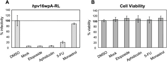
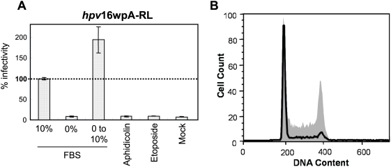
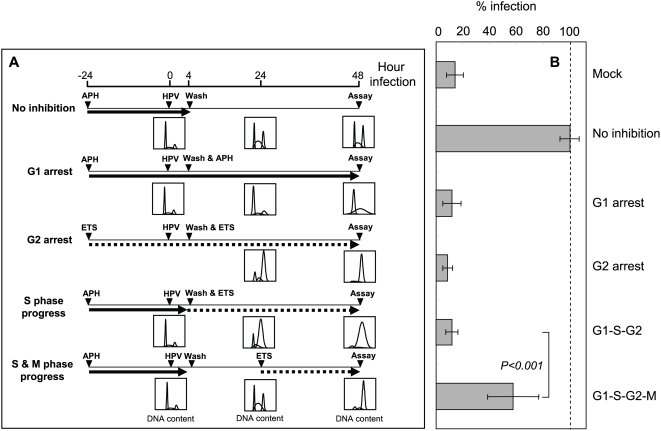
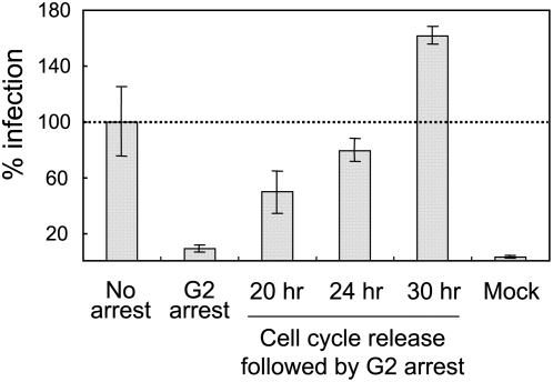
Human papillomaviruses (HPVs) are DNA viruses associated with major human cancers. As such there is a strong interest in developing new means, such as vaccines and microbicides, to prevent HPV infections. Developing the latter requires a better understanding of the infectious life cycle of HPVs. The HPV infectious life cycle is closely linked to the differentiation state of the stratified epithelium it infects, with progeny virus only made in the terminally differentiating suprabasal compartment. It has long been recognized that HPV must first establish its infection within the basal layer of stratified epithelium, but why this is the case has not been understood. In part this restriction might reflect specificity of expression of entry receptors. However, this hypothesis could not fully explain the differentiation restriction of HPV infection, since many cell types can be infected with HPVs in monolayer cell culture. Here, we used chemical biology approaches to reveal that cell cycle progression through mitosis is critical for HPV infection. Using infectious HPV16 particles containing the intact viral genome, G1-synchronized human keratinocytes as hosts, and early viral gene expression as a readout for infection, we learned that the recipient cell must enter M phase (mitosis) for HPV infection to take place. Late M phase inhibitors had no effect on infection, whereas G1, S, G2, and early M phase cell cycle inhibitors efficiently prevented infection. We conclude that host cells need to pass through early prophase for successful onset of transcription of the HPV encapsidated genes. These findings provide one reason why HPVs initially establish infections in the basal compartment of stratified epithelia. Only this compartment of the epithelium contains cells progressing through the cell cycle, and therefore it is only in these cells that HPVs can establish their infection. By defining a major condition for cell susceptibility to HPV infection, these results also have potentially important implications for HPV control.
Overview
The claims and statistics of this paper haven’t been checked yet.
Run the check — free, no account needed. It reads the open-access text and checks the sources it cites.
Integrity signals
Registry
Cites
Statistics
Figures
Verification
Claims the paper makes
The claims and statistics of this paper haven’t been checked yet.
Run the check — free, no account needed. It reads the open-access text and checks the sources it cites.
Claims other papers make about this one
Each is checked against this paper’s own text: does it say what the citing paper says it does?
In the citing paper
“Individual factors unique to the mucosal surface such as epithelial surface integrity, mucosal secretions, and immune regulation likely play a role in HPV persistence and progression to cancer (Pyeon et al., 2009; Schiffman et al., 2016).”
From Viral Infections, the Microbiome, and Probiotics (2021) · cites this paper as Pyeon et al., 2009
Self-check
Percentages, sums and reported differences re-computed from the paper’s own numbers.
The self-check needs the paper’s full text.
Citations
Cites
Primer3 on the WWW for General Users and for Biologist Programmers
Rozen, S., & Skaletsky, H. (2003) · Humana Press eBooks · doi:10.1385/1-59259-192-2:365 · Cited by 15,068
Normal keratinization in a spontaneously immortalized aneuploid human keratinocyte cell line.
Boukamp, P., Petrussevska, R. T., Breitkreutz, D., Hornung, J., Markham, A., & Fusenig, N. E. (1988) · The Journal of cell biology · doi:10.1083/jcb.106.3.761 · Cited by 4,170
Papillomaviruses and cancer: from basic studies to clinical application
Hausen, H. Z. (2002) · Nature reviews. Cancer · doi:10.1038/nrc798 · Cited by 4,031
Human papillomavirus and cervical cancer
Burd, E. M. (2003) · Clinical Microbiology Reviews · doi:10.1128/cmr.16.1.1-17.2003 · Cited by 2,420
Small Molecule Inhibitor of Mitotic Spindle Bipolarity Identified in a Phenotype-Based Screen
Mayer, T. U., Kapoor, T. M., Haggarty, S. J., King, R. W., Schreiber, S. L., & Mitchison, T. J. (1999) · Science · doi:10.1126/science.286.5441.971 · Cited by 1,752
Papillomavirus infections — a major cause of human cancers
Hausen, H. Z. (1996) · Biochimica et Biophysica Acta (BBA) - Reviews on Cancer · doi:10.1016/0304-419x(96)00020-0 · Cited by 1,532
Cdk1 is sufficient to drive the mammalian cell cycle
Santamarı́a, D., Barrière, C., Cerqueira, A., Hunt, S. L., Tardy, C., Newton, K., Cáceres, J. F., Dubus, P. R., Malumbres, M., & Barbacid, M. (2007) · Nature · doi:10.1038/nature06046 · Cited by 1,096
Structure, dynamics and functions of promyelocytic leukaemia nuclear bodies
Bernardi, R., & Pandolfi, P. P. (2007) · Nature Reviews Molecular Cell Biology · doi:10.1038/nrm2277 · Cited by 876
Exploiting Chemical Libraries, Structure, and Genomics in the Search for Kinase Inhibitors
Gray, N. S., Wodicka, L. M., Thunnissen, A. M. W. H., Norman, T., Kwon, S., Espinoza, F. H., Morgan, D. O., Barnes, G., Leclerc, S., Meijer, L., et al. (1998) · Science · doi:10.1126/science.281.5376.533 · Cited by 808
Lewis, P. F., & Emerman, M. (1994) · Journal of Virology · doi:10.1128/jvi.68.1.510-516.1994 · Cited by 797
Cited by
Sorted by citations
The Biology and Life-Cycle of Human Papillomaviruses
Doorbar, J., Quint, W. G. V., Banks, L., Bravo, I. G., Stoler, M. H., Tr, B., & Stanley, M. (2012) · Vaccine · doi:10.1016/j.vaccine.2012.06.083 · Cited by 1,633
Carcinogenic human papillomavirus infection
Schiffman, M., Doorbar, J., Wentzensen, N., de Sanjosé, S., Fakhry, C., Monk, B. J., Stanley, M. A., & Franceschi, S. (2016) · Nature Reviews Disease Primers · doi:10.1038/nrdp.2016.86 · Cited by 999
Human papillomavirus molecular biology and disease association
Doorbar, J., Egawa, N., Griffin, H. M., Kranjec, C., & Murakami, I. (2015) · Reviews in Medical Virology · doi:10.1002/rmv.1822 · Cited by 1,019
Human Papillomaviruses; Epithelial Tropisms, and the Development of Neoplasia
Egawa, N., Egawa, K., Griffin, H. M., & Doorbar, J. (2015) · Viruses · doi:10.3390/v7072802 · Cited by 585
Graham, S. V. (2017) · Clinical Science · doi:10.1042/cs20160786 · Cited by 458
Human Papillomavirus in Head and Neck Cancer: Its Role in Pathogenesis and Clinical Implications
Chung, C. H., & Gillison, M. L. (2009) · Clinical Cancer Research · doi:10.1158/1078-0432.ccr-09-0784 · Cited by 417
Human papillomaviruses: diversity, infection and host interactions
McBride, A. A. (2021) · Nature Reviews Microbiology · doi:10.1038/s41579-021-00617-5 · Cited by 372
Opportunities and challenges for human papillomavirus vaccination in cancer
Roden, R. B. S., & Stern, P. L. (2018) · Nature reviews. Cancer · doi:10.1038/nrc.2018.13 · Cited by 335
Current understanding of the mechanism of HPV infection
Schiller, J. T., Day, P. M., & Kines, R. C. (2010) · Gynecologic Oncology · doi:10.1016/j.ygyno.2010.04.004 · Cited by 332
Mucosal and Cutaneous Human Papillomavirus Infections and Cancer Biology
Gheit, T. (2019) · Frontiers in Oncology · doi:10.3389/fonc.2019.00355 · Cited by 332
McLaughlin-Drubin, M. E., Crum, C. P., & Münger, K. (2011) · Proceedings of the National Academy of Sciences · doi:10.1073/pnas.1009933108 · Cited by 337
Kines, R. C., Thompson, C. D., Lowy, D. R., Schiller, J. T., & Day, P. M. (2009) · Proceedings of the National Academy of Sciences · doi:10.1073/pnas.0908502106 · Cited by 292
L2, the minor capsid protein of papillomavirus
Wang, J. W., & Roden, R. B. S. (2013) · Virology · doi:10.1016/j.virol.2013.04.017 · Cited by 266
Zhang, L., Wu, J., Ling, M. T., Zhao, L., & Zhao, K.-N. (2015) · Molecular Cancer · doi:10.1186/s12943-015-0361-x · Cited by 264
Human papillomavirus molecular biology
Harden, M. E., & Münger, K. (2016) · Mutation Research/Reviews in Mutation Research · doi:10.1016/j.mrrev.2016.07.002 · Cited by 285
Scarth, J. A., Patterson, M. R., Morgan, E. L., & Macdonald, A. (2021) · Journal of General Virology · doi:10.1099/jgv.0.001540 · Cited by 237
Surviladze, Z., Sterk, R. T., DeHaro, S. A., & Ozbun, M. A. (2012) · Journal of Virology · doi:10.1128/jvi.02319-12 · Cited by 225
Evasion of host immune defenses by human papillomavirus
Westrich, J. A., Warren, C. J., & Pyeon, D. (2016) · Virus Research · doi:10.1016/j.virusres.2016.11.023 · Cited by 225
APOBEC3A Functions as a Restriction Factor of Human Papillomavirus
Warren, C. J., Xu, T., Guo, K., Griffin, L., Westrich, J. A., Lee, D., Lambert, P. F., Santiago, M. L., & Pyeon, D. (2014) · Journal of Virology · doi:10.1128/jvi.02383-14 · Cited by 193
Surviladze, Z., Dziduszko, A., & Ozbun, M. A. (2012) · PLoS Pathogens · doi:10.1371/journal.ppat.1002519 · Cited by 183
Viral Infections, the Microbiome, and Probiotics
Harper, A., Vijayakumar, V., Ouwehand, A. C., ter Haar, J., Obis, D., Espadaler, J., Binda, S., Desiraju, S., & Day, R. (2021) · Frontiers in Cellular and Infection Microbiology · doi:10.3389/fcimb.2020.596166 · Cited by 177
Human papillomavirus and oropharynx cancer: Biology, detection and clinical implications
Allen, C. T., Lewis, J. S., El‐Mofty, S. K., Haughey, B. H., & Nussenbaum, B. A. (2010) · The Laryngoscope · doi:10.1002/lary.20936 · Cited by 175
Pathogenesis of human papillomavirus‐associated mucosal disease
Groves, I. J., & Coleman, N. (2014) · The Journal of Pathology · doi:10.1002/path.4496 · Cited by 173
Prophylactic and Therapeutic HPV Vaccines: Current Scenario and Perspectives
Mo, Y., Ma, J., Zhang, H., Shen, J., Chen, J., Hong, J., Xu, Y., & Qian, C. (2022) · Frontiers in Cellular and Infection Microbiology · doi:10.3389/fcimb.2022.909223 · Cited by 173
Sapp, M. J., & Bienkowska‐Haba, M. (2009) · FEBS Journal · doi:10.1111/j.1742-4658.2009.07400.x · Cited by 154
How viruses access the nucleus.
Cohen, S. S., Au, S., & Panté, N. (2011) · Biochimica et Biophysica Acta · doi:10.1016/j.bbamcr.2010.12.009 · Cited by 143
Aydin, I., Weber, S., Snijder, B., Ventayol, P. S., Kühbacher, A., Becker, M., Day, P. M., Schiller, J. T., Kann, M., Pelkmans, L., et al. (2014) · PLoS Pathogens · doi:10.1371/journal.ppat.1004162 · Cited by 156
Day, P. M., Thompson, C. D., Schowalter, R. M., Lowy, D. R., & Schiller, J. T. (2013) · Journal of Virology · doi:10.1128/jvi.03222-12 · Cited by 147
Human Papillomavirus L2 Facilitates Viral Escape from Late Endosomes via Sorting Nexin 17
Bergant Marušič, M., Ozbun, M. A., Campos, S. K., Myers, M. P., & Banks, L. (2012) · Traffic · doi:10.1111/j.1600-0854.2011.01320.x · Cited by 130
Dziduszko, A., & Ozbun, M. A. (2013) · Journal of Virology · doi:10.1128/jvi.00519-13 · Cited by 150
Viral and Cellular Biomarkers in the Diagnosis of Cervical Intraepithelial Neoplasia and Cancer
Tornesello, M. L., Buonaguro, L., Rossi, P. G., & Buonaguro, F. M. (2013) · BioMed Research International · doi:10.1155/2013/519619 · Cited by 133
Mechanisms and strategies of papillomavirus replication
McBride, A. A. (2017) · Biological Chemistry · doi:10.1515/hsz-2017-0113 · Cited by 127
Popa, A., Zhang, W., Harrison, M. S., Goodner, K., Kazakov, T., Goodwin, E. C., Lipovsky, A. I., Burd, C. G., & DiMaio, D. (2015) · PLoS Pathogens · doi:10.1371/journal.ppat.1004699 · Cited by 123
Principles of Virus Uncoating: Cues and the Snooker Ball
Yamauchi, Y., & Greber, U. F. (2016) · Traffic · doi:10.1111/tra.12387 · Cited by 127
Cicchini, L., Westrich, J. A., Xu, T., Vermeer, D. W., Berger, J., Clambey, E. T., Lee, D., Song, J. I., Lambert, P. F., Greer, R. O., et al. (2016) · mBio · doi:10.1128/mbio.00270-16 · Cited by 125
Molecular mechanisms of HPV mediated neoplastic progression
Senapati, R., Senapati, N. N., & Dwibedi, B. (2016) · Infectious Agents and Cancer · doi:10.1186/s13027-016-0107-4 · Cited by 121
Histology of cervical intraepithelial neoplasia and the role of biomarkers
Martin, C. M., & O’Leary, J. J. (2011) · Best Practice & Research Clinical Obstetrics & Gynaecology · doi:10.1016/j.bpobgyn.2011.04.005 · Cited by 123
Bienkowska‐Haba, M., Williams, C., Kim, S. M., Garcea, R. L., & Sapp, M. J. (2012) · Journal of Virology · doi:10.1128/jvi.00980-12 · Cited by 116
Mechanisms by which HPV Induces a Replication Competent Environment in Differentiating Keratinocytes
Moody, C. A. (2017) · Viruses · doi:10.3390/v9090261 · Cited by 121
Oncogenic human papillomaviruses
McBride, A. A. (2017) · Philosophical Transactions of the Royal Society B Biological Sciences · doi:10.1098/rstb.2016.0273 · Cited by 112
Virus strategies for passing the nuclear envelope barrier
Kobiler, O., Drayman, N., Butin‐Israeli, V., & Oppenheim, A. (2012) · Nucleus · doi:10.4161/nucl.21979 · Cited by 116
Fay, N., & Panté, N. (2015) · Frontiers in Microbiology · doi:10.3389/fmicb.2015.00467 · Cited by 106
Aksoy, P., Gottschalk, E. Y., & Meneses, P. I. (2016) · Mutation Research/Reviews in Mutation Research · doi:10.1016/j.mrrev.2016.09.004 · Cited by 108
HPV Infection: Immunological Aspects and Their Utility in Future Therapy
Deligeoroglou, E., Giannouli, A., Athanasopoulos, N., Karountzos, V., Vatopoulou, A., Dimopoulos, K., & Creatsas, G. (2013) · Infectious Diseases in Obstetrics and Gynecology · doi:10.1155/2013/540850 · Cited by 103
Concepts of papillomavirus entry into host cells
Day, P. M., & Schelhaas, M. (2013) · Current Opinion in Virology · doi:10.1016/j.coviro.2013.11.002 · Cited by 112
Incoming human papillomavirus type 16 genome resides in a vesicular compartment throughout mitosis
DiGiuseppe, S., Łuszczek, W., Keiffer, T. R., Bienkowska‐Haba, M., Guion, L. G. M., & Sapp, M. J. (2016) · Proceedings of the National Academy of Sciences · doi:10.1073/pnas.1600638113 · Cited by 97
Wu, D., Liu, W.-S., Wang, J. C. K., Chen, C., Cheng, Y., & Lee, H. (2011) · Clinical Cancer Research · doi:10.1158/1078-0432.ccr-10-2316 · Cited by 95
Zhang, W., Kazakov, T., Popa, A., & DiMaio, D. (2014) · mBio · doi:10.1128/mbio.01777-14 · Cited by 97
Human papillomavirus infection is inhibited by host autophagy in primary human keratinocytes
Griffin, L., Cicchini, L., & Pyeon, D. (2013) · Virology · doi:10.1016/j.virol.2012.12.004 · Cited by 91
Meyers, J. M., Uberoi, A., Grace, M. R., Lambert, P. F., & Münger, K. (2017) · PLoS Pathogens · doi:10.1371/journal.ppat.1006171 · Cited by 85
RETRACTED ARTICLE: Molecular mechanisms in progression of HPV-associated cervical carcinogenesis
Gupta, S. M., & Mania‐Pramanik, J. (2019) · Journal of Biomedical Science · doi:10.1186/s12929-019-0520-2 · Cited by 91
RetractedBiology of HPV Mediated Carcinogenesis and Tumor Progression
Cosper, P. F., Bradley, S. T., Luo, Q., & Kimple, R. J. (2021) · Seminars in Radiation Oncology · doi:10.1016/j.semradonc.2021.02.006 · Cited by 83
Sp100 Provides Intrinsic Immunity against Human Papillomavirus Infection
Stepp, W. H., Meyers, J. M., & McBride, A. A. (2013) · mBio · doi:10.1128/mbio.00845-13 · Cited by 82
The Role of the DNA Damage Response throughout the Papillomavirus Life Cycle
McKinney, C. C., Hussmann, K., & McBride, A. A. (2015) · Viruses · doi:10.3390/v7052450 · Cited by 79
Warren, C. J., Griffin, L., Little, A. S., Huang, I., Farzan, M., & Pyeon, D. (2014) · PLoS ONE · doi:10.1371/journal.pone.0096579 · Cited by 79
The role of furin in papillomavirus infection
Day, P. M., & Schiller, J. T. (2009) · Future Microbiology · doi:10.2217/fmb.09.86 · Cited by 77
Identification and Validation of Human Papillomavirus Encoded microRNAs
Qian, K., Pietilä, T., Rönty, M. J., Michon, F., Frilander, M. J., Ritari, J., Tarkkanen, J., Paulin, L. G., Auvinen, P., & Auvinen, E. (2013) · PLoS ONE · doi:10.1371/journal.pone.0070202 · Cited by 76
Molecular mechanisms of human papillomavirus-related carcinogenesis in head and neck cancer
Faraji, F., Zaidi, M. A., Fakhry, C., & Gaykalova, D. A. (2017) · Microbes and Infection · doi:10.1016/j.micinf.2017.06.001 · Cited by 84
Entry Tropism of BK and Merkel Cell Polyomaviruses in Cell Culture
Schowalter, R. M., Reinhold, W. C., & Buck, C. B. (2012) · PLoS ONE · doi:10.1371/journal.pone.0042181 · Cited by 75
Griffin, H. M., Soneji, Y., van Baars, R., Arora, R., Jenkins, D., van de Sandt, M. M., Wu, Z., Quint, W. G. V., Jach, R., Okoń, K., et al. (2015) · Modern Pathology · doi:10.1038/modpathol.2015.52 · Cited by 77
DiGiuseppe, S., Keiffer, T. R., Bienkowska‐Haba, M., Łuszczek, W., Guion, L. G. M., Müller, M., & Sapp, M. J. (2015) · Journal of Virology · doi:10.1128/jvi.01588-15 · Cited by 72
Cruising the cellular highways: how human papillomavirus travels from the surface to the nucleus
DiGiuseppe, S., Bienkowska-Haba, M., Guion, L. G., & Sapp, M. (2017) · Virus Research · doi:10.1016/j.virusres.2016.10.015 · Cited by 67
Edwards, T. G., Helmus, M. J., Koeller, K. J., Bashkin, J. K., & Fisher, C. (2013) · Journal of Virology · doi:10.1128/jvi.03473-12 · Cited by 71
Epigenetic Regulation of the Human Papillomavirus Life Cycle
Mac, M., & Moody, C. A. (2020) · Pathogens · doi:10.3390/pathogens9060483 · Cited by 75
The Biology of Human Papillomaviruses
Nguyen, H. P., Ramirez‐Fort, M. K., & Rady, P. L. (2014) · Current problems in dermatology · doi:10.1159/000355959 · Cited by 70
Modulation of the DNA damage response during the life cycle of human papillomaviruses
Anacker, D. C., & Moody, C. A. (2016) · Virus Research · doi:10.1016/j.virusres.2016.11.006 · Cited by 70
Spurgeon, M. E., & Lambert, P. F. (2017) · Viruses · doi:10.3390/v9080219 · Cited by 69
Jain, M., Yadav, D., Jarouliya, U., Chavda, V. K., Yadav, A. K., Chaurasia, B. K., & Song, M. (2023) · Pathogens · doi:10.3390/pathogens12121380 · Cited by 83
HPV-associated oropharyngeal cancer: in search of surrogate biomarkers for early lesions
Lim, Y. X., & D’Silva, N. J. (2024) · Oncogene · doi:10.1038/s41388-023-02927-9 · Cited by 68
Aydin, I., Villalonga-Planells, R., Greune, L., Bronnimann, M. P., Calton, C. M., Becker, M., Lai, K.-Y., Campos, S. K., Schmidt, M. A., & Schelhaas, M. (2017) · PLoS Pathogens · doi:10.1371/journal.ppat.1006308 · Cited by 67
Stem cells in squamous head and neck cancer
Albers, A. E., Chen, C., Köberle, B., Qian, X., Klußmann, J. P., Wollenberg, B., & Kaufmann, A. M. (2011) · Critical Reviews in Oncology/Hematology · doi:10.1016/j.critrevonc.2011.03.004 · Cited by 71
Microbiome factors in HPV-driven carcinogenesis and cancers
Lin, D., Kouzy, R., Jaoude, J. A., Noticewala, S. S., Medrano, A. Y. D., Klopp, A. H., Taniguchi, C. M., & Colbert, L. E. (2020) · PLoS Pathogens · doi:10.1371/journal.ppat.1008524 · Cited by 66
Special Issue about Head and Neck Cancers: HPV Positive Cancers
Economopoulou, P., Kotsantis, I., & Psyrri, A. K. (2020) · International Journal of Molecular Sciences · doi:10.3390/ijms21093388 · Cited by 68
DiGiuseppe, S., Bienkowska‐Haba, M., Guion, L. G. M., Keiffer, T. R., & Sapp, M. J. (2017) · Journal of Virology · doi:10.1128/jvi.00537-17 · Cited by 65
Cerqueira, C., Liu, Y., Kühling, L., Chai, W., Hafezi, W., Kuppevelt, T. H. V., Kühn, J., Feizi, T., & Schelhaas, M. (2013) · Cellular Microbiology · doi:10.1111/cmi.12150 · Cited by 64
Calton, C. M., Bronnimann, M. P., Manson, A. R., Li, S., Chapman, J. A., Suarez-Berumen, M., Williamson, T. R., Molugu, S. K., Bernal, R. A., & Campos, S. K. (2017) · PLoS Pathogens · doi:10.1371/journal.ppat.1006200 · Cited by 62
Infectious Entry of Merkel Cell Polyomavirus
Becker, M., Dominguez, M., Greune, L., Soria-Martínez, L., Pfleiderer, M. M., Schowalter, R. M., Buck, C. B., Blaum, B. S., Schmidt, M. A., & Schelhaas, M. (2019) · Journal of Virology · doi:10.1128/jvi.02004-18 · Cited by 59
Tetraspanins: Host Factors in Viral Infections
New, C. S., Lee, Z.-Y., Tan, K. S., Wong, A. H., Wang, D. Y., & Tran, T. (2021) · International Journal of Molecular Sciences · doi:10.3390/ijms222111609 · Cited by 62
The Interplay between Antiviral Signalling and Carcinogenesis in Human Papillomavirus Infections
Ferreira, A., Ramalho, A. C., Marques, M., & Ribeiro, D. (2020) · Cancers · doi:10.3390/cancers12030646 · Cited by 60
DiGiuseppe, S., Bienkowska‐Haba, M., Hilbig, L., & Sapp, M. J. (2014) · Virology · doi:10.1016/j.virol.2014.04.024 · Cited by 57
Papillomavirus Infection Requires γ Secretase
Karanam, B., Peng, S., Li, T., Buck, C. B., Day, P. M., & Roden, R. B. S. (2010) · Journal of Virology · doi:10.1128/jvi.01081-10 · Cited by 57
Clinical relevance and implications of HPV-induced neoplasia in different anatomical locations
Prigge, E. S., Doeberitz, M. V. K., & Reuschenbach, M. (2016) · Mutation Research/Reviews in Mutation Research · doi:10.1016/j.mrrev.2016.06.005 · Cited by 58
Campos, S. K. (2017) · Viruses · doi:10.3390/v9120370 · Cited by 58
Alpha-Defensin HD5 Inhibits Furin Cleavage of Human Papillomavirus 16 L2 To Block Infection
Wiens, M. E., & Smith, J. G. (2014) · Journal of Virology · doi:10.1128/jvi.02901-14 · Cited by 56
Interaction of Human Tumor Viruses with Host Cell Surface Receptors and Cell Entry
Schäfer, G., Blumenthal, M. J., & Katz, A. A. (2015) · Viruses · doi:10.3390/v7052592 · Cited by 57
Biology and natural history of human papillomavirus infection
Fernandes, J. V., de Araújo, J. M. G., & Fernandes, T. A. A. D. M. (2013) · Open Access Journal of Clinical Trials · doi:10.2147/oajct.s37741 · Cited by 55
Hitchhiking on host chromatin: how papillomaviruses persist
McBride, A. A., Sakakibara, N., Stepp, W. H., & Jang, M. K. (2012) · Biochimica et Biophysica Acta (BBA) - Gene Regulatory Mechanisms · doi:10.1016/j.bbagrm.2012.01.011 · Cited by 54
Inoue, T., Zhang, P., Zhang, W., Goodner, K., Dupzyk, A., DiMaio, D., & Tsai, B. (2018) · The Journal of Cell Biology · doi:10.1083/jcb.201804171 · Cited by 54
Taylor, J. R., Fernandez, D. J., Thornton, S. M., Skeate, J. G., Lühen, K. P., Silva, D. M. D., Langen, R., & Kast, W. M. (2018) · Scientific Reports · doi:10.1038/s41598-018-30051-2 · Cited by 53
The long and winding road: human papillomavirus entry and subcellular trafficking
Ozbun, M. A., & Campos, S. K. (2021) · Current Opinion in Virology · doi:10.1016/j.coviro.2021.07.010 · Cited by 52
Kilcher, S., & Mercer, J. (2015) · Virology · doi:10.1016/j.virol.2015.01.024 · Cited by 52
The full transcription map of mouse papillomavirus type 1 (MmuPV1) in mouse wart tissues
Xue, X., Majerčiak, V., Uberoi, A., Kim, B., Gotte, D., Chen, X., Cam, M., Lambert, P. F., & Zheng, Z. (2017) · PLoS Pathogens · doi:10.1371/journal.ppat.1006715 · Cited by 52
Impact of Replication Stress in Human Papillomavirus Pathogenesis
Moody, C. A. (2018) · Journal of Virology · doi:10.1128/jvi.01012-17 · Cited by 50
Human Papillomavirus Infections and Cancer Stem Cells of Tumors from the Uterine Cervix
Lopez, J. (2012) · The Open Virology Journal · doi:10.2174/1874357901206010232 · Cited by 51
Depuydt, C., Jonckheere, J., Berth, M., Salembier, G. M., Vereecken, A. J., & Bogers, J. (2015) · Cancer Medicine · doi:10.1002/cam4.473 · Cited by 49
Superinfection Exclusion between Two High-Risk Human Papillomavirus Types during a Coinfection
Biryukov, J., & Meyers, C. (2018) · Journal of Virology · doi:10.1128/jvi.01993-17 · Cited by 49
Virion Factors That Target Daxx To Overcome Intrinsic Immunity
Schreiner, S., & Wodrich, H. (2013) · Journal of Virology · doi:10.1128/jvi.00425-13 · Cited by 47
Furin Cleavage of L2 during Papillomavirus Infection: Minimal Dependence on Cyclophilins
Bronnimann, M. P., Calton, C. M., Chiquette, S. F., Li, S., Lu, M., Chapman, J. A., Bratton, K. N., Schlegel, A. M., & Campos, S. K. (2016) · Journal of Virology · doi:10.1128/jvi.00038-16 · Cited by 47
Becker, M., Greune, L., Schmidt, M. A., & Schelhaas, M. (2018) · Journal of Virology · doi:10.1128/jvi.02106-17 · Cited by 46
YAP1 activation by human papillomavirus E7 promotes basal cell identity in squamous epithelia
Hatterschide, J., Castagnino, P. A., Kim, H. W., Sperry, S. M., Montone, K. T., Basu, D., & White, E. A. (2022) · eLife · doi:10.7554/elife.75466 · Cited by 46
Metadata
- Authors
- Pyeon, D., Pearce, S. M., Lank, S. M., Ahlquist, P., & Lambert, P. F.
- Affiliations
- University of Wisconsin–Madison (US)
- Howard Hughes Medical Institute (US)
- Corresponding author
- Ahlquist, Paul
- Lambert, Paul F.
- Venue
- PLoS Pathogens
- Publisher
- Public Library of Science (PLoS)
- Published
- 2009-02-27
- Volume
- 5
- Issue
- 2
- Pages
- e1000318
- ISSN
- 1553-7366 (ISSN-L)
- 1553-7374
- PMC
- PMC2642596
- OpenAlex
- W2074766470
- Cited by
- 348
- References
- 52
- Citations by year
- 2026: 14 · 2025: 22 · 2024: 22 · 2023: 17 · 2022: 16 · 2021: 27 · 2020: 19 · 2019: 25 · 2018: 17 · 2017: 25 · 2016: 26 · 2015: 24 · 2014: 20 · 2013: 30 · 2012: 15
- FWCI
- 9.85
- Type
- journal article
- Language
- en
- Open access
- gold
- doi.org
- Locations
- PLoS Pathogens (publishedVersion, cc-by)
- Europe PMC (PubMed Central) (publishedVersion, cc-by)
- PubMed Central (publishedVersion, cc-by)
- PubMed (publishedVersion)
- DOAJ (DOAJ: Directory of Open Access Journals) (submittedVersion, cc-by-sa)
- Figshare (submittedVersion, cc-by)
- Funders
- Howard Hughes Medical Institute
- University of Wisconsin-Madison
- National Institutes of Health (CA022443)
- National Cancer Institute (CA022443, P01CA022443)
- National Institute of Allergy and Infectious Diseases (R33AI071947, AI071947, R21AI071947)
- Topics
- Health Sciences › Medicine › Epidemiology › Cervical Cancer and HPV Research
- Life Sciences › Biochemistry, Genetics and Molecular Biology › Genetics › Virus-based gene therapy research
- Health Sciences › Medicine › Oncology › Cancer-related Molecular Pathways
- Keywords
- human papillomavirus, HPV16, cell cycle progression, mitosis, stratified epithelium, keratinocytes, G1 phase, viral gene expression, microbicides, HPV vaccine, viral entry receptor
- MeSH terms
- Antiviral Agents, Cell Cycle, Cell Division, Cell Survival, Cells, Cultured, Etoposide, Humans, Orthomyxoviridae, Prophase, Transcription, Genetic, Virion, Keratinocytes, Gene Expression Regulation, Viral, Aphidicolin, Genome, Viral, Human papillomavirus 16
About these checks
Registry, retraction, Cites and statistics results are computed, not judged, with one exception: a printed reference that no index lists is read once by a model before it counts as a citation error. Claims are extracted by a model and kept only when their quote appears verbatim in the text; each cited source is then checked, with a model’s help, for whether it says what the claim relies on. Those verdicts can be wrong — read the claim’s quote and the cited source before relying on one.
Statistics and arithmetic are recomputed, not judged.
Confidence bands are shown only for fields calibrated on our annotated test set. None is calibrated yet, so no band is shown.
What counts as a claim ›How we measure confidence ›How we check this ›
Are you an author, or do you see a problem with this page? Request a review
