

The DOI matches its Crossref record; 66 of the 66 references it cites were found in a registry or an index.
Coxsackievirus B Exits the Host Cell in Shed Microvesicles Displaying Autophagosomal Markers
Open accessAbstract
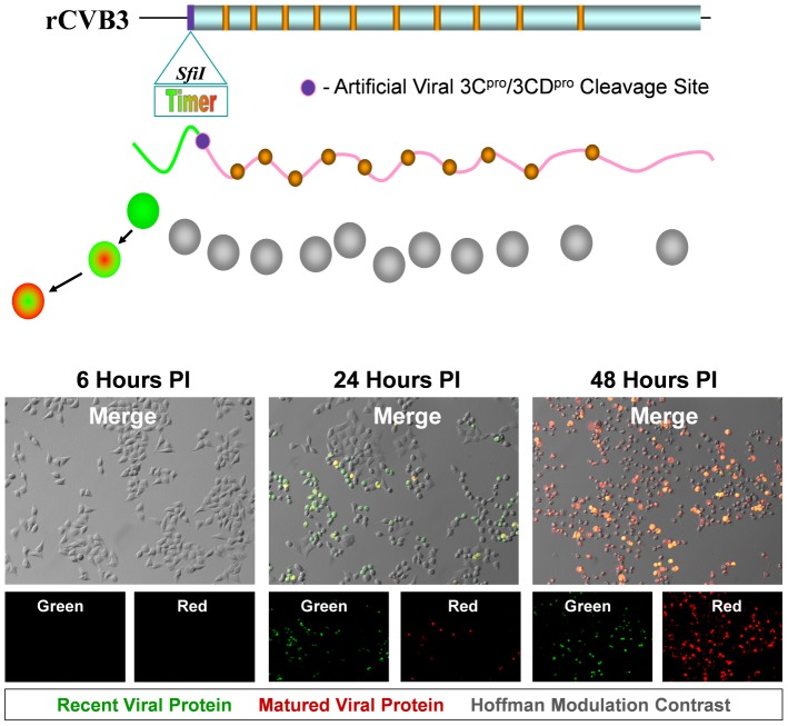
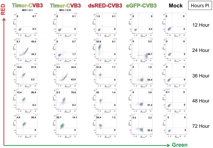
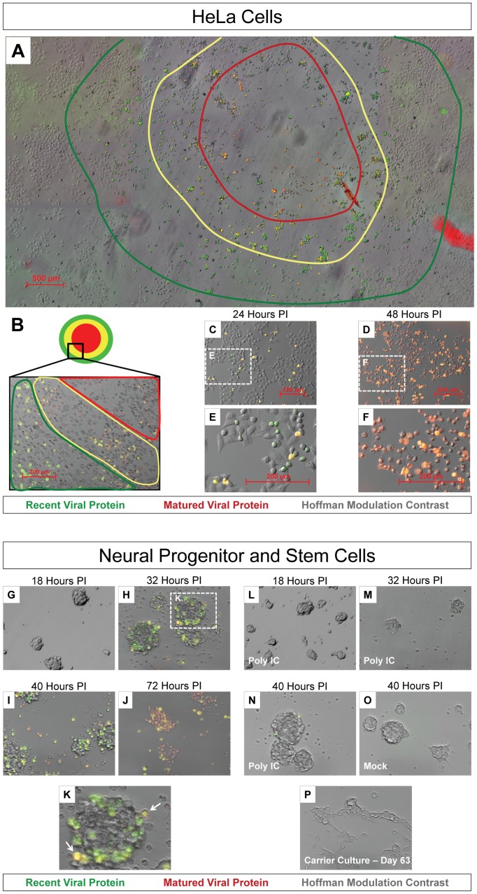
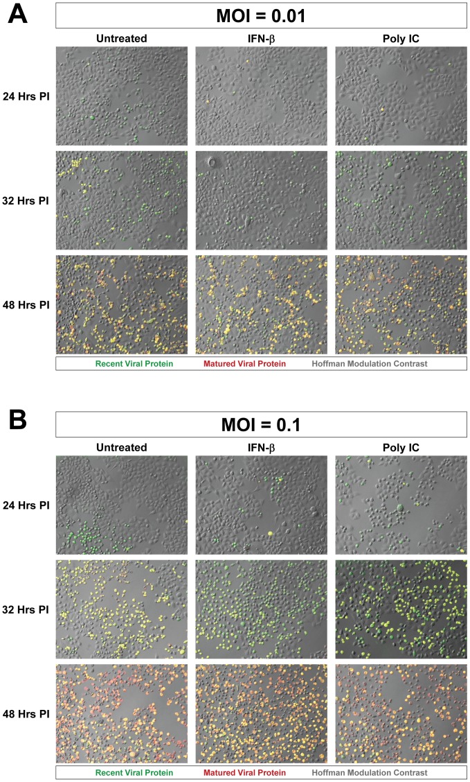
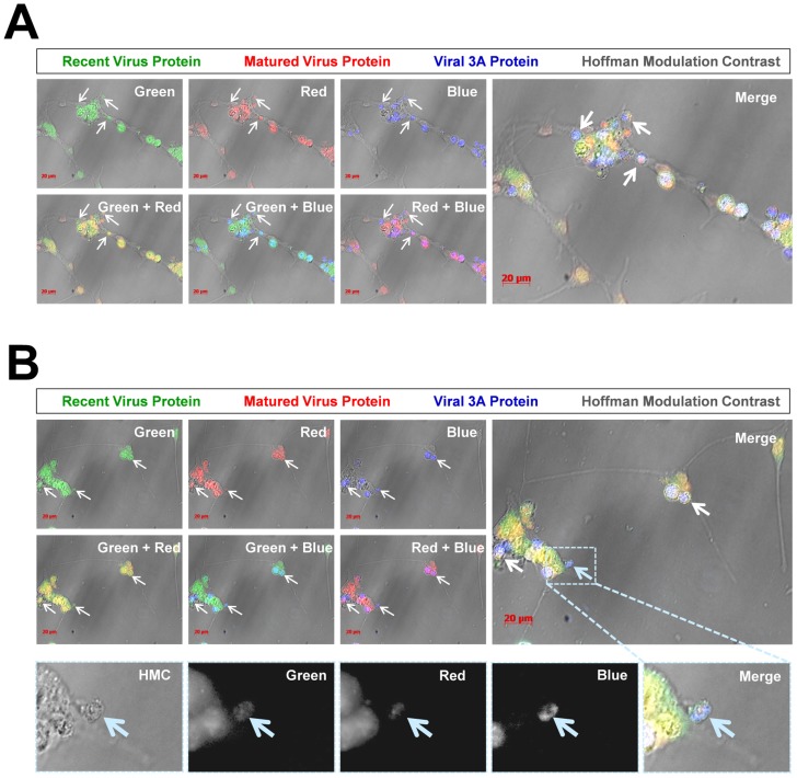
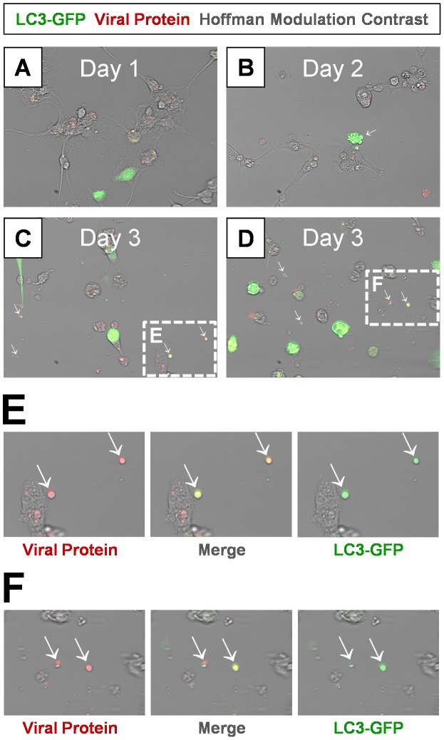
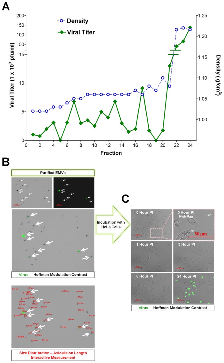
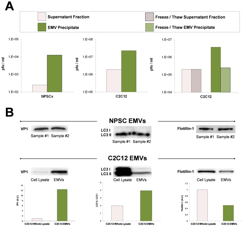
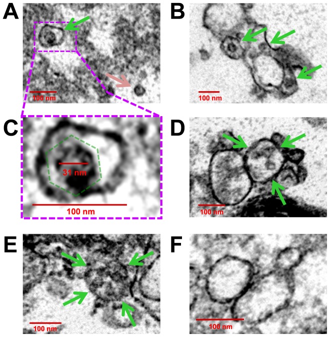
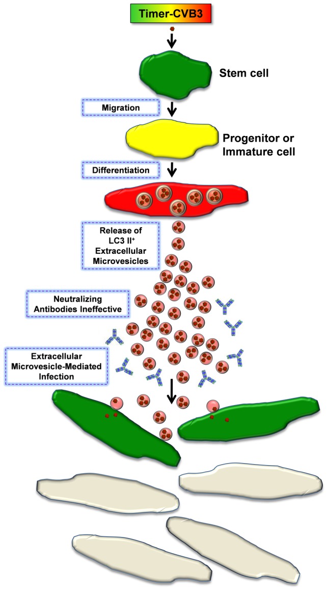
Coxsackievirus B3 (CVB3), a member of the picornavirus family and enterovirus genus, causes viral myocarditis, aseptic meningitis, and pancreatitis in humans. We genetically engineered a unique molecular marker, "fluorescent timer" protein, within our infectious CVB3 clone and isolated a high-titer recombinant viral stock (Timer-CVB3) following transfection in HeLa cells. "Fluorescent timer" protein undergoes slow conversion of fluorescence from green to red over time, and Timer-CVB3 can be utilized to track virus infection and dissemination in real time. Upon infection with Timer-CVB3, HeLa cells, neural progenitor and stem cells (NPSCs), and C2C12 myoblast cells slowly changed fluorescence from green to red over 72 hours as determined by fluorescence microscopy or flow cytometric analysis. The conversion of "fluorescent timer" protein in HeLa cells infected with Timer-CVB3 could be interrupted by fixation, suggesting that the fluorophore was stabilized by formaldehyde cross-linking reactions. Induction of a type I interferon response or ribavirin treatment reduced the progression of cell-to-cell virus spread in HeLa cells or NPSCs infected with Timer-CVB3. Time lapse photography of partially differentiated NPSCs infected with Timer-CVB3 revealed substantial intracellular membrane remodeling and the assembly of discrete virus replication organelles which changed fluorescence color in an asynchronous fashion within the cell. "Fluorescent timer" protein colocalized closely with viral 3A protein within virus replication organelles. Intriguingly, infection of partially differentiated NPSCs or C2C12 myoblast cells induced the release of abundant extracellular microvesicles (EMVs) containing matured "fluorescent timer" protein and infectious virus representing a novel route of virus dissemination. CVB3 virions were readily observed within purified EMVs by transmission electron microscopy, and infectious virus was identified within low-density isopycnic iodixanol gradient fractions consistent with membrane association. The preferential detection of the lipidated form of LC3 protein (LC3 II) in released EMVs harboring infectious virus suggests that the autophagy pathway plays a crucial role in microvesicle shedding and virus release, similar to a process previously described as autophagosome-mediated exit without lysis (AWOL) observed during poliovirus replication. Through the use of this novel recombinant virus which provides more dynamic information from static fluorescent images, we hope to gain a better understanding of CVB3 tropism, intracellular membrane reorganization, and virus-associated microvesicle dissemination within the host.
Overview
The claims and statistics of this paper haven’t been checked yet.
Run the check — free, no account needed. It reads the open-access text and checks the sources it cites.
Integrity signals
Registry
Cites
Statistics
Figures
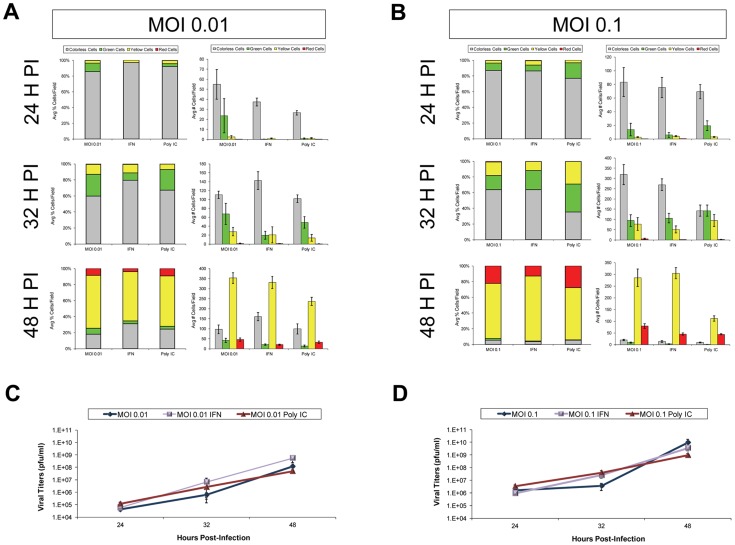
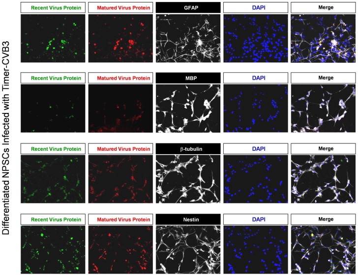
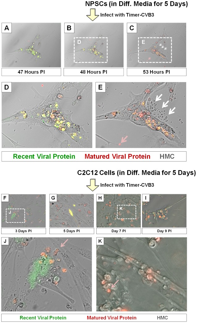
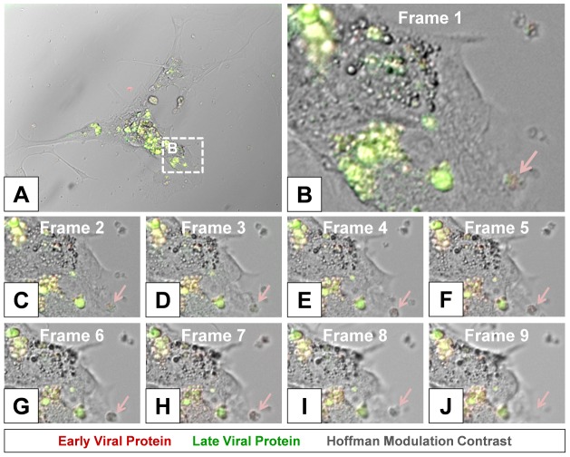
Verification
Claims the paper makes
The claims and statistics of this paper haven’t been checked yet.
Run the check — free, no account needed. It reads the open-access text and checks the sources it cites.
Claims other papers make about this one
Each is checked against this paper’s own text: does it say what the citing paper says it does?
The source backs part of the claim, not all of it — or backs it for a different population, condition or comparison.
In the citing paper
“Many viruses such as HIV, coxsackievirus, hepatitis B and C, influenza, Epstein-Barr virus, and SARS-CoV-2 highjack cellular and mitochondrial programs to enhance viral replication, package virus into EVs, and use EVs containing virus or viral components to subvert the immune response to obtain a replicative advantage in the host (Figure 1).”
From COVID-19, Myocarditis and Pericarditis (2023) · cites this paper as [45]
Experimental — accuracy not yet measured
Self-check
Percentages, sums and reported differences re-computed from the paper’s own numbers.
The self-check needs the paper’s full text.
Citations
Cites
THE USE OF LEAD CITRATE AT HIGH pH AS AN ELECTRON-OPAQUE STAIN IN ELECTRON MICROSCOPY
Reynolds, E. S. (1963) · The Journal of Cell Biology · doi:10.1083/jcb.17.1.208 · Cited by 24,938
Guidelines for the use and interpretation of assays for monitoring autophagy
Klionsky, D. J., Abdalla, F. C., Abeliovich, H., Abraham, R. T., Acevedo-Arozena, A., Adeli, K., Agholme, L., Agnello, M., Agostinis, P., Aguirre-Ghiso, J. A., et al. (2012) · Autophagy · doi:10.4161/auto.19496 · Cited by 3,996 · cited 2×
“LCII (lipidated form of LC3) studs the inner and outer autophagosome membrane and reflects autophagic activity in cells [24].”
BackgroundEditorial Office (1997) · Obesity Surgery · doi:10.1381/096089297765555359 · Cited by 3,941
The majority of microRNAs detectable in serum and saliva is concentrated in exosomes
Gallo, A., Tandon, M., Alevizos, I., & Illei, G. G. (2012) · PLoS ONE · doi:10.1371/journal.pone.0030679 · Cited by 1,060
Active Wnt proteins are secreted on exosomes
Gross, J. C., Chaudhary, V., Bartscherer, K., & Boutros, M. (2012) · Nature Cell Biology · doi:10.1038/ncb2574 · Cited by 1,025
Subversion of Cellular Autophagosomal Machinery by RNA Viruses
Jackson, W. T., Giddings, T. H., Taylor, M. P., Mulinyawe, S. B., Rabinovitch, M., Kopito, R. R., & Kirkegaard, K. (2005) · PLoS Biology · doi:10.1371/journal.pbio.0030156 · Cited by 838
A pathogenic picornavirus acquires an envelope by hijacking cellular membranes
Feng, Z., Hensley, L., McKnight, K. L., Hu, F., Madden, V. J., Ping, L., Jeong, S., Walker, C. M., Lanford, R. E., & Lemon, S. M. (2013) · Nature · doi:10.1038/nature12029 · Cited by 712
Viral Reorganization of the Secretory Pathway Generates Distinct Organelles for RNA Replication
Hsu, N.-Y., Ilnytska, O., Belov, G. A., Santiana, M., Chen, Y.-H., Takvorian, P. M., Pau, C., van der Schaar, H. M., Kaushik‐Basu, N., Balla, T., et al. (2010) · Cell · doi:10.1016/j.cell.2010.03.050 · Cited by 683
Dreux, M., Garaigorta, U., Boyd, B. S., Décembre, É., Chung, J., Whitten-Bauer, C., Wieland, S. F., & Chisari, F. V. (2012) · Cell Host & Microbe · doi:10.1016/j.chom.2012.08.010 · Cited by 496
"Fluorescent Timer": Protein That Changes Color with Time
Terskikh, A. V., Fradkov, A. F., Алексеевна, Е. Г., Zaraisky, A. G., Tan, P. B. O., Kajava, A. V., Zhao, X., Lukyanov, S., Matz, M. V., Kim, S. K., et al. (2000) · Science · doi:10.1126/science.290.5496.1585 · Cited by 402
Microvesicles and Viral Infection
Meckes, D. G., & Raab‐Traub, N. (2011) · Journal of Virology · doi:10.1128/jvi.05853-11 · Cited by 401
Autophagosome Supports Coxsackievirus B3 Replication in Host Cells
Wong, J. T., Zhang, J., Si, X., Gao, G., Mao, I., McManus, B. M., & Luo, H. (2008) · Journal of Virology · doi:10.1128/jvi.00641-08 · Cited by 388
Mukherjee, A., Morosky, S. A., Delorme‐Axford, E., Dybdahl-Sissoko, N., Oberste, M. S., Wang, T., & Coyne, C. B. (2011) · PLoS Pathogens · doi:10.1371/journal.ppat.1001311 · Cited by 316
C2C12 myoblasts release micro-vesicles containing mtDNA and proteins involved in signal transduction
Guescini, M., Guidolin, D., Vallorani, L., Casadei, L., Gioacchini, A. M., Tibollo, P., Battistelli, M., Falcieri, E., Battistin, L., Agnati, L. F., et al. (2010) · Experimental Cell Research · doi:10.1016/j.yexcr.2010.04.006 · Cited by 294 · cited 2×
“were more uniform in size (approximately 100–150 nm) and may represent traditional exosomes released by C2C12 cells [45].”
BackgroundRantakallio, P. T., Jones, P. B., Moring, J., & von Wendt, L. (1997) · International Journal of Epidemiology · doi:10.1093/ije/26.4.837 · Cited by 271
Autophagy During Cardiac Stress: Joys and Frustrations of Autophagy
Gottlieb, R. A., & Mentzer, R. M. (2010) · Annual Review of Physiology · doi:10.1146/annurev-physiol-021909-135757 · Cited by 270
Self-citationHost and virus determinants of picornavirus pathogenesis and tropism
Whitton, J. L., Cornell, C. T., & Feuer, R. (2005) · Nature Reviews Microbiology · doi:10.1038/nrmicro1284 · Cited by 249
Self-citationRepulsion of Superinfecting Virions: A Mechanism for Rapid Virus Spread
Doceul, V., Hollinshead, M. S., van der Linden, L., & Smith, G. L. (2010) · Science · doi:10.1126/science.1183173 · Cited by 230
Association of hepatitis C virus envelope proteins with exosomes
Masciopinto, F., Giovani, C., Campagnoli, S., Galli‐Stampino, L., Colombatto, P., Brunetto, M. R., Yen, T. S. B., Houghton, M., Pileri, P., & Abrignani, S. (2004) · European Journal of Immunology · doi:10.1002/eji.200424887 · Cited by 214
Enterovirus infections of the central nervous system
Rhoades, R. E., Tabor-Godwin, J. M., Tsueng, G., & Feuer, R. (2011) · Virology · doi:10.1016/j.virol.2010.12.014 · Cited by 213
Self-citationCell Cycle Status Affects Coxsackievirus Replication, Persistence, and Reactivation In Vitro
Feuer, R., Mena, I., Pagarigan, R. R., Slifka, M. K., & Whitton, J. L. (2002) · Journal of Virology · doi:10.1128/jvi.76.9.4430-4440.2002 · Cited by 183
Self-citationGBF1, a Guanine Nucleotide Exchange Factor for Arf, Is Crucial for Coxsackievirus B3 RNA Replication
Lanke, K. H. W., van der Schaar, H. M., Belov, G. A., Feng, Q., Duijsings, D., Jackson, C. L., Ehrenfeld, E., & van Kuppeveld, F. J. M. (2009) · Journal of Virology · doi:10.1128/jvi.01244-09 · Cited by 179
Horwitz, M. S., Cava, A. L., Fine, C., Rodriguez, E., Ilic, A., & Sarvetnick, N. (2000) · Nature Medicine · doi:10.1038/76277 · Cited by 166
Coxsackievirus B3 and the Neonatal CNS
Feuer, R., Mena, I., Pagarigan, R. R., Harkins, S., Hassett, D. E., & Whitton, J. L. (2003) · American Journal Of Pathology · doi:10.1016/s0002-9440(10)63496-7 · Cited by 163
Self-citationKemball, C. C., Alirezaei, M., Flynn, C. T., Wood, M. R., Harkins, S., Kiosses, W. B., & Whitton, J. L. (2010) · Journal of Virology · doi:10.1128/jvi.01417-10 · Cited by 161
Self-citationIntracellular Vesicle Acidification Promotes Maturation of Infectious Poliovirus Particles
Richards, A. L., & Jackson, W. T. (2012) · PLoS Pathogens · doi:10.1371/journal.ppat.1003046 · Cited by 141
Ornoy, A., & Tenenbaum, A. (2006) · Reproductive Toxicology · doi:10.1016/j.reprotox.2005.12.007 · Cited by 136
Extracellular Vesicles and Their Convergence with Viral Pathways
Wurdinger, T. D., Gatson, N. T., Balaj, L., Kaur, B., Breakefield, X. O., & Pegtel, D. M. (2012) · Advances in Virology · doi:10.1155/2012/767694 · Cited by 130
Enterovirus infections: Diagnosis and treatment
Sawyer, M. H. (2002) · Seminars in Pediatric Infectious Diseases · doi:10.1053/spid.2002.29756 · Cited by 130
Role of Microtubules in Extracellular Release of Poliovirus
Taylor, M. P., Burgon, T. B., Kirkegaard, K., & Jackson, W. T. (2009) · Journal of Virology · doi:10.1128/jvi.01819-08 · Cited by 106
Coxsackie virus infection of the placenta associated with neurodevelopmental delays in the newborn
Euscher, E. D. (2001) · Obstetrics and Gynecology · doi:10.1016/s0029-7844(01)01625-8 · Cited by 104
Nucleocytoplasmic Traffic Disorder Induced by Cardioviruses
Lidsky, P. V., Hato, S., Bardina, M. V., Aminev, A. G., Palmenberg, A. C., Sheval, E. V., Polyakov, V. Y., van Kuppeveld, F. J. M., & Agol, V. I. (2006) · Journal of Virology · doi:10.1128/jvi.80.6.2705-2717.2006 · Cited by 103
de Jong, A. S., Visch, H.-J., de Mattia, F., van Dommelen, M. M. T., Swarts, H. G. P., Luyten, T., Callewaert, G., Melchers, W. J. G., Willems, P. H. G. M., & van Kuppeveld, F. J. M. (2006) · Journal of Biological Chemistry · doi:10.1074/jbc.m511766200 · Cited by 98
Slifka, M. K., Pagarigan, R. R., Mena, I., Feuer, R., & Whitton, J. L. (2001) · Journal of Virology · doi:10.1128/jvi.75.5.2377-2387.2001 · Cited by 95
Self-citationPeek-a-boo: membrane hijacking and the pathogenesis of viral hepatitis
Feng, Z., & Lemon, S. M. (2013) · Trends in Microbiology · doi:10.1016/j.tim.2013.10.005 · Cited by 94
The Crystal Structure of Coxsackievirus A21 and Its Interaction with ICAM-1
Xiao, C., Bator-Kelly, C. M., Rieder, E. A., Chipman, P. R., Craig, A. G., Kuhn, R. J., Wimmer, E., & Rossmann, M. G. (2005) · Structure · doi:10.1016/j.str.2005.04.011 · Cited by 90
Coxsackievirus B4 Uses Autophagy for Replication after Calpain Activation in Rat Primary Neurons
Yoon, S. Y., Ha, Y. E., Choi, J. E., Ahn, J., Lee, H., Kweon, H., Lee, J.-Y., & Kim, D. H. (2008) · Journal of Virology · doi:10.1128/jvi.01028-08 · Cited by 88
Feuer, R., Ruller, C. M., An, N., Tabor-Godwin, J. M., Rhoades, R. E., Maciejewski, S. M., Pagarigan, R. R., Cornell, C. T., Crocker, S. J., Kiosses, W. B., et al. (2009) · Journal of Virology · doi:10.1128/jvi.02382-07 · Cited by 86
Self-citationTabor-Godwin, J. M., Ruller, C. M., Bagalso, N., An, N., Pagarigan, R. R., Harkins, S., Gilbert, P. E., Kiosses, W. B., Gude, N. A., Cornell, C. T., et al. (2010) · Journal of Neuroscience · doi:10.1523/jneurosci.1860-10.2010 · Cited by 84
Self-citationMultivesicular Bodies and Autophagy in Erythrocyte Maturation
Fader, C. M., & Colombo, M. I. (2005) · Autophagy · doi:10.4161/auto.2.2.2350 · Cited by 84
Sequences specific for enterovirus detected in spinal cord from patients with motor neurone disease
Woodall, C., Riding, M. H., Graham, D. I., & Clements, G. B. (1994) · BMJ · doi:10.1136/bmj.308.6943.1541 · Cited by 83
Enterovirus Infections of the Central Nervous System
Muir, P., & van Loon, A. M. (1997) · Intervirology · doi:10.1159/000150542 · Cited by 83
Alirezaei, M., Flynn, C. T., Wood, M. R., & Whitton, J. L. (2012) · Cell Host & Microbe · doi:10.1016/j.chom.2012.01.014 · Cited by 82
Self-citationCoxsackievirus Targets Proliferating Neuronal Progenitor Cells in the Neonatal CNS
Feuer, R., Pagarigan, R. R., Harkins, S., Liu, F., Hunziker, I. P., & Whitton, J. L. (2005) · Journal of Neuroscience · doi:10.1523/jneurosci.4517-04.2005 · Cited by 82
Self-citationViral Myocarditis: Potential Defense Mechanisms Within The Cardiomyocyte Against Virus Infection
Yajima, T. (2011) · Future Microbiology · doi:10.2217/fmb.11.40 · Cited by 79
Exosomes and retroviruses: the chicken or the egg?
Izquierdo‐Useros, N., Puertas, M. C., Borràs, F. E., Blanco, J., & Martínez‐Picado, J. (2010) · Cellular Microbiology · doi:10.1111/j.1462-5822.2010.01542.x · Cited by 78
The HTLV-1 Virological Synapse
Nejmeddine, M., & Bangham, C. R. M. (2010) · Viruses · doi:10.3390/v2071427 · Cited by 78
Cornell, C. T., Kiosses, W. B., Harkins, S., & Whitton, J. L. (2006) · Journal of Virology · doi:10.1128/jvi.02572-05 · Cited by 74
Self-citationCornell, C. T., Kiosses, W. B., Harkins, S., & Whitton, J. L. (2007) · Journal of Virology · doi:10.1128/jvi.00198-07 · Cited by 73
Self-citationA Fatal Case of Coxsackievirus B4 Meningoencephalitis
Cree, B., Bernardini, G. L., Hays, A. P., & Lowe, G. N. (2003) · Archives of Neurology · doi:10.1001/archneur.60.1.107 · Cited by 68
Wessels, E., Duijsings, D., Lanke, K. H. W., Melchers, W. J. G., Jackson, C. L., & van Kuppeveld, F. J. M. (2007) · Journal of Virology · doi:10.1128/jvi.02680-06 · Cited by 68
The role of autophagy during coxsackievirus infection of neural progenitor and stem cells
Tabor-Godwin, J. M., Tsueng, G., Sayen, M. R., Gottlieb, R. A., & Feuer, R. (2012) · Autophagy · doi:10.4161/auto.19781 · Cited by 47
Self-citationChildhood Central Nervous System Viral Infections and Adult Schizophrenia
Suvisaari, J., Mautemps, N., Haukka, J. K., Hovi, T., & Lönnqvist, J. K. (2003) · American Journal of Psychiatry · doi:10.1176/appi.ajp.160.6.1183 · Cited by 43
MRI of acute disseminated encephalomyelitis after coxsackie B infection.
David, P., Balériaux, D., Bank, W. O., Amrom, D., de Temmerman, D., Babusiaux, C., Matos, C. J. D. O., Steenwinckel, C. V., Lloret-Pastor, C., & Szliwowski, H. B. (1993) · PubMed · Cited by 39
Neural Stem Cell Depletion and CNS Developmental Defects After Enteroviral Infection
Ruller, C. M., Tabor-Godwin, J. M., Deren, D. A. V., Robinson, S. M., Maciejewski, S. M., Gluhm, S., Gilbert, P. E., An, N., Gude, N. A., Sussman, M. A., et al. (2011) · American Journal Of Pathology · doi:10.1016/j.ajpath.2011.11.016 · Cited by 37
Self-citationTsueng, G., Tabor-Godwin, J. M., Gopal, A., Ruller, C. M., Deline, S., An, N., Frausto, R. F., Milner, R., Crocker, S. J., Whitton, J. L., et al. (2011) · Journal of Virology · doi:10.1128/jvi.02261-10 · Cited by 36
Self-citationFeuer, R., & Whitton, J. L. (2008) · Springer eBooks · doi:10.1007/978-3-540-75546-3_7 · Cited by 32
Self-citationACUTE TRANSVERSE MYELITIS AND COXSACKIE A9 VIRUS INFECTION
Graber, D., Fossoud, C., Grouteau, E., Gayet-Mengelle, C., Carriere, J.-P., & Purpan, C. H. U. (1994) · The Pediatric Infectious Disease Journal · doi:10.1097/00006454-199401000-00019 · Cited by 30
Vidal, M. (2010) · Transfusion Clinique et Biologique · doi:10.1016/j.tracli.2010.05.004 · Cited by 30
CVB-induced pancreatitis and alterations in gene expression.
Ramsingh, A. (2008) · Current Topics in Microbiology and Immunology · doi:10.1007/978-3-540-75546-3_11 · Cited by 27
Puccini, J. M., Ruller, C. M., Robinson, S. M., Knopp, K. A., Buchmeier, M. J., Doran, K. S., & Feuer, R. (2014) · Laboratory Investigation · doi:10.1038/labinvest.2013.138 · Cited by 24
Self-citationInteractions between enteroviruses and autophagy in vivo
Alirezaei, M., Flynn, C. T., & Whitton, J. L. (2012) · Autophagy · doi:10.4161/auto.20160 · Cited by 23
Self-citationAlthof, N., & Whitton, J. L. (2012) · Journal of Virology · doi:10.1128/jvi.03004-12 · Cited by 22
Self-citationWoodall, C., & Graham, D. I. (2009) · European Journal of Histochemistry · doi:10.4081/877 · Cited by 20
Autophagy in coxsackievirus-infected neurons
Yoon, S.-Y., Ha, Y.-E., Choi, J.-E., Ahn, J., Lee, H., & Kim, D. (2009) · Autophagy · doi:10.4161/auto.5.3.7723 · Cited by 16
Is Autophagy an Avenue to Modulate Coxsackievirus Replication and Pathogenesis?
Luo, H., & McManus, B. M. (2012) · Future Microbiology · doi:10.2217/fmb.12.67 · Cited by 7
Research works printed but not linked: 2
- G Tsueng, 2014, Unique 2C and 3A viral protein mutations identified in hypervirulent and attenuated coxsackievirus strains isolated from persistently-infected neural progenitor cell cultures, Manuscript In Preparation
- H Luo, 2012, Is autophagy an avenue to modulate coxsackievirus replication and pathogenesis?, Future Microbiol, 7, 4
Cited by
Sorted by citations
Guidelines for the use and interpretation of assays for monitoring autophagy (4th edition) 1
Klionsky, D. J., Abdel-Aziz, A. K., Abdelfatah, S., Abdellatif, M., Abdoli, A., Abel, S., Abeliovich, H., Abildgaard, M. H., Abudu, Y. P., Acevedo-Arozena, A., et al. (2021) · Autophagy · doi:10.1080/15548627.2020.1797280 · Cited by 2,897
Self-citationExtracellular vesicles as tools and targets in therapy for diseases
Kumar, M. A., Baba, S. K., Sadida, H. Q., Al-Marzooqi, S. K., Jerobin, J., Altemani, F. H., Algehainy, N. A., Alanazi, M. A., Abou‐Samra, A., Kumar, R., et al. (2024) · Signal Transduction and Targeted Therapy · doi:10.1038/s41392-024-01735-1 · Cited by 1,545
Autophagy during viral infection — a double-edged sword
Choi, Y., Bowman, J. W., & Jung, J. U. (2018) · Nature Reviews Microbiology · doi:10.1038/s41579-018-0003-6 · Cited by 838
Autophagy-Independent Functions of the Autophagy Machinery
Galluzzi, L., & Green, D. R. (2019) · Cell · doi:10.1016/j.cell.2019.05.026 · Cited by 863
“, 2018); (3) early release of virus-loaded exosome-like microvesicles that bear autophagosomal markers (Robinson et al., 2014; Sin et al., 2017); and (4) ultimately, activation of apoptotic cell death for massive viral dissemination (Xin et al.”
BackgroundExosomes and other extracellular vesicles in host–pathogen interactions
Schorey, J. S., Cheng, Y., Singh, P., & Smith, V. L. (2014) · EMBO Reports · doi:10.15252/embr.201439363 · Cited by 752
Metadata
- Authors
- Robinson, S. M., Tsueng, G., Sin, J., Mangale, V., Rahawi, S., McIntyre, L. L., Williams, W., Kha, N., Cruz, C., Hancock, B. M., Nguyen, D. P., Sayen, M. R., Hilton, B. J., Doran, K. S., Segall, A. M., Wolkowicz, R., Cornell, C. T., Whitton, J. L., Gottlieb, R. A., & Feuer, R.
- Affiliations
- San Diego State University (US)
- Scripps Research Institute (US)
- Corresponding author
- Feuer, Ralph
- ORCID
- Venue
- PLoS Pathogens
- Publisher
- Public Library of Science (PLoS)
- Published
- 2014-04-10
- Volume
- 10
- Issue
- 4
- Pages
- e1004045
- ISSN
- 1553-7366 (ISSN-L)
- 1553-7374
- PMC
- PMC3983045
- OpenAlex
- W1994527574
- Cited by
- 322
- References
- 66
- Citations by year
- 2026: 11 · 2025: 21 · 2024: 26 · 2023: 29 · 2022: 25 · 2021: 32 · 2020: 43 · 2019: 29 · 2018: 27 · 2017: 20 · 2016: 19 · 2015: 29 · 2014: 11
- FWCI
- 18.10
- Type
- journal article
- Language
- en
- Open access
- gold
- doi.org
- Locations
- PLoS Pathogens (publishedVersion, cc-by)
- Europe PMC (PubMed Central) (publishedVersion, cc-by)
- PubMed Central (publishedVersion, cc-by)
- PubMed (publishedVersion)
- DOAJ (DOAJ: Directory of Open Access Journals) (submittedVersion, cc-by-sa)
- Figshare (submittedVersion, cc-by)
- Funders
- National Science Foundation (1046413, DBI-030829)
- San Diego State University
- National Institutes of Health (NIH-R01, HL093177, AI042314, NS054108)
- National Heart, Lung, and Blood Institute (R01HL092136, R01HL093177, T32HL116273)
- National Institute of Mental Health (R24MH065515, MH065515)
- National Institute of Allergy and Infectious Diseases (F32AI065095, R01AI042314)
- National Institute of Neurological Disorders and Stroke (R01NS054108)
- Division of Environmental Biology (1046413)
- Topics
- Health Sciences › Medicine › Infectious Diseases › Viral Infections and Immunology Research
- Life Sciences › Biochemistry, Genetics and Molecular Biology › Cell Biology › Cellular transport and secretion
- Life Sciences › Biochemistry, Genetics and Molecular Biology › Cell Biology › Autophagy in Disease and Therapy
- Keywords
- coxsackievirus B3, extracellular microvesicles, autophagy, viral dissemination, myoblasts, LC3, 3A protein, type I interferon, ribavirin, viral myocarditis, poliovirus
- MeSH terms
- Animals, Enterovirus Infections, HeLa Cells, Humans, Microtubule-Associated Proteins, Phagosomes, Viral Proteins, Virus Shedding, Enterovirus B, Human, Mice, Cell-Derived Microparticles
About these checks
Registry, retraction, Cites and statistics results are computed, not judged, with one exception: a printed reference that no index lists is read once by a model before it counts as a citation error. Claims are extracted by a model and kept only when their quote appears verbatim in the text; each cited source is then checked, with a model’s help, for whether it says what the claim relies on. Those verdicts can be wrong — read the claim’s quote and the cited source before relying on one.
Statistics and arithmetic are recomputed, not judged.
Confidence bands are shown only for fields calibrated on our annotated test set. None is calibrated yet, so no band is shown.
What counts as a claim ›How we measure confidence ›How we check this ›
Are you an author, or do you see a problem with this page? Request a review
