

The DOI matches its Crossref record; 66 of the 66 references it cites were found in a registry or an index.
Coxsackievirus B Exits the Host Cell in Shed Microvesicles Displaying Autophagosomal Markers
Open accessAbstract
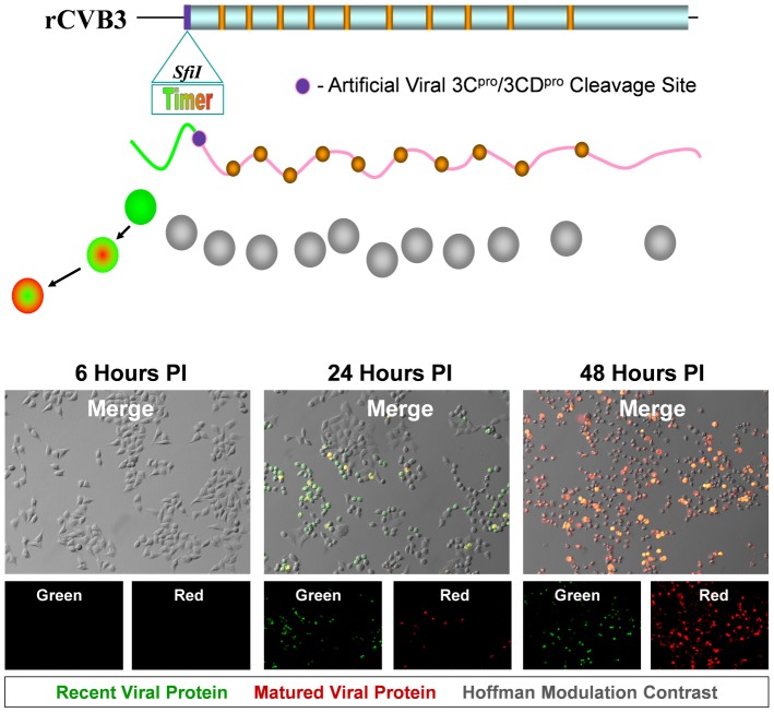
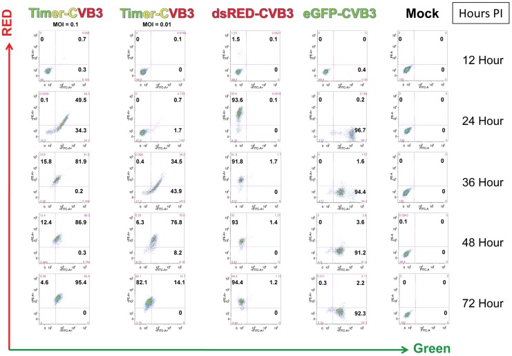
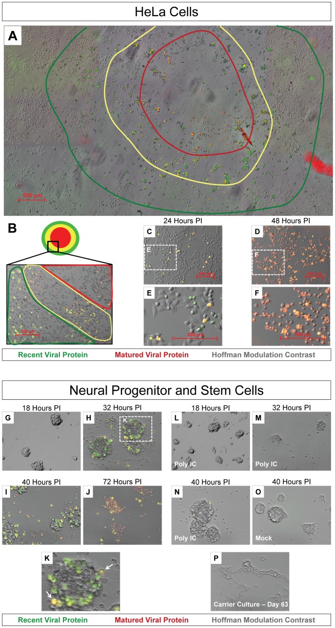
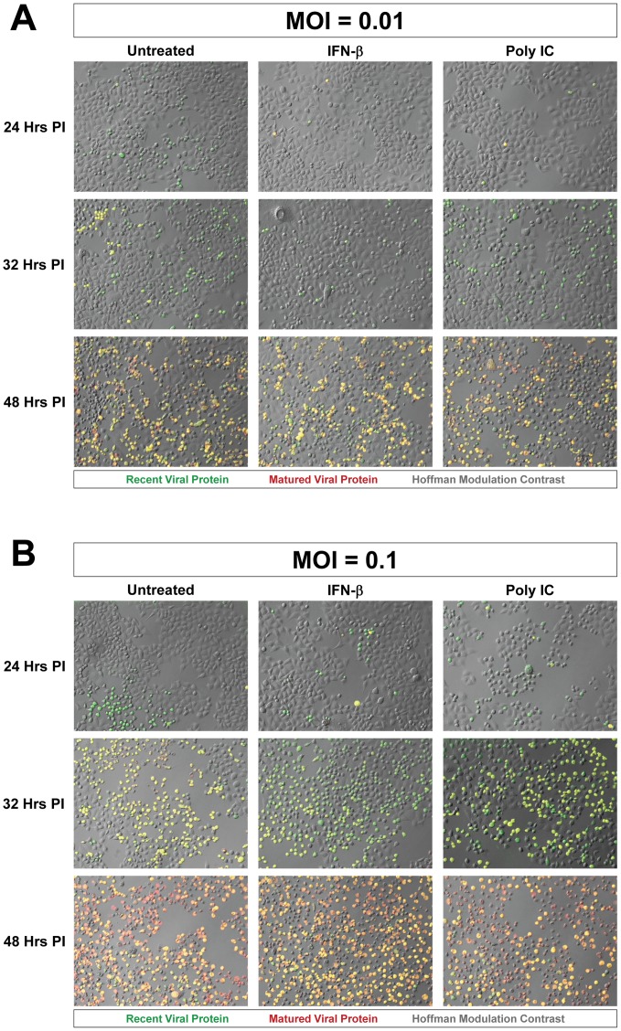
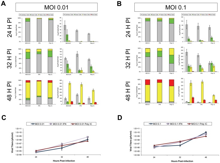
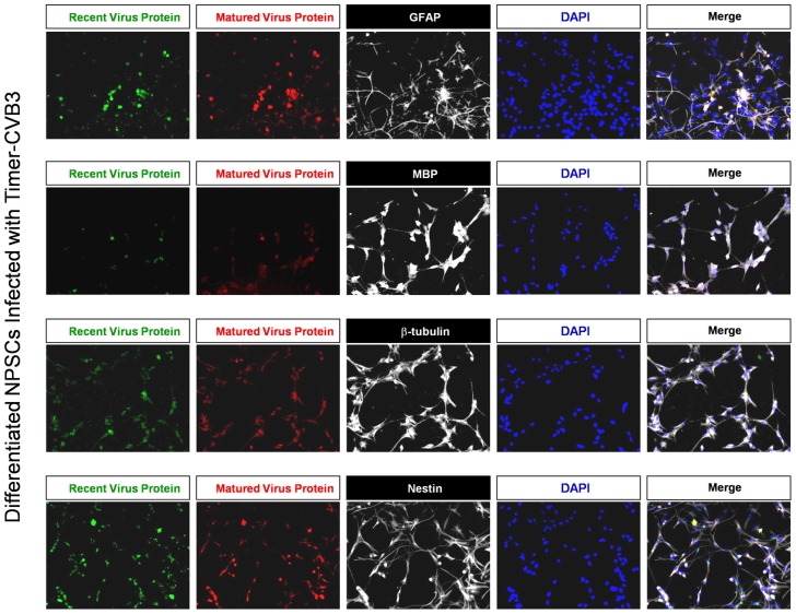
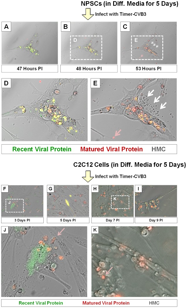
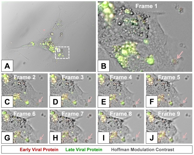
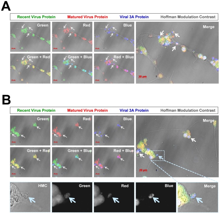
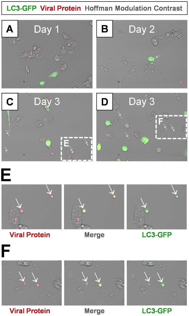
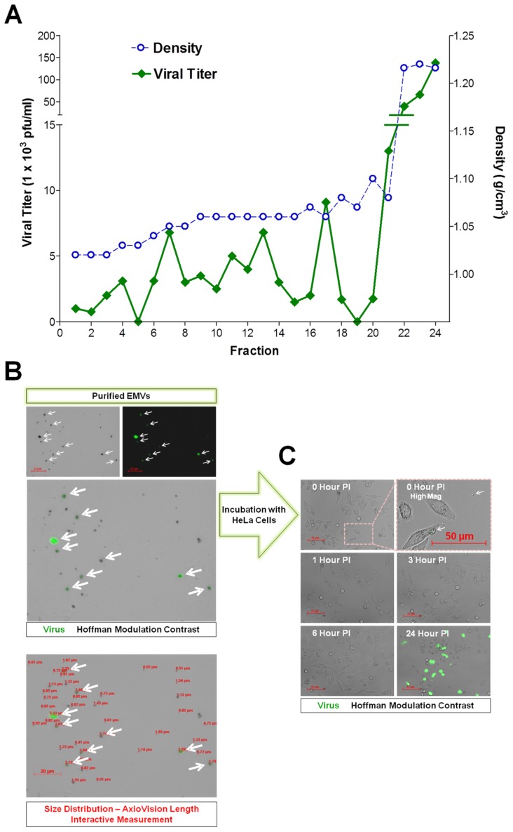
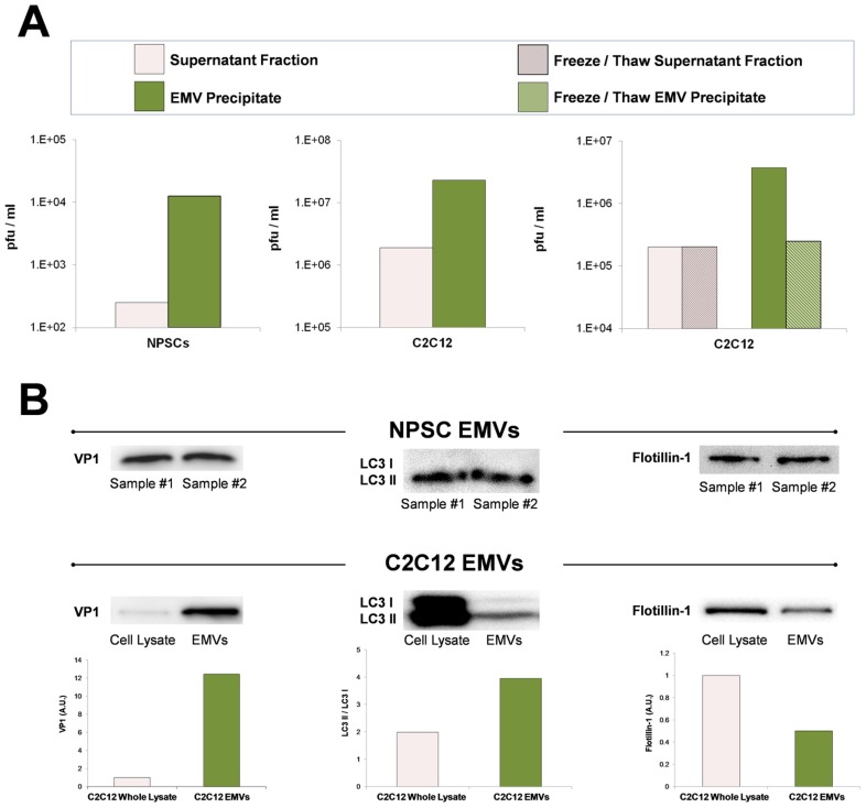
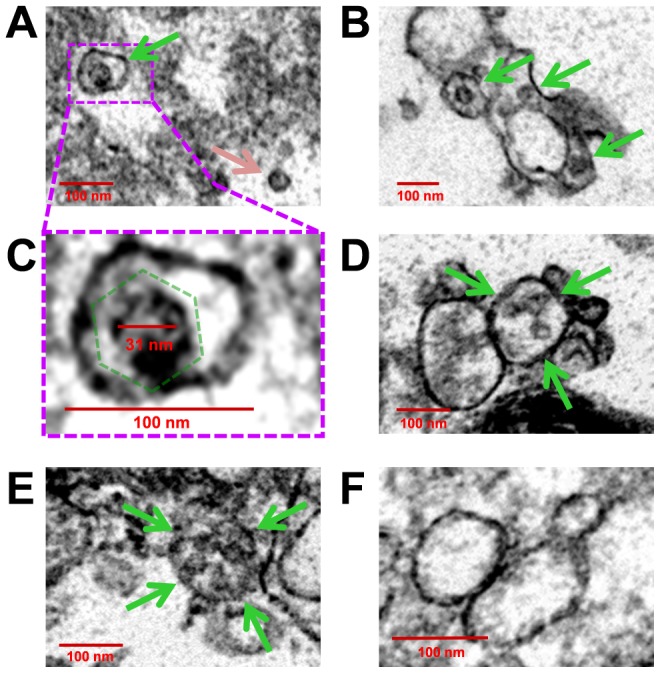
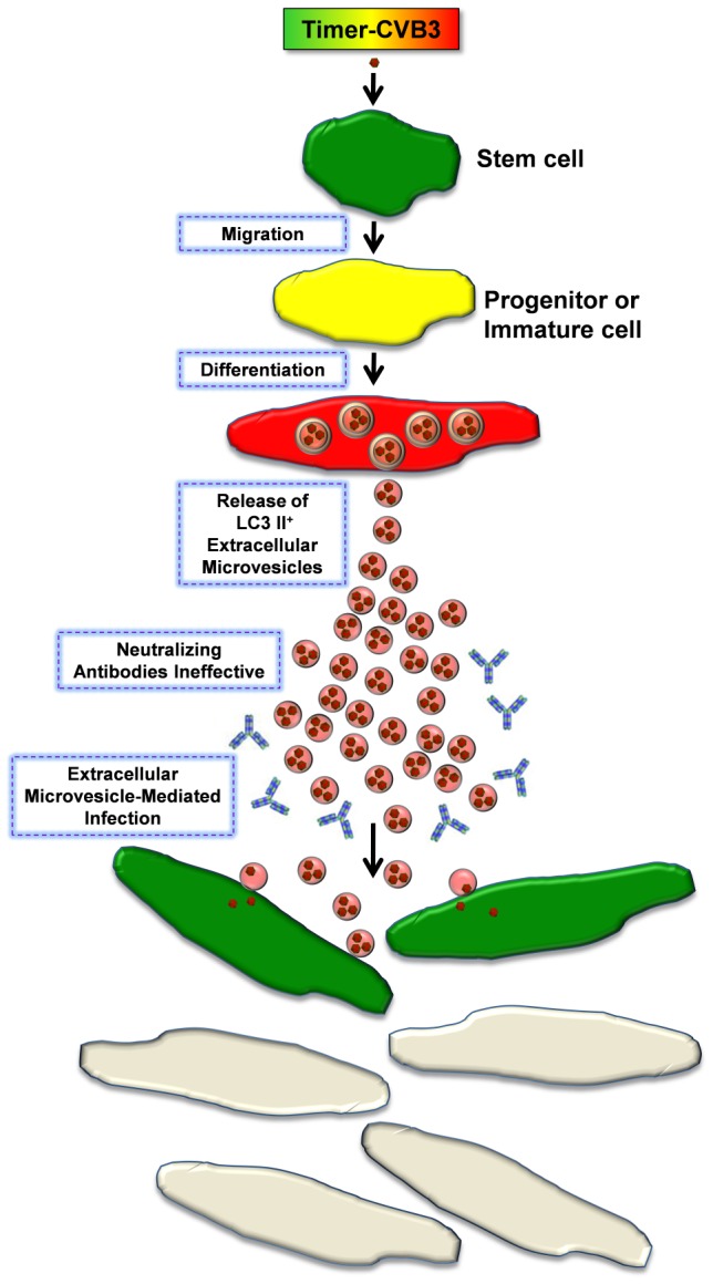
Coxsackievirus B3 (CVB3), a member of the picornavirus family and enterovirus genus, causes viral myocarditis, aseptic meningitis, and pancreatitis in humans. We genetically engineered a unique molecular marker, "fluorescent timer" protein, within our infectious CVB3 clone and isolated a high-titer recombinant viral stock (Timer-CVB3) following transfection in HeLa cells. "Fluorescent timer" protein undergoes slow conversion of fluorescence from green to red over time, and Timer-CVB3 can be utilized to track virus infection and dissemination in real time. Upon infection with Timer-CVB3, HeLa cells, neural progenitor and stem cells (NPSCs), and C2C12 myoblast cells slowly changed fluorescence from green to red over 72 hours as determined by fluorescence microscopy or flow cytometric analysis. The conversion of "fluorescent timer" protein in HeLa cells infected with Timer-CVB3 could be interrupted by fixation, suggesting that the fluorophore was stabilized by formaldehyde cross-linking reactions. Induction of a type I interferon response or ribavirin treatment reduced the progression of cell-to-cell virus spread in HeLa cells or NPSCs infected with Timer-CVB3. Time lapse photography of partially differentiated NPSCs infected with Timer-CVB3 revealed substantial intracellular membrane remodeling and the assembly of discrete virus replication organelles which changed fluorescence color in an asynchronous fashion within the cell. "Fluorescent timer" protein colocalized closely with viral 3A protein within virus replication organelles. Intriguingly, infection of partially differentiated NPSCs or C2C12 myoblast cells induced the release of abundant extracellular microvesicles (EMVs) containing matured "fluorescent timer" protein and infectious virus representing a novel route of virus dissemination. CVB3 virions were readily observed within purified EMVs by transmission electron microscopy, and infectious virus was identified within low-density isopycnic iodixanol gradient fractions consistent with membrane association. The preferential detection of the lipidated form of LC3 protein (LC3 II) in released EMVs harboring infectious virus suggests that the autophagy pathway plays a crucial role in microvesicle shedding and virus release, similar to a process previously described as autophagosome-mediated exit without lysis (AWOL) observed during poliovirus replication. Through the use of this novel recombinant virus which provides more dynamic information from static fluorescent images, we hope to gain a better understanding of CVB3 tropism, intracellular membrane reorganization, and virus-associated microvesicle dissemination within the host.
Overview
The claims and statistics of this paper haven’t been checked yet.
Run the check — free, no account needed. It reads the open-access text and checks the sources it cites.
Integrity signals
Registry
Cites
Statistics
Figures
Verification
Claims the paper makes
The claims and statistics of this paper haven’t been checked yet.
Run the check — free, no account needed. It reads the open-access text and checks the sources it cites.
Claims other papers make about this one
Each is checked against this paper’s own text: does it say what the citing paper says it does?
The source backs part of the claim, not all of it — or backs it for a different population, condition or comparison.
In the citing paper
“Many viruses such as HIV, coxsackievirus, hepatitis B and C, influenza, Epstein-Barr virus, and SARS-CoV-2 highjack cellular and mitochondrial programs to enhance viral replication, package virus into EVs, and use EVs containing virus or viral components to subvert the immune response to obtain a replicative advantage in the host (Figure 1).”
From COVID-19, Myocarditis and Pericarditis (2023) · cites this paper as [45]
Experimental — accuracy not yet measured
Self-check
Percentages, sums and reported differences re-computed from the paper’s own numbers.
The self-check needs the paper’s full text.
Citations
Cites
THE USE OF LEAD CITRATE AT HIGH pH AS AN ELECTRON-OPAQUE STAIN IN ELECTRON MICROSCOPY
Reynolds, E. S. (1963) · The Journal of Cell Biology · doi:10.1083/jcb.17.1.208 · Cited by 24,938
Guidelines for the use and interpretation of assays for monitoring autophagy
Klionsky, D. J., Abdalla, F. C., Abeliovich, H., Abraham, R. T., Acevedo-Arozena, A., Adeli, K., Agholme, L., Agnello, M., Agostinis, P., Aguirre-Ghiso, J. A., et al. (2012) · Autophagy · doi:10.4161/auto.19496 · Cited by 3,996 · cited 2×
“LCII (lipidated form of LC3) studs the inner and outer autophagosome membrane and reflects autophagic activity in cells [24].”
BackgroundEditorial Office (1997) · Obesity Surgery · doi:10.1381/096089297765555359 · Cited by 3,941
The majority of microRNAs detectable in serum and saliva is concentrated in exosomes
Gallo, A., Tandon, M., Alevizos, I., & Illei, G. G. (2012) · PLoS ONE · doi:10.1371/journal.pone.0030679 · Cited by 1,060
Active Wnt proteins are secreted on exosomes
Gross, J. C., Chaudhary, V., Bartscherer, K., & Boutros, M. (2012) · Nature Cell Biology · doi:10.1038/ncb2574 · Cited by 1,025
Subversion of Cellular Autophagosomal Machinery by RNA Viruses
Jackson, W. T., Giddings, T. H., Taylor, M. P., Mulinyawe, S. B., Rabinovitch, M., Kopito, R. R., & Kirkegaard, K. (2005) · PLoS Biology · doi:10.1371/journal.pbio.0030156 · Cited by 838
A pathogenic picornavirus acquires an envelope by hijacking cellular membranes
Feng, Z., Hensley, L., McKnight, K. L., Hu, F., Madden, V. J., Ping, L., Jeong, S., Walker, C. M., Lanford, R. E., & Lemon, S. M. (2013) · Nature · doi:10.1038/nature12029 · Cited by 712
Viral Reorganization of the Secretory Pathway Generates Distinct Organelles for RNA Replication
Hsu, N.-Y., Ilnytska, O., Belov, G. A., Santiana, M., Chen, Y.-H., Takvorian, P. M., Pau, C., van der Schaar, H. M., Kaushik‐Basu, N., Balla, T., et al. (2010) · Cell · doi:10.1016/j.cell.2010.03.050 · Cited by 683
Dreux, M., Garaigorta, U., Boyd, B. S., Décembre, É., Chung, J., Whitten-Bauer, C., Wieland, S. F., & Chisari, F. V. (2012) · Cell Host & Microbe · doi:10.1016/j.chom.2012.08.010 · Cited by 496
"Fluorescent Timer": Protein That Changes Color with Time
Terskikh, A. V., Fradkov, A. F., Алексеевна, Е. Г., Zaraisky, A. G., Tan, P. B. O., Kajava, A. V., Zhao, X., Lukyanov, S., Matz, M. V., Kim, S. K., et al. (2000) · Science · doi:10.1126/science.290.5496.1585 · Cited by 402
Research works printed but not linked: 2
- G Tsueng, 2014, Unique 2C and 3A viral protein mutations identified in hypervirulent and attenuated coxsackievirus strains isolated from persistently-infected neural progenitor cell cultures, Manuscript In Preparation
- H Luo, 2012, Is autophagy an avenue to modulate coxsackievirus replication and pathogenesis?, Future Microbiol, 7, 4
Cited by
Sorted by citations
Guidelines for the use and interpretation of assays for monitoring autophagy (4th edition) 1
Klionsky, D. J., Abdel-Aziz, A. K., Abdelfatah, S., Abdellatif, M., Abdoli, A., Abel, S., Abeliovich, H., Abildgaard, M. H., Abudu, Y. P., Acevedo-Arozena, A., et al. (2021) · Autophagy · doi:10.1080/15548627.2020.1797280 · Cited by 2,897
Self-citationExtracellular vesicles as tools and targets in therapy for diseases
Kumar, M. A., Baba, S. K., Sadida, H. Q., Al-Marzooqi, S. K., Jerobin, J., Altemani, F. H., Algehainy, N. A., Alanazi, M. A., Abou‐Samra, A., Kumar, R., et al. (2024) · Signal Transduction and Targeted Therapy · doi:10.1038/s41392-024-01735-1 · Cited by 1,545
Autophagy during viral infection — a double-edged sword
Choi, Y., Bowman, J. W., & Jung, J. U. (2018) · Nature Reviews Microbiology · doi:10.1038/s41579-018-0003-6 · Cited by 838
Autophagy-Independent Functions of the Autophagy Machinery
Galluzzi, L., & Green, D. R. (2019) · Cell · doi:10.1016/j.cell.2019.05.026 · Cited by 863
“, 2018); (3) early release of virus-loaded exosome-like microvesicles that bear autophagosomal markers (Robinson et al., 2014; Sin et al., 2017); and (4) ultimately, activation of apoptotic cell death for massive viral dissemination (Xin et al.”
BackgroundExosomes and other extracellular vesicles in host–pathogen interactions
Schorey, J. S., Cheng, Y., Singh, P., & Smith, V. L. (2014) · EMBO Reports · doi:10.15252/embr.201439363 · Cited by 752
Autophagy and autophagy-related proteins in the immune system
Shibutani, S., Saitoh, T., Nowag, H., Münz, C., & Yoshimori, T. (2015) · Nature Immunology · doi:10.1038/ni.3273 · Cited by 593
Ponpuak, M., Mandell, M. A., Kimura, T., Chauhan, S., Cleyrat, C., & Deretic, V. P. (2015) · Current Opinion in Cell Biology · doi:10.1016/j.ceb.2015.04.016 · Cited by 527
The life cycle of non-polio enteroviruses and how to target it
Baggen, J., Thibaut, H. J., Strating, J. R. P. M., & van Kuppeveld, F. J. M. (2018) · Nature Reviews Microbiology · doi:10.1038/s41579-018-0005-4 · Cited by 500
Phosphatidylserine Vesicles Enable Efficient En Bloc Transmission of Enteroviruses
Chen, Y.-H., Du, W., Hagemeijer, M. C., Takvorian, P. M., Pau, C., Cali, A., Brantner, C. A., Stempinski, E., Connelly, P. S., Ma, H.-C., et al. (2015) · Cell · doi:10.1016/j.cell.2015.01.032 · Cited by 470
Vesicle-Cloaked Virus Clusters Are Optimal Units for Inter-organismal Viral Transmission
Santiana, M., Ghosh, S., Ho, B. A., Rajasekaran, V., Du, W., Mutsafi, Y., Jesús-Díaz, D. A. D., Sosnovtsev, S. V., Levenson, E. A., Parra, G. I., et al. (2018) · Cell Host & Microbe · doi:10.1016/j.chom.2018.07.006 · Cited by 270
Viral effects on the content and function of extracellular vesicles
Raab‐Traub, N., & Dittmer, D. P. (2017) · Nature Reviews Microbiology · doi:10.1038/nrmicro.2017.60 · Cited by 270
Extracellular Vesicles Exploit Viral Entry Routes for Cargo Delivery
van Dongen, H. M., Masoumi, N., Witwer, K. W., & Pegtel, D. M. (2016) · Microbiology and Molecular Biology Reviews · doi:10.1128/mmbr.00063-15 · Cited by 271
Coxsackievirus B3 Replication and Pathogenesis
Garmaroudi, F. S., Marchant, D., Hendry, R. G., Luo, H., Yang, D., Ye, X., Shi, J., & McManus, B. M. (2015) · Future Microbiology · doi:10.2217/fmb.15.5 · Cited by 232
Autophagy-dependent secretion: mechanism, factors secreted, and disease implications
New, J., & Thomas, S. M. (2019) · Autophagy · doi:10.1080/15548627.2019.1596479 · Cited by 234
The role of autophagy in viral infections
Chen, T., Tu, S., Ding, L., Jin, M., Chen, H., & Zhou, H. (2023) · Journal of Biomedical Science · doi:10.1186/s12929-023-00899-2 · Cited by 235
Viruses and the autophagy pathway
Jackson, W. T. (2015) · Virology · doi:10.1016/j.virol.2015.03.042 · Cited by 215
Picornaviridae—the ever-growing virus family
Zell, R. (2017) · Archives of Virology · doi:10.1007/s00705-017-3614-8 · Cited by 196
COVID-19, Myocarditis and Pericarditis
Fairweather, D., Beetler, D. J., Di Florio, D. N., Musigk, N., Heidecker, B., & Cooper, L. T. (2023) · Circulation Research · doi:10.1161/circresaha.123.321878 · Cited by 194
Mechanisms and Concepts in RNA Virus Population Dynamics and Evolution
Dolan, P., Whitfield, Z. J., & Andino, R. (2018) · Annual Review of Virology · doi:10.1146/annurev-virology-101416-041718 · Cited by 178
Viral and host heterogeneity and their effects on the viral life cycle
Jones, J. E., Sage, V. L., & Lakdawala, S. S. (2020) · Nature Reviews Microbiology · doi:10.1038/s41579-020-00449-9 · Cited by 173
Autophagy Beyond Intracellular MHC Class II Antigen Presentation
Münz, C. (2016) · Trends in Immunology · doi:10.1016/j.it.2016.08.017 · Cited by 158
Replication and Inhibitors of Enteroviruses and Parechoviruses
van der Linden, L., Wolthers, K. C., & van Kuppeveld, F. J. M. (2015) · Viruses · doi:10.3390/v7082832 · Cited by 162
Extracellular vesicles are the Trojan horses of viral infection
Altan‐Bonnet, N. (2016) · Current Opinion in Microbiology · doi:10.1016/j.mib.2016.05.004 · Cited by 139
Fu, Y., Zhang, L., Zhang, F., Tang, T. F., Zhou, Q. G., Feng, C., Jin, Y., & Wu, Z. (2017) · PLoS Pathogens · doi:10.1371/journal.ppat.1006611 · Cited by 137
Mohamud, Y., Shi, J., Qu, J., Poon, T. K. C., Xue, Y. C., Deng, H., Zhang, J., & Luo, H. (2018) · Cell Reports · doi:10.1016/j.celrep.2018.02.090 · Cited by 138
Lipid Tales of Viral Replication and Transmission
Altan‐Bonnet, N. (2016) · Trends in Cell Biology · doi:10.1016/j.tcb.2016.09.011 · Cited by 132
Mao, J., Lin, E., He, L., Yu, J., Tan, P., & Zhou, Y. (2019) · Advances in experimental medicine and biology · doi:10.1007/978-981-15-0606-2_5 · Cited by 141
Corona, A. K., Saulsbery, H. M., Velazquez, A. C., & Jackson, W. T. (2018) · Cell Reports · doi:10.1016/j.celrep.2018.03.003 · Cited by 130
Virus Impact on Lipids and Membranes
Ketter, E., & Randall, G. C. (2019) · Annual Review of Virology · doi:10.1146/annurev-virology-092818-015748 · Cited by 124
Opportunistic intruders: how viruses orchestrate ER functions to infect cells
Ravindran, M. S., Bagchi, P., Cunningham, C. N., & Tsai, B. (2016) · Nature Reviews Microbiology · doi:10.1038/nrmicro.2016.60 · Cited by 117
Dorobantu, C. M., Albulescu, L., Harak, C., Feng, Q., van Kampen, M., Strating, J. R. P. M., Gorbalenya, A. E., Lohmann, V., van der Schaar, H. M., & van Kuppeveld, F. J. M. (2015) · PLoS Pathogens · doi:10.1371/journal.ppat.1005185 · Cited by 119
Mitochondrial quality control: Easy come, easy go
Stotland, A. B., & Gottlieb, R. A. (2015) · Biochimica et Biophysica Acta (BBA) - Molecular Cell Research · doi:10.1016/j.bbamcr.2014.12.041 · Cited by 123
Self-citationAutophagy and Mammalian Viruses
Paul, P., & Münz, C. (2016) · Advances in virus research · doi:10.1016/bs.aivir.2016.02.002 · Cited by 119
This old heart: Cardiac aging and autophagy
Linton, P., Gurney, M. A., Sengstock, D. M., Mentzer, R. M., & Gottlieb, R. A. (2014) · Journal of Molecular and Cellular Cardiology · doi:10.1016/j.yjmcc.2014.12.017 · Cited by 115
Self-citationvan der Schaar, H. M., Dorobantu, C. M., Albulescu, L., Strating, J. R. P. M., & van Kuppeveld, F. J. M. (2016) · Trends in Microbiology · doi:10.1016/j.tim.2016.02.017 · Cited by 118
Enteroviral proteases: structure, host interactions and pathogenicity
Laitinen, O. H., Svedin, E., Kapell, S., Nurminen, A., Hytönen, V. P., & Flodström‐Tullberg, M. (2016) · Reviews in Medical Virology · doi:10.1002/rmv.1883 · Cited by 110
Autophagy proteins in antigen processing for presentation on MHC molecules
Münz, C. (2016) · Immunological Reviews · doi:10.1111/imr.12422 · Cited by 121
Recent progress in understanding coxsackievirus replication, dissemination, and pathogenesis
Sin, J., Mangale, V., Thienphrapa, W., Gottlieb, R. A., & Feuer, R. (2015) · Virology · doi:10.1016/j.virol.2015.06.006 · Cited by 111
Self-citationCoxsackievirus B Escapes the Infected Cell in Ejected Mitophagosomes
Sin, J., McIntyre, L., Stotland, A., Feuer, R., & Gottlieb, R. A. (2017) · Journal of Virology · doi:10.1128/jvi.01347-17 · Cited by 103
Self-citationProtein composition of the hepatitis A virus quasi-envelope
McKnight, K. L., Xie, L., González‐López, O., Rivera-Serrano, E. E., Chen, X., & Lemon, S. M. (2017) · Proceedings of the National Academy of Sciences · doi:10.1073/pnas.1619519114 · Cited by 105
Viral journeys on the intracellular highways
Robinson, M., Schor, S., Barouch‐Bentov, R., & Einav, S. (2018) · Cellular and Molecular Life Sciences · doi:10.1007/s00018-018-2882-0 · Cited by 101
Extracellular Vesicles in Viral Infections: Two Sides of the Same Coin?
Martins, S. D. T., & Alves, L. R. (2020) · Frontiers in Cellular and Infection Microbiology · doi:10.3389/fcimb.2020.593170 · Cited by 98
Cellular entry and uncoating of naked and quasi-enveloped human hepatoviruses
Rivera-Serrano, E. E., González‐López, O., Das, A., & Lemon, S. M. (2019) · eLife · doi:10.7554/elife.43983 · Cited by 96
Enteroviral Infection: The Forgotten Link to Amyotrophic Lateral Sclerosis?
Xue, Y. C., Feuer, R., Cashman, N. R., & Luo, H. (2018) · Frontiers in Molecular Neuroscience · doi:10.3389/fnmol.2018.00063 · Cited by 97
Self-citationRichardson, S. J., & Morgan, N. G. (2018) · Current Opinion in Pharmacology · doi:10.1016/j.coph.2018.07.006 · Cited by 96
Role of autophagy in IL-1β export and release from cells
Claude‐Taupin, A., Bissa, B., Jia, J., Gu, Y., & Deretic, V. P. (2018) · Seminars in Cell and Developmental Biology · doi:10.1016/j.semcdb.2018.03.012 · Cited by 89
Extracellular vesicles: Vehicles of en bloc viral transmission
Altan‐Bonnet, N., Perales, C., & Domingo, E. (2019) · Virus Research · doi:10.1016/j.virusres.2019.03.023 · Cited by 82
A New Infectious Unit: Extracellular Vesicles Carrying Virus Populations
Kerviel, A., Zhang, M. Y., & Altan‐Bonnet, N. (2021) · Annual Review of Cell and Developmental Biology · doi:10.1146/annurev-cellbio-040621-032416 · Cited by 85
Differential and convergent utilization of autophagy components by positive-strand RNA viruses
Abernathy, E., Mateo, R., Majzoub, K., van Buuren, N. J., Bird, S. W., Carette, J. E., & Kirkegaard, K. (2019) · PLoS Biology · doi:10.1371/journal.pbio.2006926 · Cited by 87
A time to reap, a time to sow: Mitophagy and biogenesis in cardiac pathophysiology
Andres, A. M., Stotland, A., Queliconi, B. B., & Gottlieb, R. A. (2015) · Journal of Molecular and Cellular Cardiology · doi:10.1016/j.yjmcc.2014.10.003 · Cited by 78
Self-citationRole of Microvesicles in the Spread of Herpes Simplex Virus 1 in Oligodendrocytic Cells
Bello-Morales, R., Praena, B., de la Nuez, C., Rejas, M. T., Guerra, M., Galán-Ganga, M., Izquierdo, M., Calvo, V., Krummenacher, C., & López‐Guerrero, J. A. (2018) · Journal of Virology · doi:10.1128/jvi.00088-18 · Cited by 79
Extracellular Vesicles in Viral Spread and Antiviral Response
Bello‐Morales, R., Ripa, I., & López‐Guerrero, J. A. (2020) · Viruses · doi:10.3390/v12060623 · Cited by 76
CALCOCO2/NDP52 and SQSTM1/p62 differentially regulate coxsackievirus B3 propagation
Mohamud, Y., Qu, J., Xue, Y. C., Liu, H., Deng, H., & Luo, H. (2018) · Cell Death and Differentiation · doi:10.1038/s41418-018-0185-5 · Cited by 76
Jiang, W., Ma, P., Deng, L., Liu, Z., Wang, X., Liu, X., & Long, G. (2020) · Journal of Extracellular Vesicles · doi:10.1080/20013078.2020.1716513 · Cited by 74
Catch Me If You Can: The Link between Autophagy and Viruses
Lennemann, N. J., & Coyne, C. B. (2015) · PLoS Pathogens · doi:10.1371/journal.ppat.1004685 · Cited by 75
The autophagic machinery ensures nonlytic transmission of mycobacteria
Gerstenmaier, L., Pilla, R., Herrmann, L., Herrmann, H., Prado, M., Villafano, G. J., Kolonko, M., Reimer, R., Soldati, T., King, J., et al. (2015) · Proceedings of the National Academy of Sciences · doi:10.1073/pnas.1423318112 · Cited by 79
Arzt, J., Belsham, G. J., Lohse, L., Bøtner, A. G., & Stenfeldt, C. (2018) · mSphere · doi:10.1128/msphere.00365-18 · Cited by 74
Escape of non-enveloped virus from intact cells
Bird, S. W., & Kirkegaard, K. (2015) · Virology · doi:10.1016/j.virol.2015.03.044 · Cited by 74
Mao, L., Wu, J., Shen, L., Yang, J., Chen, J., & Xu, H. (2016) · Virus Genes · doi:10.1007/s11262-016-1292-3 · Cited by 72
Enterovirus 71 infection of motor neuron-like NSC-34 cells undergoes a non-lytic exit pathway
Too, I. H. K., Yeo, H., Sessions, O. M., Yan, B., Libau, E. A., Howe, J. L. C., Lim, Z. Q., Shalini, S.-M., Ong, W., Chua, K. B., et al. (2016) · Scientific Reports · doi:10.1038/srep36983 · Cited by 72
Autophagy, EVs, and Infections: A Perfect Question for a Perfect Time
Pleet, M. L., Branscome, H., DeMarino, C., Pinto, D. O., Zadeh, M. A., Rodriguez, M., Sariyer, I. K., El‐Hage, N., & Kashanchi, F. (2018) · Frontiers in Cellular and Infection Microbiology · doi:10.3389/fcimb.2018.00362 · Cited by 73
Viral cell-to-cell spread: Conventional and non-conventional ways
Cifuentes-Muñoz, N., Najjar, F. E., & Dutch, R. (2020) · Advances in virus research · doi:10.1016/bs.aivir.2020.09.002 · Cited by 71
Enterovirus Transmission by Secretory Autophagy
Mutsafi, Y., & Altan‐Bonnet, N. (2018) · Viruses · doi:10.3390/v10030139 · Cited by 67
van der Grein, S. G., Defourny, K. A. Y., Rabouw, H. H., Galiveti, C. R., Langereis, M. A., Wauben, M. H. M., Arkesteijn, G. J. A., van Kuppeveld, F. J. M., & Hoen, E. N. M. N. (2019) · PLoS Pathogens · doi:10.1371/journal.ppat.1007594 · Cited by 68
Extracellular vesicles in virus infection and pathogenesis
McNamara, R. P., & Dittmer, D. P. (2020) · Current Opinion in Virology · doi:10.1016/j.coviro.2020.07.014 · Cited by 69
Lyn kinase regulates egress of flaviviruses in autophagosome-derived organelles
Li, M. Y., Naik, T. S., Siu, L. Y.-L., Acuto, O., Spooner, E., Wang, P., Yang, X., Lin, Y., Bruzzone, R., Ashour, J., et al. (2020) · Nature Communications · doi:10.1038/s41467-020-19028-w · Cited by 67
Zhang, S., Yi, C., Li, C., Zhang, F., Peng, J., Wang, Q., Liu, X., Ye, X., Li, P., Wu, M., et al. (2019) · Antiviral Research · doi:10.1016/j.antiviral.2019.104547 · Cited by 64
Cell entry and release of quasi-enveloped human hepatitis viruses
Das, A., Rivera-Serrano, E. E., Yin, X., Walker, C. M., Feng, Z., & Lemon, S. M. (2023) · Nature Reviews Microbiology · doi:10.1038/s41579-023-00889-z · Cited by 64
RIP3 Regulates Autophagy and Promotes Coxsackievirus B3 Infection of Intestinal Epithelial Cells
Harris, K. G., Morosky, S. A., Drummond, C., Patel, M., Kim, C., Stolz, D. B., Bergelson, J. M., Cherry, S. R., & Coyne, C. B. (2015) · Cell Host & Microbe · doi:10.1016/j.chom.2015.07.007 · Cited by 64
Manipulation of autophagy by (+) RNA viruses
Wong, H. H., & Sanyal, S. (2019) · Seminars in Cell and Developmental Biology · doi:10.1016/j.semcdb.2019.07.013 · Cited by 62
MitoTimer: a novel protein for monitoring mitochondrial turnover in the heart
Gottlieb, R. A., & Stotland, A. B. (2014) · Journal of Molecular Medicine · doi:10.1007/s00109-014-1230-6 · Cited by 66
Self-citationAtg8ylation as a general membrane stress and remodeling response
Kumar, S. G., Jia, J., & Deretic, V. P. (2021) · Cell Stress · doi:10.15698/cst2021.09.255 · Cited by 63
The PI3K/Akt/mTOR pathway is involved in CVB3-induced autophagy of HeLa cells
Chang, H., Li, X., Cai, Q., Li, C., Tian, L., Chen, J., Xing, X., Gan, Y., Ouyang, W., & Yang, Z. (2017) · International Journal of Molecular Medicine · doi:10.3892/ijmm.2017.3008 · Cited by 62
Extracellular vesicles provide a capsid‐free vector for oncolytic adenoviral DNA delivery
Saari, H., Turunen, T. A., Lõhmus, A., Turunen, M. P., Jalasvuori, M., Butcher, S. J., Ylä‐Herttuala, S., Viitala, T., Cerullo, V., Siljander, P. R. M., et al. (2020) · Journal of Extracellular Vesicles · doi:10.1080/20013078.2020.1747206 · Cited by 65
Autophagy’s secret life: secretion instead of degradation
Claude‐Taupin, A., Jia, J., Mudd, M. H., & Deretic, V. P. (2017) · Essays in Biochemistry · doi:10.1042/ebc20170024 · Cited by 62
Viral Aggregation: The Knowns and Unknowns
Pradhan, S., Varsani, A., Leff, C. L., Swanson, C. J., & Hariadi, R. F. (2022) · Viruses · doi:10.3390/v14020438 · Cited by 60
The role of autophagy in intracellular pathogen nutrient acquisition
Steele, S. P., Brunton, J., & Kawula, T. H. (2015) · Frontiers in Cellular and Infection Microbiology · doi:10.3389/fcimb.2015.00051 · Cited by 54
Lin, P., Chu, M., & Liu, H. (2020) · The Kaohsiung Journal of Medical Sciences · doi:10.1002/kjm2.12299 · Cited by 53
Extracellular Vesicles in Herpes Viral Spread and Immune Evasion
Bello‐Morales, R., & López‐Guerrero, J. A. (2018) · Frontiers in Microbiology · doi:10.3389/fmicb.2018.02572 · Cited by 53
Parechovirus A Pathogenesis and the Enigma of Genotype A-3
Sridhar, A., Karelehto, E., Brouwer, L., Pajkrt, D., & Wolthers, K. C. (2019) · Viruses · doi:10.3390/v11111062 · Cited by 53
Why viruses sometimes disperse in groups†
Sanjuán, R., & Thoulouze, M. (2019) · Virus Evolution · doi:10.1093/ve/vez014 · Cited by 57
Membrane-assisted assembly and selective secretory autophagy of enteroviruses
Dahmane, S., Kerviel, A., Morado, D. R., Shankar, K., Ahlman, B., Lazarou, M., Altan‐Bonnet, N., & Carlson, L. A. (2022) · Nature Communications · doi:10.1038/s41467-022-33483-7 · Cited by 59
Autophagy during Early Virus–Host Cell Interactions
Viret, C., Rozières, A., & Faure, M. (2018) · Journal of Molecular Biology · doi:10.1016/j.jmb.2018.04.018 · Cited by 52
Mitochondrial Dynamics Imbalance: A Strategy for Promoting Viral Infection
Ren, Z., Zhang, X., Ding, T., Zhong, Z., Hu, H., Xu, Z., & Deng, J. (2020) · Frontiers in Microbiology · doi:10.3389/fmicb.2020.01992 · Cited by 50
The Virus-Host Interplay: Biogenesis of +RNA Replication Complexes
Reid, C. R., Airo, A. M., & Hobman, T. C. (2015) · Viruses · doi:10.3390/v7082825 · Cited by 50
The Autophagic Machinery in Viral Exocytosis
Münz, C. (2017) · Frontiers in Microbiology · doi:10.3389/fmicb.2017.00269 · Cited by 52
Rhinovirus replication and innate immunity in highly differentiated human airway epithelial cells
Warner, S., Wiehler, S., Michi, A. N., & Proud, D. (2019) · Respiratory Research · doi:10.1186/s12931-019-1120-0 · Cited by 50
van der Schaar, H. M., Melia, C. E., van Bruggen, J. A. C., Strating, J. R. P. M., van Geenen, M. E. D., Koster, A. J., Bárcena, M., & van Kuppeveld, F. J. M. (2016) · mSphere · doi:10.1128/msphere.00104-16 · Cited by 49
Knyazev, E. N., Nersisyan, S. A., & Tonevitsky, A. (2021) · Frontiers in Immunology · doi:10.3389/fimmu.2021.636966 · Cited by 47
Plaques Formed by Mutagenized Viral Populations Have Elevated Coinfection Frequencies
Aguilera, E. R., Erickson, A. K., Jesudhasan, P. R. R., Robinson, C. M., & Pfeiffer, J. K. (2017) · mBio · doi:10.1128/mbio.02020-16 · Cited by 50
van der Grein, S. G., Defourny, K. A. Y., Slot, E. F. J., & Hoen, E. N. M. N. (2018) · Seminars in Immunopathology · doi:10.1007/s00281-018-0678-9 · Cited by 52
Cooperation between different variants: A unique potential for virus evolution
Shirogane, Y., Watanabe, S., & Yanagi, Y. (2019) · Virus Research · doi:10.1016/j.virusres.2019.02.015 · Cited by 50
BK Polyomavirus Hijacks Extracellular Vesicles for En Bloc Transmission
Handala, L., Blanchard, E., Raynal, P. I., Roingeard, P., Morel, V., Descamps, V., Castelain, S., François, C., Duverlie, G., Brochot, É., et al. (2020) · Journal of Virology · doi:10.1128/jvi.01834-19 · Cited by 48
Yang, J. E., Rossignol, E. D., Chang, D. K., Zaia, J., Forrester, I., Raja, K., Saulsbery, H. M., Nicastro, D., Jackson, W. T., & Bullitt, E. (2020) · Scientific Reports · doi:10.1038/s41598-020-64531-1 · Cited by 48
Viral proteases activate the CARD8 inflammasome in the human cardiovascular system
Nadkarni, R., Chu, W., Lee, C. Q. E., Mohamud, Y., Yap, L., Toh, G. A., Beh, S., Lim, R., Fan, Y. M., Zhang, Y. L., et al. (2022) · The Journal of Experimental Medicine · doi:10.1084/jem.20212117 · Cited by 50
Antiviral Effects of Menthol on Coxsackievirus B
Taylor, D. J. R., Hamid, S. M., Andres, A. M., Saadaeijahromi, H., Piplani, H., Germano, J. D. F., Song, Y., Sawaged, S., Feuer, R., Pandol, S. J., et al. (2020) · Viruses · doi:10.3390/v12040373 · Cited by 46
Self-citationVirus, Exosome, and MicroRNA: New Insights into Autophagy
Nahand, J. S., Salmaninejad, A., Mollazadeh, S., Zadeh, S. S. T., Rezaee, M., Sheida, A., Sadoughi, F., Dana, P. M., Rafiyan, M., Zamani, M., et al. (2022) · Advances in experimental medicine and biology · doi:10.1007/5584_2022_715 · Cited by 45
Poliovirus-induced changes in cellular membranes throughout infection
Jackson, W. T. (2014) · Current Opinion in Virology · doi:10.1016/j.coviro.2014.09.007 · Cited by 45
The role of autophagy in microbial infection and immunity
Sun, J., Desai, M. M., & Fang, R. (2015) · ImmunoTargets and Therapy · doi:10.2147/itt.s76720 · Cited by 45
Is Autophagy Involved in the Diverse Effects of Antidepressants?
Rein, T. (2019) · Cells · doi:10.3390/cells8010044 · Cited by 45
Metadata
- Authors
- Robinson, S. M., Tsueng, G., Sin, J., Mangale, V., Rahawi, S., McIntyre, L. L., Williams, W., Kha, N., Cruz, C., Hancock, B. M., Nguyen, D. P., Sayen, M. R., Hilton, B. J., Doran, K. S., Segall, A. M., Wolkowicz, R., Cornell, C. T., Whitton, J. L., Gottlieb, R. A., & Feuer, R.
- Affiliations
- San Diego State University (US)
- Scripps Research Institute (US)
- Corresponding author
- Feuer, Ralph
- ORCID
- Venue
- PLoS Pathogens
- Publisher
- Public Library of Science (PLoS)
- Published
- 2014-04-10
- Volume
- 10
- Issue
- 4
- Pages
- e1004045
- ISSN
- 1553-7366 (ISSN-L)
- 1553-7374
- PMC
- PMC3983045
- OpenAlex
- W1994527574
- Cited by
- 322
- References
- 66
- Citations by year
- 2026: 11 · 2025: 21 · 2024: 26 · 2023: 29 · 2022: 25 · 2021: 32 · 2020: 43 · 2019: 29 · 2018: 27 · 2017: 20 · 2016: 19 · 2015: 29 · 2014: 11
- FWCI
- 18.10
- Type
- journal article
- Language
- en
- Open access
- gold
- doi.org
- Locations
- PLoS Pathogens (publishedVersion, cc-by)
- Europe PMC (PubMed Central) (publishedVersion, cc-by)
- PubMed Central (publishedVersion, cc-by)
- PubMed (publishedVersion)
- DOAJ (DOAJ: Directory of Open Access Journals) (submittedVersion, cc-by-sa)
- Figshare (submittedVersion, cc-by)
- Funders
- National Science Foundation (1046413, DBI-030829)
- San Diego State University
- National Institutes of Health (NIH-R01, HL093177, AI042314, NS054108)
- National Heart, Lung, and Blood Institute (R01HL092136, R01HL093177, T32HL116273)
- National Institute of Mental Health (R24MH065515, MH065515)
- National Institute of Allergy and Infectious Diseases (F32AI065095, R01AI042314)
- National Institute of Neurological Disorders and Stroke (R01NS054108)
- Division of Environmental Biology (1046413)
- Topics
- Health Sciences › Medicine › Infectious Diseases › Viral Infections and Immunology Research
- Life Sciences › Biochemistry, Genetics and Molecular Biology › Cell Biology › Cellular transport and secretion
- Life Sciences › Biochemistry, Genetics and Molecular Biology › Cell Biology › Autophagy in Disease and Therapy
- Keywords
- coxsackievirus B3, extracellular microvesicles, autophagy, viral dissemination, myoblasts, LC3, 3A protein, type I interferon, ribavirin, viral myocarditis, poliovirus
- MeSH terms
- Animals, Enterovirus Infections, HeLa Cells, Humans, Microtubule-Associated Proteins, Phagosomes, Viral Proteins, Virus Shedding, Enterovirus B, Human, Mice, Cell-Derived Microparticles
About these checks
Registry, retraction, Cites and statistics results are computed, not judged, with one exception: a printed reference that no index lists is read once by a model before it counts as a citation error. Claims are extracted by a model and kept only when their quote appears verbatim in the text; each cited source is then checked, with a model’s help, for whether it says what the claim relies on. Those verdicts can be wrong — read the claim’s quote and the cited source before relying on one.
Statistics and arithmetic are recomputed, not judged.
Confidence bands are shown only for fields calibrated on our annotated test set. None is calibrated yet, so no band is shown.
What counts as a claim ›How we measure confidence ›How we check this ›
Are you an author, or do you see a problem with this page? Request a review
