Student or educator? Sign up with your school email and get 6 months of Pro free.
Cited by 167

The DOI matches its Crossref record; 2 of the 2 references it cites were found in a registry or an index.

The effect of tau antisense oligonucleotides on neurite formation of cultured cerebellar macroneurons

Caceres, A, Potrebic, S, Kosik, KS·1991·The Journal of Neuroscience

Abstract

Tau, a microtubule-associated protein (MAP) enriched in axons, may have a role in the generation and maintenance of an axonal morphology. Neurons from embryonic day 15 rat cerebellum in culture elaborate two morphologically distinct neurite populations--one with nontapering, elongated axonlike neurites and the other with tapered dendritelike neurites that branch frequently and are selectively stained with antibodies to MAP2. Tau antisense oligonucleotides were utilized in two ways: (1) continuous application of antisense every 24 hr for variable periods of time or (2) application of antisense that was delayed until neurite differentiation was underway. In both cases, 24 hr after the administration of the antisense, tau protein was not detected immunocytochemically. When the antisense was given continuously directly after plating, the neurites persisted as simple minor outgrowths. When antisense was added 72 hr after plating, axonlike neurites were lost, while the remaining neurites continued to grow and increase in complexity. We concluded that the initial establishment of an elongated axonlike neurite is a prerequisite for further neurite maturation; however, once the axon is established, the remaining neurites are able to grow independently and assume a tapered dendritelike appearance.

Overview

The claims and statistics of this paper haven’t been checked yet.

Run the check — free, no account needed. It reads the open-access text and checks the sources it cites.

A few free checks a day · papers already verified open instantly · results are public

Integrity signals

Registry

Crossref · 10.1523/jneurosci.11-06-01515.1991

Cites

2 of 2 references verified0 citation errors · 0 not checkable

Statistics

Figures

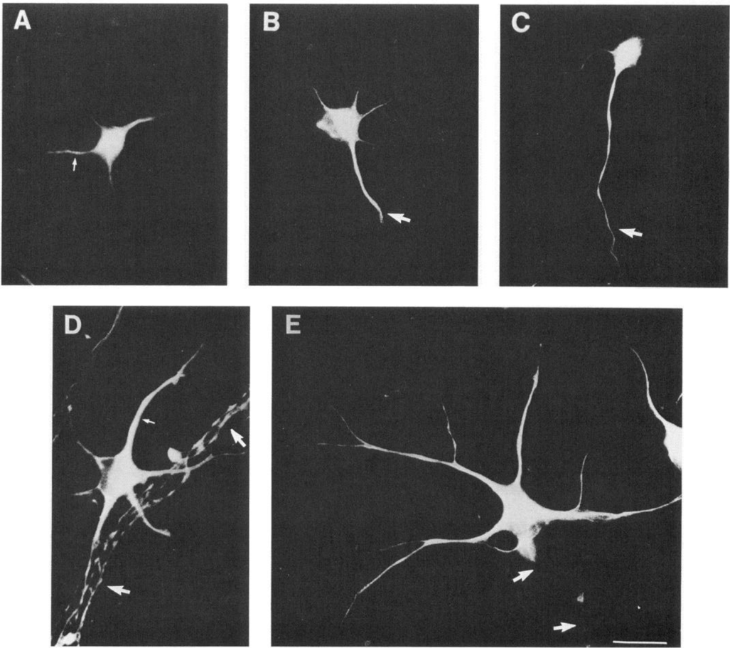
Figure 1 Development of cerebellar macroneurons labeled with tau monoclonal antibody tau 1 (A-C) or MAP2 (R4) antibody (D, E). A, Stage II cell with minor neurites (arrow) after 12 hr in culture. B, Transition to stage III after 24 hr in culture; the arrow shows an elongating neurite. C, Stage III cell with a single elongated neurite (arrow) and very-fine-caliber remaining minor neurites after 24 hr in culture. At this stage, the remaining minor neurites become thinner. D, Stage IV neuron after 4 d in culture, stained with MAP2 antibody, at which time the caliber of the minor neurites increases and many neurites appear tapered (small arrows); the large arrows point to a bundle of axons, which at this time point are still reactive with the MAP2 antibody. E, Seven days after plating, only the dendritelike neurites are labeled with the MAP2 antibody.The arrows point to the trajectory of an axon apparent only by phase contrast. Scale bar, 10 μm.
Figure 1 Development of cerebellar macroneurons labeled with tau monoclonal antibody tau 1 (A-C) or MAP2 (R4) antibody (D, E). A, Stage II cell with minor neurites (arrow) after 12 hr in culture. B, Transition to stage III after 24 hr in culture; the arrow shows an elongating neurite. C, Stage III cell with a single elongated neurite (arrow) and very-fine-caliber remaining minor neurites after 24 hr in culture. At this stage, the remaining minor neurites become thinner. D, Stage IV neuron after 4 d in culture, stained with MAP2 antibody, at which time the caliber of the minor neurites increases and many neurites appear tapered (small arrows); the large arrows point to a bundle of axons, which at this time point are still reactive with the MAP2 antibody. E, Seven days after plating, only the dendritelike neurites are labeled with the MAP2 antibody.The arrows point to the trajectory of an axon apparent only by phase contrast. Scale bar, 10 μm.CC-BY-NC-SA · From the PDF
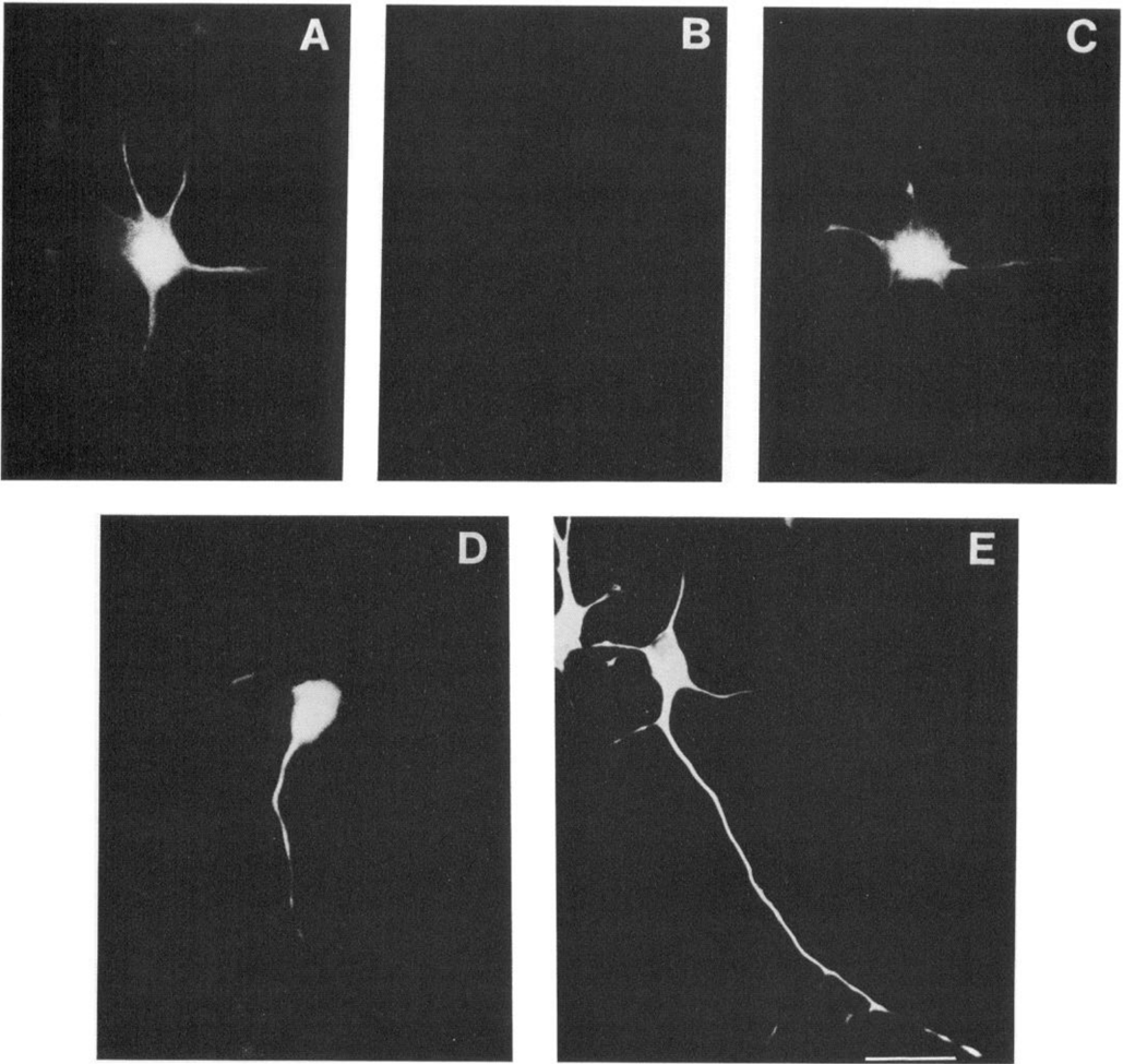
Figure 2 Continuous exposure to tau antisense given 1, 24, and 48 hr after plating and fixed at 72 hr. A and B are double-labeled neurons stained with MAP2 antibody R4 (A) and tau antibody tau 1 (B). Only trace tau immunoreactivity is detectable. C is an identically treated plate of cells sai s t t ing cells stained with tau 1, 2 (D) and 3 (E) d following the cessation of antisense administration. Scale bar, 15 μm.
Figure 2 Continuous exposure to tau antisense given 1, 24, and 48 hr after plating and fixed at 72 hr. A and B are double-labeled neurons stained with MAP2 antibody R4 (A) and tau antibody tau 1 (B). Only trace tau immunoreactivity is detectable. C is an identically treated plate of cells sai s t t ing cells stained with tau 1, 2 (D) and 3 (E) d following the cessation of antisense administration. Scale bar, 15 μm.CC-BY-NC-SA · From the PDF
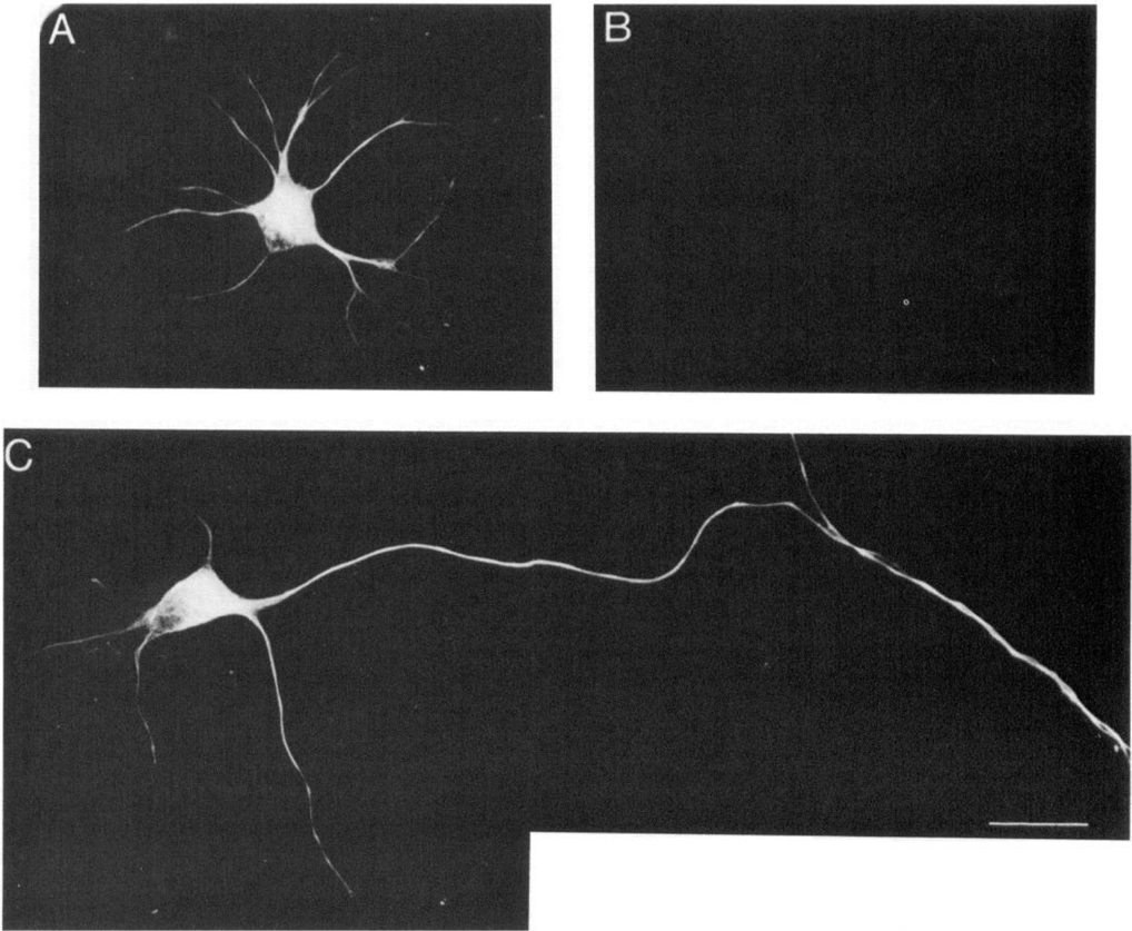
Fi s i a a n nr later and double labeled with tau antibody 5E2 (1:20) and MAP2 polyclonal antibody R4 (1:500). A and B represent the tau antisense, and C, the sense control. Tau immunoreactivity is demonstrated in B and C, and MAP2 immunoreactivity, in A. Scale bar, 10 μm.
Fi s i a a n nr later and double labeled with tau antibody 5E2 (1:20) and MAP2 polyclonal antibody R4 (1:500). A and B represent the tau antisense, and C, the sense control. Tau immunoreactivity is demonstrated in B and C, and MAP2 immunoreactivity, in A. Scale bar, 10 μm.CC-BY-NC-SA · From the PDF
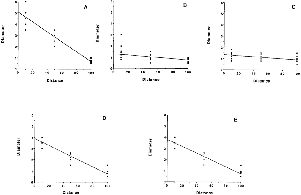
Figure 4 Relationship of the neurite diameter to the distance from the celi body for dendritelike processes after 7 d in culture (A), axonlike processes after 7 d in culture (B), minor neurites after 1 d in culture (C), neurites after 4 d in culture (D), and neurites treated with tau antisense given at 72 hr after plating and fixed 24 hr later (E). Diameters are in microns, and the distance from the cell body is standardized for the variable lengths of cultures À-E by setting the neurite tip at 100 and measuring the diameter at three points that represent percentages of the total length. The correlation coefficients (r) with length are A, 0.92; B, 0.24; C, 0.24; D, 0.87; and E, 0.92. The analysis utilized primary neurites that did not undergo further branching. Twenty cells with an average of 2.5 neurites per cell were drawn for each graph.
Figure 4 Relationship of the neurite diameter to the distance from the celi body for dendritelike processes after 7 d in culture (A), axonlike processes after 7 d in culture (B), minor neurites after 1 d in culture (C), neurites after 4 d in culture (D), and neurites treated with tau antisense given at 72 hr after plating and fixed 24 hr later (E). Diameters are in microns, and the distance from the cell body is standardized for the variable lengths of cultures À-E by setting the neurite tip at 100 and measuring the diameter at three points that represent percentages of the total length. The correlation coefficients (r) with length are A, 0.92; B, 0.24; C, 0.24; D, 0.87; and E, 0.92. The analysis utilized primary neurites that did not undergo further branching. Twenty cells with an average of 2.5 neurites per cell were drawn for each graph.CC-BY-NC-SA · From the PDF
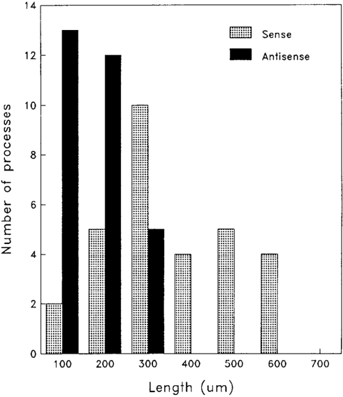
Figure 6 The left shift to shorter neurites is shown in the antisensetreated population. The mean value for the longest sense-treated neurite is 302.0 μm, with a range of 45.7–551.3 μm. The mean value for the longest antisense-treated neurite is 130.5, with a range of 27.1–298.3 μm. This difference was highly significant (p < 0.0003) using the MannWhitney U test for the nonparametric distribution of the histogram.
Figure 6 The left shift to shorter neurites is shown in the antisensetreated population. The mean value for the longest sense-treated neurite is 302.0 μm, with a range of 45.7–551.3 μm. The mean value for the longest antisense-treated neurite is 130.5, with a range of 27.1–298.3 μm. This difference was highly significant (p < 0.0003) using the MannWhitney U test for the nonparametric distribution of the histogram.CC-BY-NC-SA · From the PDF
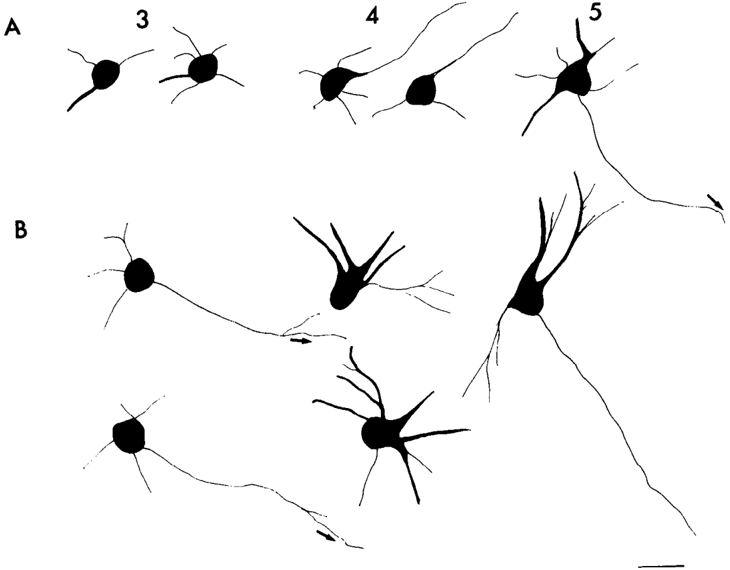
Figure 7 Camera lucida drawings of anti-tubulin-labeled neurons treated with either continuous or delayed exposure to tau antisense oligonucleotides for the number of days in culture indicated along the top. Row A cells were continuously exposed to tau antisense from the time of plating (see Materials and Methods). Row B cells received a single dose of tau antisense 72 hr after plating and were fixed 24 hr later. At 3 d with daily exposure to antisense since the time of plating, only short minor neurites are evident, whereas untreated cells (row B) at 3 d show an elongated process. With the cessation of daily antisense treatment, the cells begin to show recovery at 4 d, whereas delayed antisense administration results in the loss of the axonlike neurite and continued growth of the other neurites. By 5 d, cells that were initially exposed to daily antisense administration continue to recover and look similar to untreated cells at day 3. With a single dose of antisense at day 3, recovery of the axon by day 5 is apparent. Arrows correspond to. neurites that extend beyond the figure.
Figure 7 Camera lucida drawings of anti-tubulin-labeled neurons treated with either continuous or delayed exposure to tau antisense oligonucleotides for the number of days in culture indicated along the top. Row A cells were continuously exposed to tau antisense from the time of plating (see Materials and Methods). Row B cells received a single dose of tau antisense 72 hr after plating and were fixed 24 hr later. At 3 d with daily exposure to antisense since the time of plating, only short minor neurites are evident, whereas untreated cells (row B) at 3 d show an elongated process. With the cessation of daily antisense treatment, the cells begin to show recovery at 4 d, whereas delayed antisense administration results in the loss of the axonlike neurite and continued growth of the other neurites. By 5 d, cells that were initially exposed to daily antisense administration continue to recover and look similar to untreated cells at day 3. With a single dose of antisense at day 3, recovery of the axon by day 5 is apparent. Arrows correspond to. neurites that extend beyond the figure.CC-BY-NC-SA · From the PDF

Verification

Claims the paper makes

The claims and statistics of this paper haven’t been checked yet.

Run the check — free, no account needed. It reads the open-access text and checks the sources it cites.

A few free checks a day · papers already verified open instantly · results are public

Claims other papers make about this one

Each is checked against this paper’s own text: does it say what the citing paper says it does?

Self-check

Percentages, sums and reported differences re-computed from the paper’s own numbers.

The self-check needs the paper’s full text.

Citations

Cited by

See all 167

Metadata

Authors
Caceres, A., Potrebic, S., & Kosik, K.
Venue
The Journal of Neuroscience
Published
1991-06-01
Volume
11
Issue
6
Pages
1515-1523
ISSN
  • 0270-6474
  • 1529-2401
Cited by
167
References
2
Language
en

About these checks

Registry, retraction, Cites and statistics results are computed, not judged, with one exception: a printed reference that no index lists is read once by a model before it counts as a citation error. Claims are extracted by a model and kept only when their quote appears verbatim in the text; each cited source is then checked, with a model’s help, for whether it says what the claim relies on. Those verdicts can be wrong — read the claim’s quote and the cited source before relying on one.

Statistics and arithmetic are recomputed, not judged.

Confidence bands are shown only for fields calibrated on our annotated test set. None is calibrated yet, so no band is shown.

What counts as a claim ›How we measure confidence ›How we check this ›

Are you an author, or do you see a problem with this page? Request a review

How to cite

Cite CoChat for the checks on this page, and the paper for its own findings.

This page
CoChat. (2026). Citation check: The effect of tau antisense oligonucleotides on neurite formation of cultured cerebellar macroneurons. https://app.cochat.ai/papers/10.1523/jneurosci.11-06-01515.1991
The paper
Caceres, A., Potrebic, S., & Kosik, K. (1991). The effect of tau antisense oligonucleotides on neurite formation of cultured cerebellar macroneurons. The Journal of Neuroscience. https://doi.org/10.1523/jneurosci.11-06-01515.1991

Data sources

Bibliographic data: Semantic Scholar, OpenAlex, Crossref.

Contains information from the Semantic Scholar Academic Graph Datasets, made available under the ODC Attribution License (ODC-BY 1.0).

Semantic Scholar’s data is described in: Kinney, R., et al. (2023). The Semantic Scholar Open Data Platform. arXiv:2301.10140.