

The DOI matches its Crossref record; 4 of the 4 references it cites were found in a registry or an index.
Age-dependent neuronal and synaptic degeneration in mice transgenic for the C terminus of the amyloid precursor protein.
Abstract
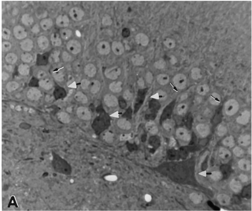
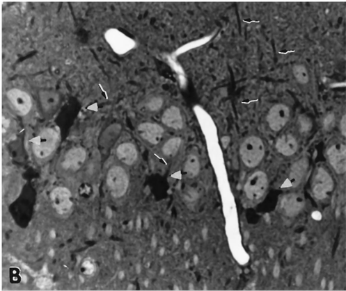
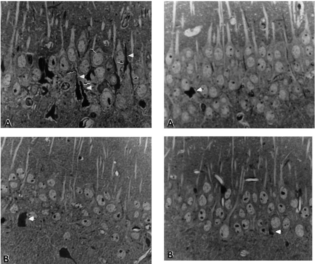
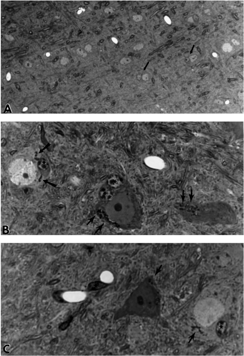
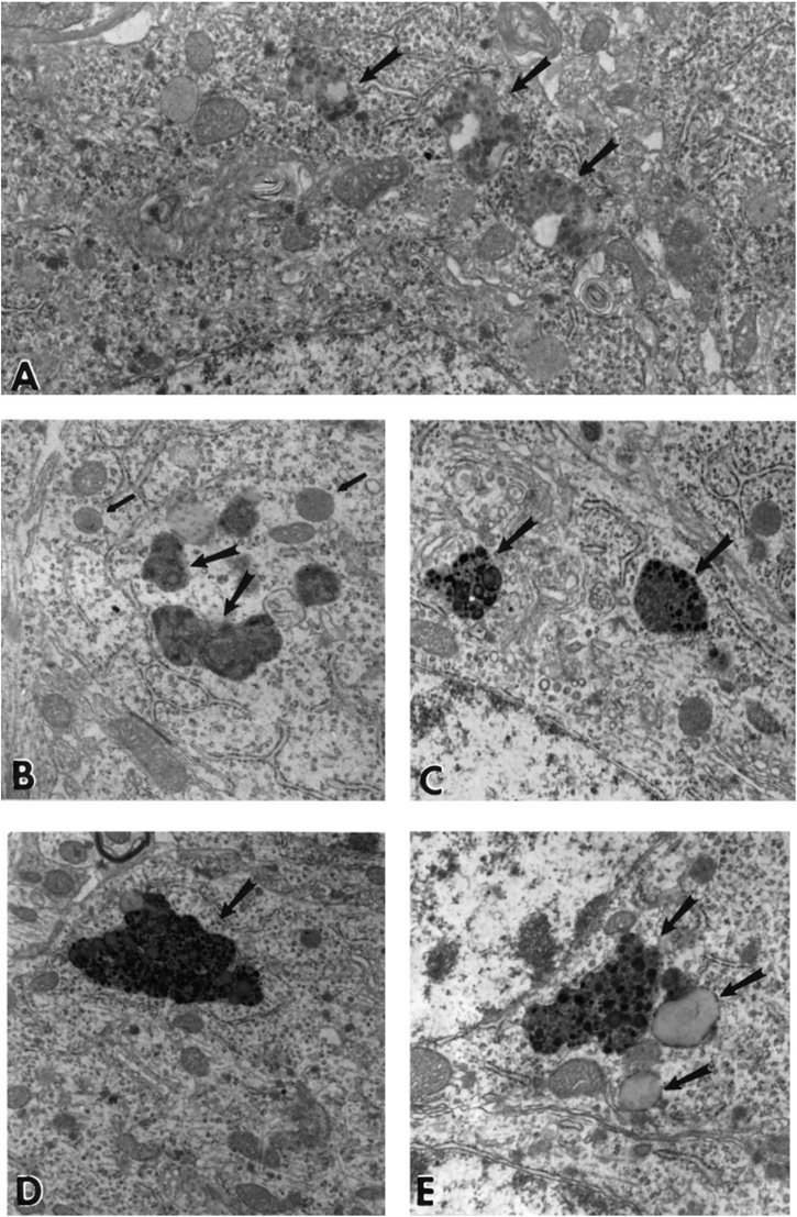
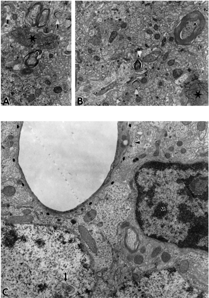
The molecular basis for the degeneration of neurons and the deposition of amyloid in plaques and in the cerebrovasculature in Alzheimer’s disease (AD) is incompletely understood. We have proposed that one molecule common to these abnormal processes is a fragment of the Alzheimer amyloid precursor protein (APP) comprising the C-terminal 100 amino acids of this molecule (APP-C100). We tested this hypothesis by creating transgenic mice expressing APP-C100 in the brain. We report here that aging (18–28 month) APP-C100 transgenic mice exhibit profound degeneration of neurons and synapses in Ammon’s horn and the dentate gyrus of the hippocampal formation. Of the 106 transgenic mice between 8 and 28 months of age that were examined, all of those older than 18 months displayed severe hippocampal degeneration. The numerous degenerating axonal profiles contained increased numbers of neurofilaments, whorls of membrane, and accumulations of debris resembling secondary lysosomes near the cell body. The dendrites of degenerating granule and pyramidal cells contained disorganized, wavy microtubules. Cerebral blood vessels had thickened refractile basal laminae, and microglia laden with debris lay adjacent to larger venous vessels. Mice transgenic for Flag-APP-C100 (in which the hydrophilic Flag tag was fused to the N terminus of APP-C100) showed a similar degree of neurodegeneration in the hippocampal formation as early as 12 months of age. The 45 control mice displayed only occasional necrotic cells and no extensive cell degeneration in the same brain regions. These findings show that APP-C100 is capable of causing some of the neuropathological features of AD.
Overview
The claims and statistics of this paper haven’t been checked yet.
Run the check — free, no account needed. It reads the open-access text and checks the sources it cites.
Integrity signals
Registry
Cites
Statistics
Figures
Verification
Claims the paper makes
The claims and statistics of this paper haven’t been checked yet.
Run the check — free, no account needed. It reads the open-access text and checks the sources it cites.
Claims other papers make about this one
Each is checked against this paper’s own text: does it say what the citing paper says it does?
The source backs part of the claim, not all of it — or backs it for a different population, condition or comparison.
In the citing paper
“many studies using genetic or pharmacological approaches to increase C99 levels in animal models consistently demonstrate the pathogenic properties of C99 linked with AD phenotypes”
From Emerging Alternative Proteinases in APP Metabolism and Alzheimer’s Disease Pathogenesis: A Focus on MT1-MMP and MT5-MMP (2019) · cites this paper as Oster-Granite et al., 1996
Experimental — accuracy not yet measured
Self-check
Percentages, sums and reported differences re-computed from the paper’s own numbers.
The self-check needs the paper’s full text.
Citations
Cites
Alzheimer-type neuropathology in transgenic mice overexpressing V717F β-amyloid precursor protein
Games, D., Adams, D., Alessandrini, R., Barbour, R., Borthelette, P., Blackwell, C., Carr, T., Clemens, J., Donaldson, T., Gillespie, F., et al. (1995) · Nature · doi:10.1038/373523a0 · Cited by 2,516
Synapse loss in frontal cortex biopsies in Alzheimer's disease: Correlation with cognitive severity
DeKosky, S. T., & Scheff, S. W. (1990) · Annals of Neurology · doi:10.1002/ana.410270502 · Cited by 2,376
The Alzheimer's Aβ peptide induces neurodegeneration and apoptotic cell death in transgenic mice
LaFerla, F. M., Tinkle, B. T., Bieberich, C. J., Haudenschild, C. C., & Jay, G. (1995) · Nature Genetics · doi:10.1038/ng0195-21 · Cited by 513
Quantitation of cerebral atrophy in preclinical and end-stage Alzheimer’s disease
de la Monte, S. M. (1989) · Annals of Neurology · doi:10.1002/ana.410250506 · Cited by 229
Research works printed but not linked: 35
- 10.1016/0014-4886(89)90156-8 · DOI
- 10.1073/pnas.87.15.6003 · DOI
- 10.1097/00005072-199311000-00007 · DOI · Paper page
- 10.1016/0006-8993(94)91858-9 · DOI · Paper page
- 10.1016/0306-4522(92)90265-4 · DOI · Paper page
- 10.1016/S0006-291X(05)80126-3 · DOI
- 10.1016/0304-3940(93)90192-N · DOI
- 10.1006/exnr.1994.1101 · DOI
- 10.1093/hmg/1.7.505 · DOI
- Hamos, 1989, Synaptic loss in Alzheimer’s disease and other dementias., Neurology, 39, 355, 10.1212/WNL.39.3.355 · DOI · Paper page
- 10.1111/j.1471-4159.1993.tb03194.x · DOI
- 10.1126/science.1542796 · DOI
- Kalaria, 1992, The blood-brain barrier and cerebral microcirculation in Alzheimer disease., Cerebrovasc Brain Metab Rev, 4, 226
- 10.1073/pnas.89.22.10857 · DOI
- 10.1523/JNEUROSCI.12-05-01679.1992 · DOI
- 10.1016/0006-8993(94)01400-C · DOI
- 10.1097/00005072-199311000-00009 · DOI · Paper page
- 10.1007/BF00296182 · DOI
- Neve RL Boyce FM (1996) Construction and analysis of transgenic mice expressing amyloidogenic fragments of the Alzheimer amyloid protein precursor. in Paradigms of neural injury, methods in neurosciences, Vol 30, ed Perez-Polo JR (Academic, San Diego), pp 298–314. · DOI
- 10.1016/0169-328X(86)90033-1 · DOI
- 10.1016/0169-328X(87)90012-X · DOI
- 10.1073/pnas.89.8.3448 · DOI
- 10.1002/cne.901690404 · DOI
- 10.1002/jemt.1070280305 · DOI
- Prickett, 1989, A calcium-dependent antibody for identification and purification of recombinant proteins., Biotechniques, 7, 580
- 10.1038/352239a0 · DOI
- 10.1073/pnas.85.19.7341 · DOI
- 10.1016/0169-328X(94)90092-2 · DOI
- 10.1002/ana.410100209 · DOI · Paper page
- Terry, 1990, Physical basis of cognitive alterations in Alzheimer’s disease: synapse loss is the major correlate of cognitive impairment., Ann Neurol, 30, 572, 10.1002/ana.410300410 · DOI · Paper page
- 10.1016/0306-4522(94)90310-7 · DOI
- 10.1126/science.1857970 · DOI
- 10.1126/science.3083509 · DOI
- 10.1126/science.2474201 · DOI · Paper page
- 10.1038/359064a0 · DOI
Cited by
Sorted by citations
Pathways towards and Away from Alzheimer’s Disease
Mattson, M. P. (2004) · Nature · doi:10.1038/nature02621 · Cited by 3,052
A subset of NSAIDs lower amyloidogenic Aβ42 independently of cyclooxygenase activity
Weggen, S., Eriksen, J. L., Das, P., Sagi, S. A., Wang, R., Pietrzik, C. U., Findlay, K. A., Smith, T. E., Murphy, M. P., Bulter, T., et al. (2001) · Nature · doi:10.1038/35102591 · Cited by 1,388
Moechars, D., Dewachter, I., Lorent, K., Reversé, D., Baekelandt, V., Naidu, A., Tesseur, I., Spittaels, K., Haute, C. V. D., Checler, F., et al. (1999) · Journal of Biological Chemistry · doi:10.1074/jbc.274.10.6483 · Cited by 668
D'Andrea, M. R., Nagele, R. G., Wang, H., Peterson, P. A., & Lee, D. H. S. (2001) · Histopathology · doi:10.1046/j.1365-2559.2001.01082.x · Cited by 390
Effects of aging and sensory loss on glial cells in mouse visual and auditory cortices
Tremblay, M., Zettel, M. L., Ison, J. R., Allen, P. D., & Majewska, A. K. (2012) · Glia · doi:10.1002/glia.22287 · Cited by 309
Lauritzen, I., Pardossi-Piquard, R., Bauer, C., Brigham, E., Abraham, J.-D., Ranaldi, S., Fraser, P., St-George-Hyslop, P., Le Thuc, O., Espin, V., et al. (2012) · The Journal of Neuroscience · doi:10.1523/jneurosci.2775-12.2012 · Cited by 201
“Apart from being the substrate for the generation of amyloidogenic fragments, A and AICD, C99 may also have a more direct role in AD pathogenesis (Oster-Granite et al., 1996; Nalbantoglu et al., 1997; Suh, 1997; Choi et al., 2001; Bittner et al., 2009; Mitani et al., 2012; Tamayev et al., 2012).”
BackgroundGarcía-González, L., Pilat, D., Baranger, K., & Rivera, S. (2019) · Frontiers in Aging Neuroscience · doi:10.3389/fnagi.2019.00244 · Cited by 88
Mahar, I., Albuquerque, M. S., Mondragon-Rodriguez, S., Cavanagh, C., Davoli, M. A., Chabot, J.-G., Williams, S., Mechawar, N., Quirion, R., & Krantic, S. (2017) · Frontiers in Aging Neuroscience · doi:10.3389/fnagi.2016.00327 · Cited by 63
Ayton, S., & Bush, A. I. (2021) · Ageing Research Reviews · doi:10.1016/j.arr.2020.101212 · Cited by 45
Metadata
- Authors
- Oster-Granite, M. L., McPhie, D. L., Greenan, J., & Neve, R. L.
- Venue
- The Journal of Neuroscience
- Publisher
- Society for Neuroscience
- Published
- 1996-11-01
- Volume
- 16
- Issue
- 21
- Pages
- 6732-6741
- ISSN
- 0270-6474
- 1529-2401
- Cited by
- 162
- References
- 4
- Language
- en
About these checks
Registry, retraction, Cites and statistics results are computed, not judged, with one exception: a printed reference that no index lists is read once by a model before it counts as a citation error. Claims are extracted by a model and kept only when their quote appears verbatim in the text; each cited source is then checked, with a model’s help, for whether it says what the claim relies on. Those verdicts can be wrong — read the claim’s quote and the cited source before relying on one.
Statistics and arithmetic are recomputed, not judged.
Confidence bands are shown only for fields calibrated on our annotated test set. None is calibrated yet, so no band is shown.
What counts as a claim ›How we measure confidence ›How we check this ›
Are you an author, or do you see a problem with this page? Request a review
