

The DOI matches its Crossref record; 56 of the 56 references it cites were found in a registry or an index.
TLR8 signaling enhances tumor immunity by preventing tumor‐induced T‐cell senescence
Open accessAbstract
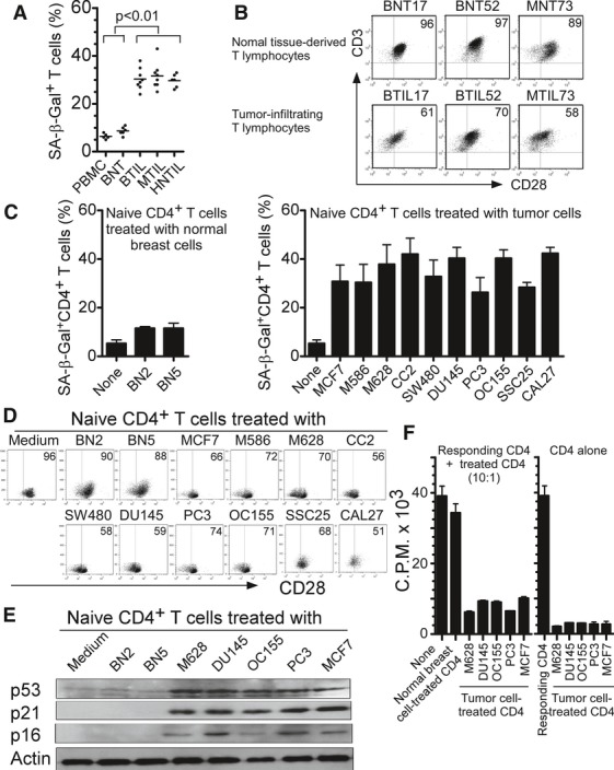
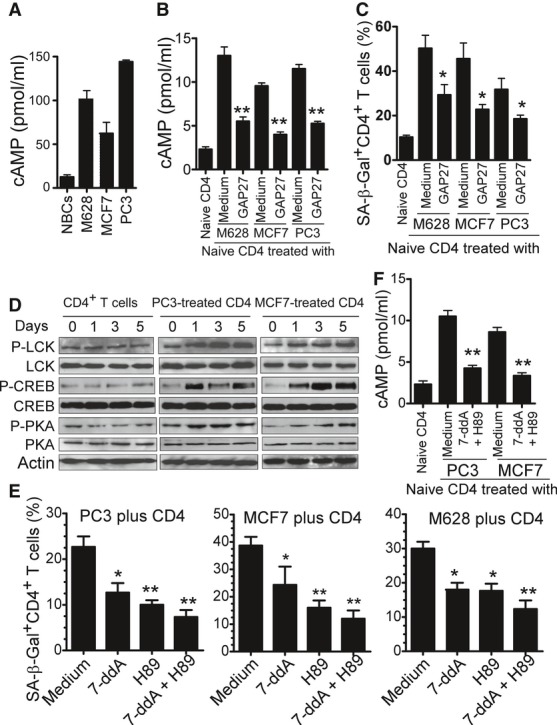
Accumulating evidence suggests the immunosuppressive microenvironments created by malignant tumors represent a major obstacle for effective anti‐tumor immunity. A better understanding of the suppressive mechanisms mediated by tumor microenvironments and the development of strategies to reverse the immune suppression are major challenges for the success of tumor immunotherapy. Here, we report that human tumor cells can induce senescence in naïve/effector T cells, exhibiting potent suppressive function in vitro and in vivo . We further show that tumor‐derived endogenous cyclic adenosine monophosphate (cAMP) is responsible for the induction of T‐cell senescence. Importantly, activation of TLR8 signaling in tumor cells can block the induction and reverse the suppression of senescent naïve and tumor‐specific T cells in vitro and in vivo , resulting in enhanced anti‐tumor immunity. These studies identify a novel mechanism of human tumor‐mediated immune suppression and provide a new strategy to reverse tumor immunosuppressive effects for tumor immunotherapy.
Overview
The claims and statistics of this paper haven’t been checked yet.
Run the check — free, no account needed. It reads the open-access text and checks the sources it cites.
Integrity signals
Registry
Cites
Statistics
Figures
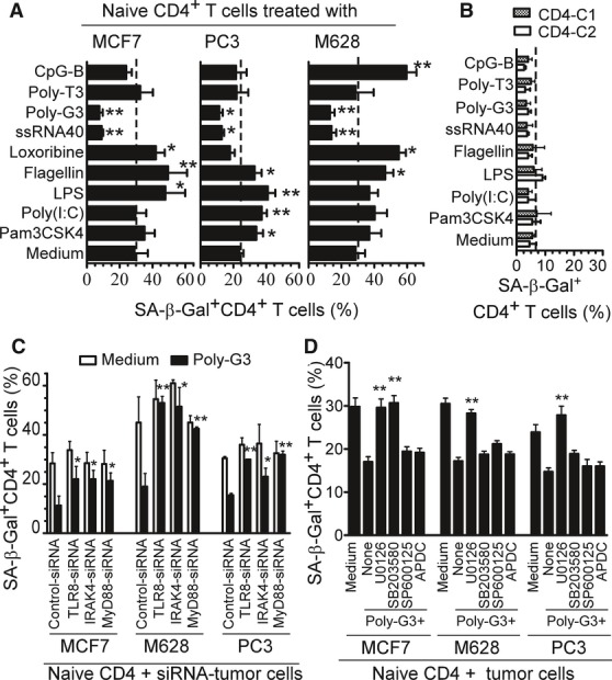
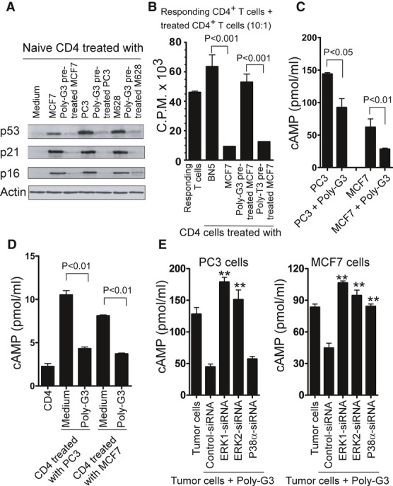
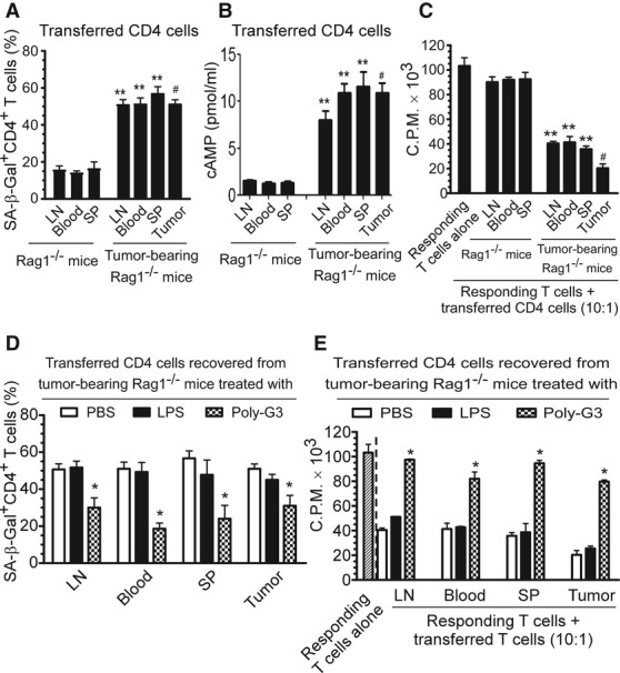
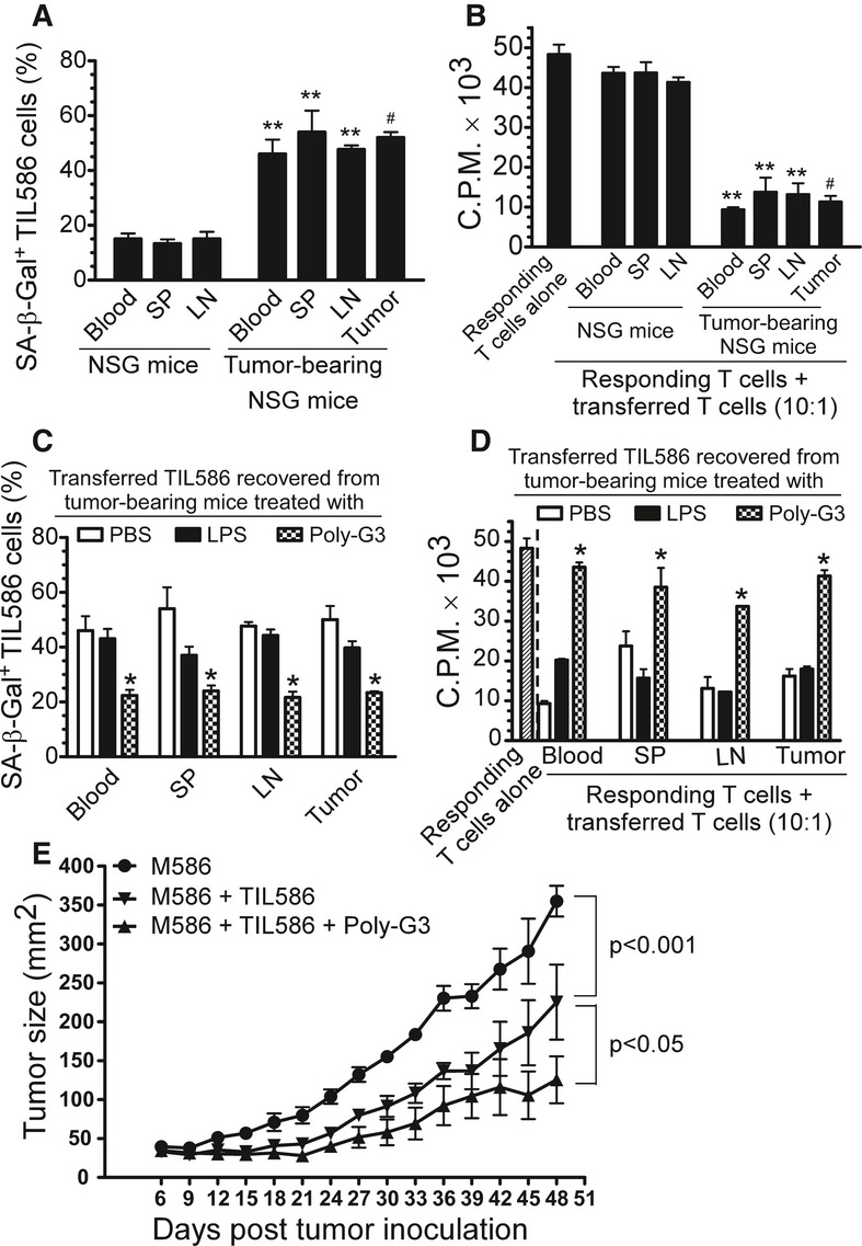
Verification
Claims the paper makes
The claims and statistics of this paper haven’t been checked yet.
Run the check — free, no account needed. It reads the open-access text and checks the sources it cites.
Claims other papers make about this one
Each is checked against this paper’s own text: does it say what the citing paper says it does?
The cited source says what the claim attributes to it.
In the citing paper
“Furthermore, the activation of Toll-like receptor 8 signaling in tumor cells can enhance antitumor immunity by blocking the induction of senescent T cells and tumor-specific T cells in vitro and in vivo and reversing their inhibitory effect [63].”
From Immunosenescence: a key player in cancer development (2020) · cites this paper as [63]
In the citing paper
“In addition, tumor cells activate the PKA-CREB and P38 pathways by producing cAMP, thereby causing DNA damage and senescence of T cells [51, 53, 58–63].”
From Immunosenescence: a key player in cancer development (2020) · cites this paper as [63]
Self-check
Percentages, sums and reported differences re-computed from the paper’s own numbers.
The self-check needs the paper’s full text.
Citations
Cites
The limited in vitro lifetime of human diploid cell strains
Hayflick, L. (1965) · Experimental Cell Research · doi:10.1016/0014-4827(65)90211-9 · Cited by 5,876
Tumor-associated B7-H1 promotes T-cell apoptosis: A potential mechanism of immune evasion
Dong, H., Strome, S. E., Salomao, D. R., Tamura, H., Hirano, F., Flies, D. B., Roche, P. C., Lu, J., Zhu, G., Tamada, K., et al. (2002) · Nature Medicine · doi:10.1038/nm730 · Cited by 5,025
Cellular senescence: when bad things happen to good cells
Campisi, J., & d'Adda di Fagagna, F. (2007) · Nature Reviews Molecular Cell Biology · doi:10.1038/nrm2233 · Cited by 4,441
Cancer immunotherapy: moving beyond current vaccines
Rosenberg, S. A., Yang, J. C.-H., & Restifo, N. P. (2004) · Nature Medicine · doi:10.1038/nm1100 · Cited by 2,980
The tumor microenvironment and its role in promoting tumor growth
Whiteside, T. L. (2008) · Oncogene · doi:10.1038/onc.2008.271 · Cited by 2,376
Uyttenhove, C., Pilotte, L., Théate, I., Stroobant, V., Colau, D., Parmentier, N., Boon, T., & Van den Eynde, B. J. (2003) · Nature Medicine · doi:10.1038/nm934 · Cited by 2,305
Persistent DNA damage signalling triggers senescence-associated inflammatory cytokine secretion
Rodier, F., Coppé, J.-P., Patil, C. K., Hoeijmakers, W. A. M., Muñoz, D. P., Raza, S. R., Freund, A., Campeau, E., Davalos, A. R., & Campisi, J. (2009) · Nature Cell Biology · doi:10.1038/ncb1909 · Cited by 2,232
Interleukin‐10‐secreting type 1 regulatory T cells in rodents and humans
Roncarolo, M. G., Gregori, S., Battaglia, M., Bacchetta, R., Fleischhauer, K., & Levings, M. K. (2006) · Immunological Reviews · doi:10.1111/j.0105-2896.2006.00420.x · Cited by 1,206
A2A adenosine receptor protects tumors from antitumor T cells
Ohta, A., Gorelik, E., Prasad, S. J., Ronchese, F., Lukashev, D. E., Wong, M. K. K., Huang, X., Caldwell, S. A., Liu, K., Smith, P. T., et al. (2006) · Proceedings of the National Academy of Sciences · doi:10.1073/pnas.0605251103 · Cited by 1,095
Innate Immunity Gone Awry: Linking Microbial Infections to Chronic Inflammation and Cancer
Karin, M., Lawrence, T., & Nizet, V. (2006) · Cell · doi:10.1016/j.cell.2006.02.016 · Cited by 938
Cited by
Sorted by citations
Immunosenescence: a key player in cancer development
Lian, J., Yue, Y., Yu, W., & Zhang, Y. (2020) · Journal of Hematology & Oncology · doi:10.1186/s13045-020-00986-z · Cited by 510
Self-citationTargeting Adenosine in Cancer Immunotherapy to Enhance T-Cell Function
Viganó, S., Alatzoglou, D., Irving, M. B., Ménétrier‐Caux, C., Caux, C., Romero, P. J., & Coukos, G. (2019) · Frontiers in Immunology · doi:10.3389/fimmu.2019.00925 · Cited by 434
Exhaustion and senescence: two crucial dysfunctional states of T cells in the tumor microenvironment
Zhao, Y., Shao, Q., & Peng, G. (2020) · Cellular & Molecular Immunology · doi:10.1038/s41423-019-0344-8 · Cited by 382
Self-citationReprogramming lipid metabolism prevents effector T cell senescence and enhances tumor immunotherapy
Liu, X., Hartman, C. L., Li, L., Albert, C. J., Si, F., Gao, A., Huang, L. J., Zhao, Y., Lin, W., Hsueh, E. C., et al. (2021) · Science Translational Medicine · doi:10.1126/scitranslmed.aaz6314 · Cited by 287
Self-citationT cell senescence and CAR-T cell exhaustion in hematological malignancies
Kasakovski, D., Xu, L., & Li, Y. (2018) · Journal of Hematology & Oncology · doi:10.1186/s13045-018-0629-x · Cited by 282
Li, L., Liu, X., Sanders, K. L., Edwards, J. L., Ye, J., Si, F., Gao, A., Huang, L., Hsueh, E. C., Ford, D. A., et al. (2018) · Cell Metabolism · doi:10.1016/j.cmet.2018.09.020 · Cited by 248
Self-citationTrial Watch: Toll-like receptor agonists in cancer immunotherapy
Smith, M. M., García-Martínez, E., Pitter, M. R., Fucikova, J. P., Špíšek, R., Zitvogel, L., Kroemer, G., & Galluzzi, L. (2018) · OncoImmunology · doi:10.1080/2162402x.2018.1526250 · Cited by 237
Regulatory T cells trigger effector T cell DNA damage and senescence caused by metabolic competition
Liu, X., Mo, W., Ye, J., Li, L., Zhang, Y., Hsueh, E. C., Hoft, D. F., & Peng, G. (2018) · Nature Communications · doi:10.1038/s41467-017-02689-5 · Cited by 224
Self-citationTargeting innate immune pathways for cancer immunotherapy
Cao, L. L., & Kagan, J. C. (2023) · Immunity · doi:10.1016/j.immuni.2023.07.018 · Cited by 234
The Evolving Role of CD8+CD28− Immunosenescent T Cells in Cancer Immunology
Huff, W. X., Kwon, J. H., Henriquez, M., Fetcko, K., & Dey, M. (2019) · International Journal of Molecular Sciences · doi:10.3390/ijms20112810 · Cited by 217
Cellular senescence and the tumour microenvironment
Takasugi, M., Yoshida, Y., & Ohtani, N. (2022) · Molecular Oncology · doi:10.1002/1878-0261.13268 · Cited by 190
Senescent T cells: a potential biomarker and target for cancer therapy
Zhang, J., He, T., Xue, L., & Guo, H. (2021) · EBioMedicine · doi:10.1016/j.ebiom.2021.103409 · Cited by 175
Huang, L., Xu, H., & Peng, G. (2018) · Cellular and Molecular Immunology · doi:10.1038/cmi.2018.4 · Cited by 170
Self-citationFigueiredo, C. R., Kalirai, H., Sacco, J. J., Azevedo, R., Duckworth, A. D., Slupsky, J. R., Coulson, J. M., & Coupland, S. E. (2020) · The Journal of Pathology · doi:10.1002/path.5384 · Cited by 151
Context-dependent functions of pattern recognition receptors in cancer
Man, S. M., & Jenkins, B. J. (2022) · Nature reviews. Cancer · doi:10.1038/s41568-022-00462-5 · Cited by 143
Senolytics for Cancer Therapy: Is All that Glitters Really Gold?
Carpenter, V. J., Saleh, T., & Gewirtz, D. A. (2021) · Cancers · doi:10.3390/cancers13040723 · Cited by 124
Wei, F., Wang, D., Wei, J., Tang, N., Tang, L. M., Xiong, F., Guo, C. J., Zhou, M., Li, X., Li, G., et al. (2020) · Cellular and Molecular Life Sciences · doi:10.1007/s00018-020-03581-0 · Cited by 128
Baicalein and baicalin inhibit colon cancer using two distinct fashions of apoptosis and senescence
Dou, J., Wang, Z., Ma, L., Peng, B., Mao, K., Li, C., Su, M., Zhou, C., & Peng, G. (2018) · Oncotarget · doi:10.18632/oncotarget.24015 · Cited by 123
Self-citationLiu, X., Hoft, D. F., & Peng, G. (2020) · Journal of Clinical Investigation · doi:10.1172/jci133679 · Cited by 116
Self-citationTargeting Toll-Like Receptors for Cancer Therapy
Braunstein, M. J., Kucharczyk, J. R., & Adams, S. (2018) · Targeted Oncology · doi:10.1007/s11523-018-0589-7 · Cited by 114
Reshaping of the tumor microenvironment by cellular senescence: An opportunity for senotherapies
D’Ambrosio, M., & Gil, J. (2023) · Developmental Cell · doi:10.1016/j.devcel.2023.05.010 · Cited by 111
Li, H., Su, Q., Li, B., Lan, L., Wang, C., Li, W., Wang, G., Chen, W., He, Y., & Zhang, C. (2020) · Journal of Cellular and Molecular Medicine · doi:10.1111/jcmm.15104 · Cited by 106
Liu, X., Si, F., Bagley, D., Ma, F., Zhang, Y., Tao, Y., Shaw, E. C., & Peng, G. (2022) · Journal for ImmunoTherapy of Cancer · doi:10.1136/jitc-2022-005020 · Cited by 104
Self-citationToll-Like Receptor-Based Strategies for Cancer Immunotherapy
Pahlavanneshan, S., Sayadmanesh, A., Ebrahimiyan, H., & Basiri, M. (2021) · Journal of Immunology Research · doi:10.1155/2021/9912188 · Cited by 96
Ma, F., Liu, X., Zhang, Y., Tao, Y., Zhao, L., Abusalamah, H., Huffman, C., Harbison, R. A., Puram, S. V., Wang, Y., et al. (2025) · Science Translational Medicine · doi:10.1126/scitranslmed.adm7269 · Cited by 106
Self-citationTargeting cellular senescence in cancer and aging: roles of p53 and its isoforms
Beck, J. A., Turnquist, C., Horikawa, I., & Harris, C. C. (2020) · Carcinogenesis · doi:10.1093/carcin/bgaa071 · Cited by 97
Huang, F., Lei, J., Sun, Y., Yan, F., Chen, B., Zhang, L., Lu, Z., Cao, R. Y., Lin, Y., Wang, C.-C., et al. (2018) · OncoImmunology · doi:10.1080/2162402x.2018.1446720 · Cited by 90
Suzuki, J., Yamada, T., Inoue, K., Nabe, S., Kuwahara, M., Takemori, N., Takemori, A., Matsuda, S., Kanoh, M., Imai, Y., et al. (2018) · Nature Communications · doi:10.1038/s41467-018-05854-6 · Cited by 89
Zhang, F., Guo, J., Yu, S., Zheng, Y., Duan, M., Zhao, L., Wang, Y., Yang, Z., & Jiang, X. (2024) · 癌症:英文版 · doi:10.1002/cac2.12591 · Cited by 100
Tumor-derived ILT4 induces T cell senescence and suppresses tumor immunity
Gao, A., Liu, X., Lin, W., Wang, J., Wang, S., Si, F., Huang, L. J., Zhao, Y., Sun, Y., & Peng, G. (2021) · Journal for ImmunoTherapy of Cancer · doi:10.1136/jitc-2020-001536 · Cited by 90
Self-citationMa, C., Wang, F., Han, B., Zhong, X., Si, F., Ye, J., Hsueh, E. C., Robbins, L., Kiefer, S. M., Zhang, Y., et al. (2018) · Molecular Cancer · doi:10.1186/s12943-018-0824-y · Cited by 79
Self-citationTan, T. C. J., Knight, J. R. P., Sbarrato, T., Dudek, K., Willis, A. E., & Zamoyska, R. (2017) · Proceedings of the National Academy of Sciences · doi:10.1073/pnas.1700939114 · Cited by 76
NK and NKT cells have distinct properties and functions in cancer
Liu, X., Li, L., Si, F., Huang, L., Zhao, Y., Zhang, C., Hoft, D. F., & Peng, G. (2021) · Oncogene · doi:10.1038/s41388-021-01880-9 · Cited by 85
Self-citationWu, Z., Uhl, B., Gires, O., & Reichel, C. A. (2023) · Journal of Biomedical Science · doi:10.1186/s12929-023-00915-5 · Cited by 83
Controlling T cell senescence in the tumor microenvironment for tumor immunotherapy
Ye, J., & Peng, G. (2015) · OncoImmunology · doi:10.4161/2162402x.2014.994398 · Cited by 78
Self-citationThe senescence journey in cancer immunoediting
Zingoni, A., Antonangeli, F., Sozzani, S., Santoni, A., Cippitelli, M., & Soriani, A. (2024) · Molecular Cancer · doi:10.1186/s12943-024-01973-5 · Cited by 77
Xiong, Y., Xiong, Y., Zhang, H., Zhao, Y., Han, K., Zhang, J., Zhao, D., Yu, Z., Geng, Z., Wang, L., et al. (2021) · Frontiers in Immunology · doi:10.3389/fimmu.2021.780897 · Cited by 70
Silva, C. G. D., Camps, M. G. M., Li, T. M. W. Y., Zerrillo, L., Löwik, C. W. G. M., OSSENDORP, F. A., & Cruz, L. J. (2019) · Theranostics · doi:10.7150/thno.34429 · Cited by 67
Senescent stromal cells: roles in the tumor microenvironment
Gabai, Y., Assouline, B., & Ben‐Porath, I. (2022) · Trends in cancer · doi:10.1016/j.trecan.2022.09.002 · Cited by 67
Senescence in the Development and Response to Cancer with Immunotherapy: A Double-Edged Sword
Battram, A. M., Bachiller, M., & Martín-Antonio, B. (2020) · International Journal of Molecular Sciences · doi:10.3390/ijms21124346 · Cited by 66
Toll-like receptor-targeted anti-tumor therapies: Advances and challenges
Yang, Y., Li, H., Fotopoulou, C., Cunnea, P., & Zhao, X. (2022) · Frontiers in Immunology · doi:10.3389/fimmu.2022.1049340 · Cited by 61
Citrate Promotes Excessive Lipid Biosynthesis and Senescence in Tumor Cells for Tumor Therapy
Zhao, Y., Liu, X., Si, F., Huang, L., Gao, A., Lin, W., Hoft, D. F., Shao, Q., & Peng, G. (2021) · Advanced Science · doi:10.1002/advs.202101553 · Cited by 59
Self-citationSenescence and tumor suppression
Hinds, P. W., & Pietruska, J. R. (2017) · F1000Research · doi:10.12688/f1000research.11671.1 · Cited by 56
Knowns and Unknowns about CAR-T Cell Dysfunction
Titov, A., Kaminskiy, Y., Ganeeva, I., Zmievskaya, E., Valiullina, A., Rakhmatullina, A., Petukhov, A., Рифкатовна, М. Р., Rizvanov, A. A., & Bulatov, E. (2022) · Cancers · doi:10.3390/cancers14041078 · Cited by 56
ILT4 functions as a potential checkpoint molecule for tumor immunotherapy
Gao, A., Sun, Y., & Peng, G. (2018) · Biochimica et Biophysica Acta (BBA) - Reviews on Cancer · doi:10.1016/j.bbcan.2018.04.001 · Cited by 56
Self-citationT-cell abnormalities in common variable immunodeficiency: the hidden defect
Wong, G. K. Y., & Huissoon, A. P. (2016) · Journal of Clinical Pathology · doi:10.1136/jclinpath-2015-203351 · Cited by 53
Baicalein and Baicalin Promote Melanoma Apoptosis and Senescence via Metabolic Inhibition
Huang, L., Peng, B., Nayak, Y. R., Wang, C., Si, F., Liu, X., Dou, J., Xu, H., & Peng, G. (2020) · Frontiers in Cell and Developmental Biology · doi:10.3389/fcell.2020.00836 · Cited by 51
Self-citationSenescent cells in cancer therapy: why and how to remove them
Zhang, J., Zhang, D., & Yu, B. (2021) · Cancer Letters · doi:10.1016/j.canlet.2021.07.002 · Cited by 52
T-cell senescence accelerates angiotensin II-induced target organ damage
Pan, X., Wu, F., Chen, X.-H., Chen, D.-R., Chen, H., Kong, L., Ruan, C., & Gao, P. (2020) · Cardiovascular Research · doi:10.1093/cvr/cvaa032 · Cited by 50
The P2X7 Receptor in Tumor Immunity
Grassi, F., & Conti, B. D. P. (2021) · Frontiers in Cell and Developmental Biology · doi:10.3389/fcell.2021.694831 · Cited by 49
Toll-Like Receptors in Adaptive Immunity
Kumar, V. (2021) · Handbook of experimental pharmacology · doi:10.1007/164_2021_543 · Cited by 46
Litak, J., Grochowski, C., Litak, J., Osuchowska, I., Gosik, K., Radzikowska, E., Kamieniak, P., & Roliński, J. (2020) · International Journal of Molecular Sciences · doi:10.3390/ijms21093114 · Cited by 47
Cellular senescence and hematological malignancies: From pathogenesis to therapeutics
Hu, D., Yuan, S., Zhong, J., Liu, Z., Wang, Y., Liu, L., Li, J., Wen, F., Liu, J., & Zhang, J. (2021) · Pharmacology & Therapeutics · doi:10.1016/j.pharmthera.2021.107817 · Cited by 47
Tumor microenvironment metabolites directing T cell differentiation and function
Liu, X., Hoft, D. F., & Peng, G. (2021) · Trends in Immunology · doi:10.1016/j.it.2021.12.004 · Cited by 45
Self-citationRamello, M. C., Núñez, N. G., Tosello, J., Bossio, S., Canale, F. P., Abrate, C., Ponce, N. E., del Castillo, A., Ledesma, M., Viel, S., et al. (2021) · Frontiers in Immunology · doi:10.3389/fimmu.2021.713132 · Cited by 46
Age-related decline in CD8+ tissue resident memory T cells compromises antitumor immunity
Pei, S., Deng, X., Yang, R., Wang, H., Shi, J., Wang, X., Huang, J., Tian, Y., Wang, R., Zhang, S., et al. (2024) · Nature Aging · doi:10.1038/s43587-024-00746-5 · Cited by 43
Self-citationHuo, Y., Zhou, Y., Zheng, J.-H., Jin, G., Tao, L., Yao, H., Zhang, J., Sun, Y., Liu, Y., & Hu, L.-P. (2022) · Frontiers in Immunology · doi:10.3389/fimmu.2022.983116 · Cited by 38
Immunotherapeutic strategies in head and neck cancer: challenges and opportunities
Liu, X., Harbison, R. A., Varvares, M. A., Puram, S. V., & Peng, G. (2025) · Journal of Clinical Investigation · doi:10.1172/jci188128 · Cited by 38
Self-citationT cell senescence: a new perspective on immunotherapy in lung cancer
Huang, M., Wang, Y., Fang, L., Liu, C., Feng, F., Liu, L., & Sun, C. (2024) · Frontiers in Immunology · doi:10.3389/fimmu.2024.1338680 · Cited by 37
TLR7 Signaling Regulates Th17 Cells and Autoimmunity: Novel Potential for Autoimmune Therapy
Ye, J., Wang, Y., Liu, X., Li, L., Opejin, A., Hsueh, E. C., Luo, H., Wang, T., Hawiger, D., & Peng, G. (2017) · The Journal of Immunology · doi:10.4049/jimmunol.1601890 · Cited by 36
Self-citationBiomechanics of T Cell Dysfunctions in Chronic Diseases
Gunasinghe, S. D., Peres, N. G., Goyette, J., & Gaus, K. (2021) · Frontiers in Immunology · doi:10.3389/fimmu.2021.600829 · Cited by 33
The role of novel immunotherapies in non-Hodgkin lymphoma
Pishko, A. M., & Nasta, S. D. (2017) · Translational Cancer Research · doi:10.21037/tcr.2017.01.08 · Cited by 33
Si, F., Liu, X., Tao, Y., Zhang, Y., Ma, F., Hsueh, E. C., Puram, S. V., & Peng, G. (2024) · Journal for ImmunoTherapy of Cancer · doi:10.1136/jitc-2023-008219 · Cited by 31
Self-citationMetabolic reprogramming in T cell senescence: a novel strategy for cancer immunotherapy
Liu, L., Hao, Z., Yang, X., Li, Y., Wang, S., & Li, L. (2025) · Cell Death Discovery · doi:10.1038/s41420-025-02468-y · Cited by 33
Zhong, Z., Hong, M. H., Chen, X., Xi, Y., Xu, Y., Kong, D., Deng, J., Li, Y., Hu, R., Sun, C., et al. (2020) · BMC Medical Genomics · doi:10.1186/s12920-020-0707-0 · Cited by 30
Xiong, Y., WANG, Y., Zhang, J., Zhao, N., Zhang, A., Zhang, A., Zhao, D., Yu, Z., Yin, Y., Song, L., et al. (2020) · Stem Cell Research & Therapy · doi:10.1186/s13287-020-01993-0 · Cited by 29
Harnessing Metabolic Reprogramming to Improve Cancer Immunotherapy
Liang, Y., Tan, Y., Chen, G., Fan, J., & Zhang, J. (2021) · International Journal of Molecular Sciences · doi:10.3390/ijms221910268 · Cited by 29
T-cell Senescence in the Tumor Microenvironment
Huang, L., Zhang, C., Jiang, A., Lin, A., Zhu, L., Mou, W., Zeng, D., Liu, Z., Tang, B., Zhang, J., et al. (2025) · Cancer Immunology Research · doi:10.1158/2326-6066.cir-24-0894 · Cited by 29
Hu, M. Y., Zhang, J., Yu, Y., Tu, K., Yang, T., Wang, Y., Hu, Q., Kong, L., & Zhang, Z. (2020) · Small · doi:10.1002/smll.202004905 · Cited by 26
Tumor Secretome to Adoptive Cellular Immunotherapy: Reduce Me Before I Make You My Partner
Etxebeste-Mitxeltorena, M., del Rincón-Loza, I., & Martín-Antonio, B. (2021) · Frontiers in Immunology · doi:10.3389/fimmu.2021.717850 · Cited by 26
A Critical Appraisal of the Utility of Targeting Therapy-Induced Senescence for Cancer Treatment
Saleh, T., Greenberg, E. F., Faber, A. C., Harada, H., & Gewirtz, D. A. (2025) · Cancer Research · doi:10.1158/0008-5472.can-24-2219 · Cited by 28
Fu, T., Jin, X., He, M., Chen, Y.-Y., Yang, Y., Chen, L., Zhang, H.-Y.-L., Fan, L., Wu, J., Wang, Z.-H., et al. (2025) · Science Translational Medicine · doi:10.1126/scitranslmed.adj7808 · Cited by 26
Emerging role of RNA sensors in tumor microenvironment and immunotherapy
Yang, R. Y., Yu, S., Xu, T., Zhang, J., & Wu, S. (2022) · Journal of Hematology & Oncology · doi:10.1186/s13045-022-01261-z · Cited by 27
Torres, F., Leon, S., Ummarino, A., Redín, E., Allavena, P., Serrano, D., Anfray, C., & Calvo, A. (2022) · Biomedicines · doi:10.3390/biomedicines10071590 · Cited by 24
Senescent Stromal Cells in the Tumor Microenvironment: Victims or Accomplices?
Ye, M., Huang, X., Wu, Q., & Liu, F. (2023) · Cancers · doi:10.3390/cancers15071927 · Cited by 27
T cell exhaustion and senescence for ovarian cancer immunotherapy
Zhao, J., Wang, Z., Tian, Y., Ning, J., & Ye, H. (2024) · Seminars in Cancer Biology · doi:10.1016/j.semcancer.2024.07.001 · Cited by 26
Senescent Tumor CD8+ T Cells: Mechanisms of Induction and Challenges to Immunotherapy
Liu, W., Stachura, P., Xu, H. C., Bhatia, S., Borkhardt, A., Lang, P. A., & Pandyra, A. A. (2020) · Cancers · doi:10.3390/cancers12102828 · Cited by 27
Liu, Y., Hawkins, O. E., Vilgelm, A. E., Pawlikowski, J. S., Ecsedy, J., Sosman, J. A., Kelley, M. C., & Richmond, A. J. (2015) · Clinical Cancer Research · doi:10.1158/1078-0432.ccr-15-0293 · Cited by 26
Toll-like receptors in breast cancer immunity and immunotherapy
Zhou, J., Zhang, L., Liu, S., DeRubeis, D., & Zhang, D. (2024) · Frontiers in Immunology · doi:10.3389/fimmu.2024.1418025 · Cited by 23
Self-citationVeld, R. V. H. I. ‘., Silva, C. G. D., Jager, M. J., Cruz, L. J., & OSSENDORP, F. A. (2021) · Pharmaceutics · doi:10.3390/pharmaceutics13091470 · Cited by 24
Gut microbiota and immunosenescence in cancer
Xu, Y., He, C., Xi, Y., Zhang, Y., & Bai, Y. (2024) · Seminars in Cancer Biology · doi:10.1016/j.semcancer.2024.07.004 · Cited by 23
Self-citationImmunosenescence and age-related immune cells: causes of age-related diseases
Kim, N., Sim, S.-J., Han, H.-G., Yoon, J.-H., & Han, Y. (2024) · Archives of Pharmacal Research · doi:10.1007/s12272-024-01529-7 · Cited by 22
Towers, R., Trombello, L., Fusenig, M., Tunger, A., Baumann, A.-L., Savoldelli, R., Wehner, R., Faßlrinner, F., Arndt, C., Dazzi, F., et al. (2024) · Cancer Immunology Immunotherapy · doi:10.1007/s00262-023-03594-1 · Cited by 21
Immunosenescence: A new direction in anti-aging research
Li, H., Lin, S., Wang, Y., Shi, Y., Fang, X., Wang, J., Cui, H., Bian, Y., & Qi, X. (2024) · International Immunopharmacology · doi:10.1016/j.intimp.2024.112900 · Cited by 21
Angrini, M., Varthaman, A., & Cremer, I. (2020) · Advances in experimental medicine and biology · doi:10.1007/978-3-030-44518-8_9 · Cited by 20
Accumulation of CD28null Senescent T-Cells Is Associated with Poorer Outcomes in COVID19 Patients
Coleman, M. J., Zimmerly, K. M., & Yang, X. O. (2021) · Biomolecules · doi:10.3390/biom11101425 · Cited by 20
Topically Applied Resiquimod versus Imiquimod as a Potential Adjuvant in Melanoma Treatment
Tambunlertchai, S., Geary, S. M., & Salem, A. K. (2022) · Pharmaceutics · doi:10.3390/pharmaceutics14102076 · Cited by 19
Liu, J., Zheng, R., Zhang, Y., Jia, S., He, Y., & Liu, J. (2023) · Cancers · doi:10.3390/cancers15092640 · Cited by 20
Self-citationImplications of Senescent T Cells for Cancer Immunotherapy
Kasamatsu, T. (2023) · Cancers · doi:10.3390/cancers15245835 · Cited by 20
From infection to immune exhaustion: The Epstein-Barr virus and its contribution to Immunosenescence
Hu, L., Zhu, T., Long, J., Luo, Q., & Lyu, X. (2025) · Biochimica et Biophysica Acta (BBA) - Reviews on Cancer · doi:10.1016/j.bbcan.2025.189421 · Cited by 20
de Nałęcz, A. K., Ciszak, L., Usnarska‐Zubkiewicz, L., Frydecka, I., Pawlak, E., Szmyrka−Kaczmarek, M., & Kosmaczewska, A. (2021) · International Journal of Molecular Sciences · doi:10.3390/ijms22179298 · Cited by 18
Decoding T cell senescence in cancer: Is revisiting required?
Magkouta, S., Markaki, E., Evangelou, K., Petty, R., Verginis, P., & Gorgoulis, V. G. (2024) · Seminars in Cancer Biology · doi:10.1016/j.semcancer.2024.11.003 · Cited by 17
Fungi, immunosenescence and cancer
Xu, B., Luo, Z., Niu, X., Li, Z., Lu, Y., & Li, J. (2025) · Seminars in Cancer Biology · doi:10.1016/j.semcancer.2025.01.002 · Cited by 17
Shukla, S., Kumari, S., Bal, S. K., Mónaco, D. C., Ribeiro, S. P., Sékaly, R., & Sharma, A. (2021) · Current Opinion in HIV and AIDS · doi:10.1097/coh.0000000000000692 · Cited by 17
MM-BMSCs induce naïve CD4+ T lymphocytes dysfunction through fibroblast activation protein α
Wu, X., Wang, Y., Xu, J., Luo, T., Deng, J., & Hu, Y. (2017) · Oncotarget · doi:10.18632/oncotarget.17538 · Cited by 17
Yun, S. J., Lee, B., Komori, K., Lee, M. J., Lee, B. G., Kim, K. H., & Park, S. (2018) · Molecular Immunology · doi:10.1016/j.molimm.2018.12.006 · Cited by 19
Recent Advances in the Aging Microenvironment of Breast Cancer
Jiang, X., Shen, H., Xi, S., Fang, J., Lu, Y., Lu, Y., Zheng, J., & Fu, P. (2022) · Cancers · doi:10.3390/cancers14204990 · Cited by 17
The Tumor Microenvironment Reprograms Immune Cells
Cao, H., Gao, S., Jogani, R., & Sugimura, R. (2022) · Cellular Reprogramming · doi:10.1089/cell.2022.0047 · Cited by 17
Participation of T cells in generating immune protection against cancers
Deo, A. S., Shrijana, Sruthika, S. U., Karun, S., Bisaria, K., & Sarkar, K. (2024) · Pathology - Research and Practice · doi:10.1016/j.prp.2024.155534 · Cited by 18
Neoadjuvant chemotherapy modulates exhaustion of T cells in breast cancer patients
Rodríguez, I. J., Bernal-Estévez, D. A., Llano-León, M., Bonilla, C., & Parra-López, C. (2023) · PLoS ONE · doi:10.1371/journal.pone.0280851 · Cited by 16
Metadata
- Authors
- Ye, J., Ma, C., Hsueh, E. C., Dou, J., Mo, W., Liu, S., Han, B., Huang, Y., Zhang, Y., Varvares, M. A., Hoft, D. F., & Peng, G.
- Affiliations
- Saint Louis University (US)
- Corresponding author
- Peng, Guangyong
- ORCID
- Venue
- EMBO Molecular Medicine
- Published
- 2014-09-17
- Volume
- 6
- Issue
- 10
- Pages
- 1294-1311
- ISSN
- 1757-4676 (ISSN-L)
- 1757-4684
- PMC
- PMC4287933
- OpenAlex
- W2000656984
- Cited by
- 161
- References
- 56
- Citations by year
- 2026: 6 · 2025: 24 · 2024: 24 · 2023: 14 · 2022: 16 · 2021: 27 · 2020: 22 · 2019: 6 · 2018: 13 · 2017: 5 · 2016: 1 · 2015: 3
- FWCI
- 1.37
- Type
- journal article
- Language
- en
- Open access
- gold
- doi.org
- Locations
- EMBO Molecular Medicine (publishedVersion, cc-by)
- PubMed Central (publishedVersion, cc-by)
- PubMed (publishedVersion)
- DOAJ (DOAJ: Directory of Open Access Journals) (submittedVersion)
- Funders
- American Cancer Society (RSG‐10‐160‐01‐LIB)
- Melanoma Research Alliance
- National Institutes of Health
- Topics
- Life Sciences › Immunology and Microbiology › Immunology › T-cell and B-cell Immunology
- Life Sciences › Immunology and Microbiology › Immunology › Immunotherapy and Immune Responses
- Health Sciences › Medicine › Oncology › Cancer Immunotherapy and Biomarkers
- Keywords
- T cell senescence, tumor immunosuppression, tumor microenvironment, cyclic AMP, antitumor immunity, cancer immunotherapy, tumor-specific T cells
- MeSH terms
- Cyclic AMP, Animals, Cells, Cultured, Flow Cytometry, Humans, Neoplasms, T-Lymphocytes, Blotting, Western, Signal Transduction, CD4-Positive T-Lymphocytes, Immunotherapy, Adoptive, Mice, SCID, Mice, Inbred NOD, Cellular Senescence, Mice, Knockout, Coculture Techniques, Xenograft Model Antitumor Assays, RNA Interference, Cell Line, Tumor, Toll-Like Receptor 8, Interleukin Receptor Common gamma Subunit, Tumor Microenvironment, MCF-7 Cells
About these checks
Registry, retraction, Cites and statistics results are computed, not judged, with one exception: a printed reference that no index lists is read once by a model before it counts as a citation error. Claims are extracted by a model and kept only when their quote appears verbatim in the text; each cited source is then checked, with a model’s help, for whether it says what the claim relies on. Those verdicts can be wrong — read the claim’s quote and the cited source before relying on one.
Statistics and arithmetic are recomputed, not judged.
Confidence bands are shown only for fields calibrated on our annotated test set. None is calibrated yet, so no band is shown.
What counts as a claim ›How we measure confidence ›How we check this ›
Are you an author, or do you see a problem with this page? Request a review
