

The DOI matches its Crossref record; 1 of the 1 references it cites were found in a registry or an index.
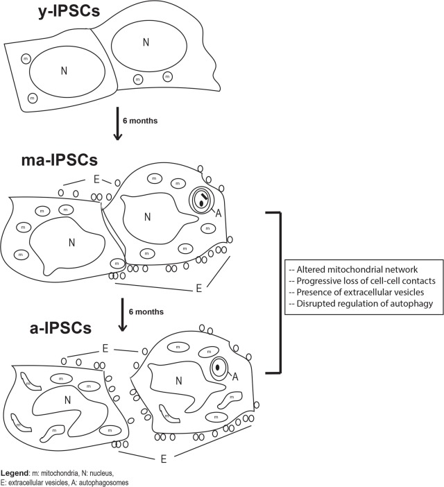
Senescence-associated ultrastructural features of long-term cultures of induced pluripotent stem cells (iPSCs)
Overview
The claims and statistics of this paper haven’t been checked yet.
Run the check — free, no account needed. It reads the open-access text and checks the sources it cites.
Integrity signals
Registry
Cites
Statistics
Figures
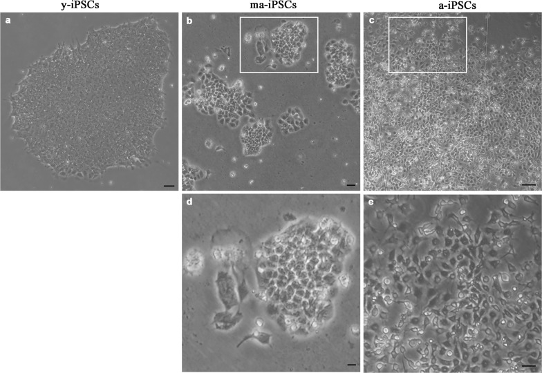
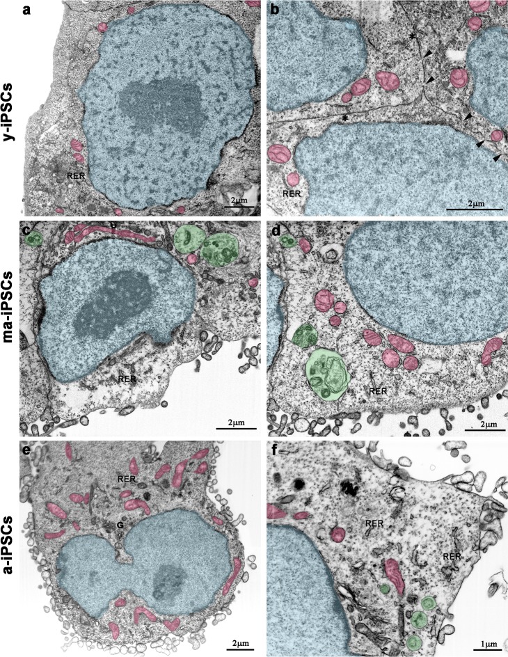
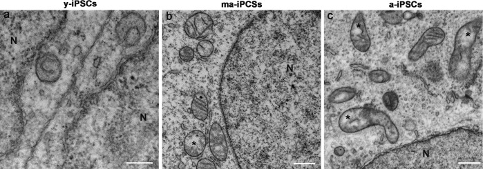
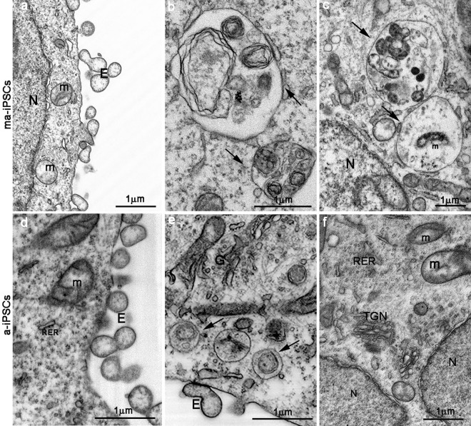
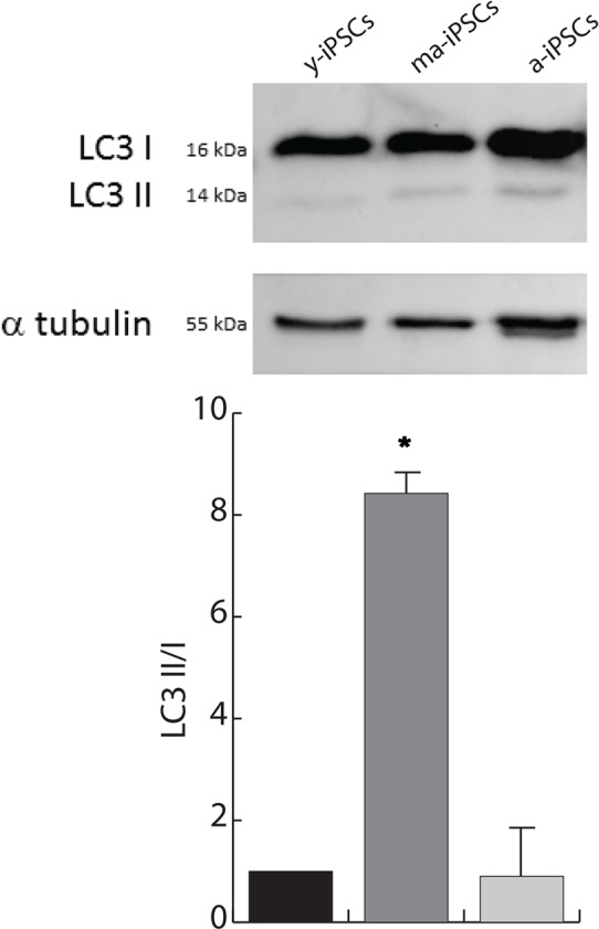
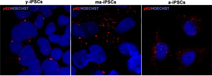
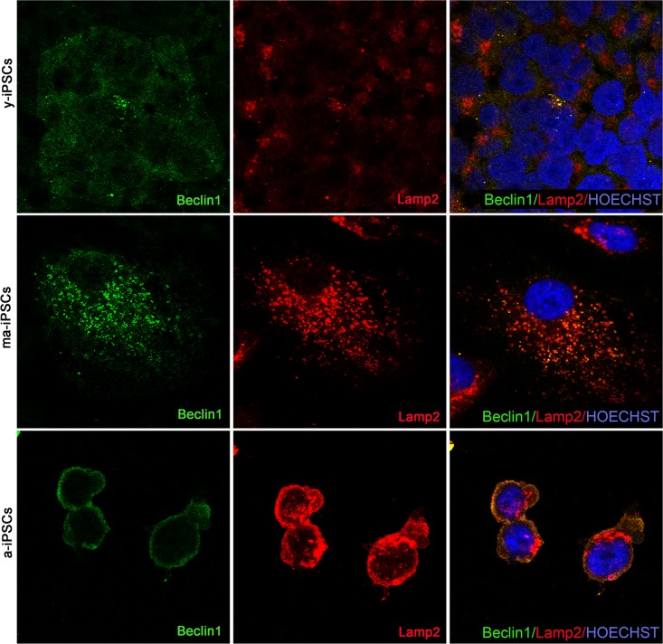
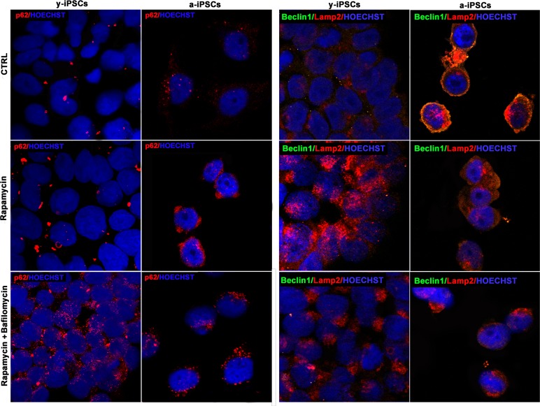
Verification
Claims the paper makes
The claims and statistics of this paper haven’t been checked yet.
Run the check — free, no account needed. It reads the open-access text and checks the sources it cites.
Claims other papers make about this one
Each is checked against this paper’s own text: does it say what the citing paper says it does?
In the citing paper
“another method to consider for monitoring autophagy in yeast (and potentially in plants) is to count autophagic bodies by TEM using at least two time points [149].The advantage of this approach is that it can provide accurate information on flux even when the autophagosomes are abnormally small [150,151].”
From Guidelines for the use and interpretation of assays for monitoring autophagy (4th edition) 1 (2021) · cites this paper as [150]
In the citing paper
“Accordingly, particular mutants may display complete maturation of prApe1 under autophagy-inducing conditions, but may still have a defect in other types of selective autophagy, as well as being unable to induce a normal level of nonselective autophagy [150].”
From Guidelines for the use and interpretation of assays for monitoring autophagy (4th edition) 1 (2021) · cites this paper as [150]
Self-check
Percentages, sums and reported differences re-computed from the paper’s own numbers.
The self-check needs the paper’s full text.
Citations
Cites
Feedback between p21 and reactive oxygen production is necessary for cell senescence
Passos, J. F., Nelson, G., Wang, C., Richter, T., Simillion, C., Proctor, C. J., Miwa, S., Olijslagers, S., Hallinan, J., Wipat, A., et al. (2010) · Molecular Systems Biology · doi:10.1038/msb.2010.5 · Cited by 1,019 · cited 1×
“These results are in line with previous data from ours and other Authors’ groups reporting. increased mitochondrial biogenesis [7, 23], and are possibly due to adaptation of the cells to the culture environment (5% CO 2 and 21% O 2 ).”
Research works printed but not linked: 36
- Yamanaka, 2006, Induction of pluripotent stem cells from mouse embryonic and adult fibroblast cultures by defined factors., Cell, 12, 663, 10.1016/j.cell.2006.07.024 · DOI · Paper page
- Bertini, 2014, In vitro neurogenesis: development and functional implications of iPSC technology., Cell Mol Life Sci, 7, 1623, 10.1007/s00018-013-1511-1 · DOI
- Prigione, 2014, Mitochondrial function in pluripotent stem cells and cellular reprogramming., Gerontology, 6, 174, 10.1159/000355050 · DOI
- Zavazava, 2015, Human iPS cell-derived insulin producing cells form vascularized organoids under the kidney capsules of diabetic mice., PLoS One, 1, e0116582, 10.1371/journal.pone.0116582 · DOI
- Meier, 2016, Ultrastructural demonstration of Cx43 gap junctions in induced pluripotent stem cells from human cord blood., Histochem Cell Biol, 14, 529, 10.1007/s00418-016-1469-9 · DOI
- Zhang, 2013, Genetic and epigenetic variations in iPSCs: potential causes and implications for application., Cell Stem Cell, 1, 149, 10.1016/j.stem.2013.07.001 · DOI
- Compagnucci, 2014, Aged iPSCs display an uncommon mitochondrial appearance and fail to undergo in vitro neurogenesis., Aging (Albany NY), 6, 1094, 10.18632/aging.100708 · DOI
- Compagnucci, 2017, Aged induced pluripotent stem cell (iPSCs) as a new cellular model for studying premature aging., Aging (Albany NY), 9, 1453, 10.18632/aging.101248 · DOI
- Subramaniam, 2015, Focused ion beams in biology., Nat Methods, 1, 1021, 10.1038/nmeth.3623 · DOI
- Humbel, 2014, Focused ion beam scanning electron microscopy in biology., J Microsc, 25, 109, 10.1111/jmi.12127 · DOI
- Stoorvogel, 2013, Extracellular vesicles: exosomes, microvesicles, and friends., J Cell Biol, 20, 373, 10.1083/jcb.201211138 · DOI · Paper page
- Johansen, 2007, p62/SQSTM1 binds directly to Atg8/LC3 to facilitate degradation of ubiquitinated protein aggregates by autophagy., J Biol Chem, 28, 24131, 10.1074/jbc.M702824200 · DOI · Paper page
- Yamanaka, 2016, A decade of transcription factor-mediated reprogramming to pluripotency., Nat Rev Mol Cell Biol, 1, 183, 10.1038/nrm.2016.8 · DOI
- Chhabra, 2017, Derivation of Human Induced Pluripotent Stem Cell (iPSC) Lines and Mechanism of Pluripotency: Historical Perspective and Recent Advances., Stem Cell Rev, 10.1007/s12015-017-9766-9 · DOI
- Martin, 2017, Genome stability of programmed stem cell products., Adv Drug Deliv Rev, 10.1016/j.addr.2017.09.004 · DOI
- Cibelli, 2010, Mitochondrial rejuvenation after induced pluripotency., PLoS One, 5, e14095, 10.1371/journal.pone.0014095 · DOI
- Romani, 2012, Functional structure of antennal sensilla in the myrmecophilous beetle Paussus favieri (Coleoptera, Carabidae, Paussini)., Micron, 4, 705, 10.1016/j.micron.2011.10.013 · DOI
- Romani, 2015, Functional anatomy of the explosive defensive system of bombardier beetles (Coleoptera, Carabidae, Brachininae)., Arthropod Struct Dev, 4, 468, 10.1016/j.asd.2015.08.013 · DOI
- Tang, 2015, Exosomes/microvesicles from induced pluripotent stem cells deliver cardioprotective miRNAs and prevent cardiomyocyte apoptosis in the ischemic myocardium., Int J Cardiol, 19, 61, 10.1016/j.ijcard.2015.05.020 · DOI
- Yang, 2017, Exosomes Generated From iPSC-Derivatives: New Direction for Stem Cell Therapy in Human Heart Diseases., Circ Res, 12, 407, 10.3892/ol.2017.5824 · DOI
- Jiang, 2017, Role of stem cell-derived exosomes in cancer., Oncol Lett, 1, 2855, 10.1161/CIRCRESAHA.116.309307 · DOI
- Brenner, 2007, Mitochondria in stem cells., Mitochondrion, 7, 289, 10.1016/j.mito.2007.05.002 · DOI
- Evans, 2012, The metabolome of induced pluripotent stem cells reveals metabolic changes occurring in somatic cell reprogramming., Cell Res, 2, 168, 10.1038/cr.2011.177 · DOI
- Teitell, 2012, Metabolic regulation in pluripotent stem cells during reprogramming and self-renewal., Cell Stem Cell, 1, 589, 10.1016/j.stem.2012.10.005 · DOI
- Narendra, 2011, Mechanisms of mitophagy., Nat Rev Mol Cell Biol, 1, 9, 10.1038/nrm3028 · DOI · Paper page
- Popa-Wagner, 2017, Autophagy in aging and disease., Rom J Morphol Embryol, 5, 27
- Ramalho-Santos, 2011, Open chromatin in pluripotency and reprogramming., Nat Rev Mol Cell Biol, 1, 36, 10.1038/nrm3036 · DOI
- Menendez, 2016, Mitophagy-driven mitochondrial rejuvenation regulates stem cell fate., Aging (Albany NY), 8, 1330, 10.18632/aging.100976 · DOI
- Wong, 2016, Study of mitochondrial respiratory defects on reprogramming to human induced pluripotent stem cells., Aging (Albany NY), 8, 945, 10.18632/aging.100950 · DOI
- Pébay, 2017, Mitochondrial replacement in an iPSC model of Leber’s hereditary optic neuropathy., Aging (Albany NY), 9, 1341, 10.18632/aging.101231 · DOI
- Nakatsuji, 2017, Friedreich’s ataxia induced pluripotent stem cell-derived cardiomyocytes display electrophysiological abnormalities and calcium handling deficiency., Aging (Albany NY), 9, 1440, 10.18632/aging.101247 · DOI
- Fambrough, 1997, Different steady state subcellular distributions of the three splice variants of lysosome-associated membrane protein LAMP-2 are determined largely by the COOH-terminal amino acid residue., J Cell Biol, 13, 1161, 10.1083/jcb.137.5.1161 · DOI
- Bonifacino, 2005, Role of the endocytic machinery in the sorting of lysosome-associated membrane proteins., Mol Biol Cell, 1, 4231, 10.1091/mbc.E05-03-0213 · DOI
- Kalraiya, 2005, Altered melanoma cell surface glycosylation mediates organ specific adhesion and metastasis via lectin receptors on the lung vascular endothelium., Clin Exp Metastasis, 2, 11, 10.1007/s10585-005-2036-2 · DOI
- Kiyama, 2017, Three-dimensional analysis of somatic mitochondrial dynamics in fission-deficient injured motor neurons using FIB/SEM., J Comp Neurol, 52, 2535, 10.1002/cne.24213 · DOI
- Nath, 2014, Three-dimensional imaging of HIV-1 virological synapses reveals membrane architectures involved in virus transmission., J Virol, 8, 10327, 10.1128/JVI.00788-14 · DOI
Cited by
Sorted by citations
Guidelines for the use and interpretation of assays for monitoring autophagy (4th edition) 1
Klionsky, D. J., Abdel-Aziz, A. K., Abdelfatah, S., Abdellatif, M., Abdoli, A., Abel, S., Abeliovich, H., Abildgaard, M. H., Abudu, Y. P., Acevedo-Arozena, A., et al. (2021) · Autophagy · doi:10.1080/15548627.2020.1797280 · Cited by 2,897
Self-citation
Metadata
- Authors
- Colasuonno, F., Borghi, R., Niceforo, A., Muzzi, M., Bertini, E., Di Giulio, A., Moreno, S., & Compagnucci, C.
- Affiliations
- Department of Science, LIME, University "Roma Tre", Rome 00146, Italy
- Department of Neuroscience, Unit of Neuromuscular and Neurodegenerative Diseases, Laboratory of Molecular Medicine, Bambino Gesu' Children's Research Hospital, IRCCS, Rome 00146, Italy
- Venue
- Aging
- Publisher
- Impact Journals, LLC
- Published
- 2017-10-23
- Volume
- 9
- Issue
- 10
- Pages
- 2209-2222
- ISSN
- 1945-4589
- PMC
- PMC5680563
- Cited by
- 13
- References
- 1
- Language
- en
About these checks
Registry, retraction, Cites and statistics results are computed, not judged, with one exception: a printed reference that no index lists is read once by a model before it counts as a citation error. Claims are extracted by a model and kept only when their quote appears verbatim in the text; each cited source is then checked, with a model’s help, for whether it says what the claim relies on. Those verdicts can be wrong — read the claim’s quote and the cited source before relying on one.
Statistics and arithmetic are recomputed, not judged.
Confidence bands are shown only for fields calibrated on our annotated test set. None is calibrated yet, so no band is shown.
What counts as a claim ›How we measure confidence ›How we check this ›
Are you an author, or do you see a problem with this page? Request a review
