

The DOI matches its Crossref record; 1 of the 1 references it cites were found in a registry or an index.
Mast cells promote melanoma colonization of lungs
Overview
The claims and statistics of this paper haven’t been checked yet.
Run the check — free, no account needed. It reads the open-access text and checks the sources it cites.
Integrity signals
Registry
Cites
Statistics
Figures
Verification
Claims the paper makes
The claims and statistics of this paper haven’t been checked yet.
Run the check — free, no account needed. It reads the open-access text and checks the sources it cites.
Claims other papers make about this one
Each is checked against this paper’s own text: does it say what the citing paper says it does?
In the citing paper
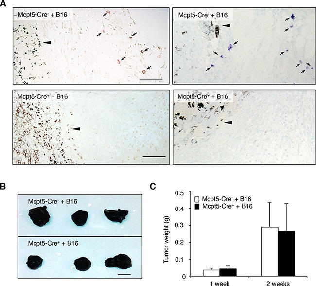
“Indeed, subcutaneous growth of B16 melanoma is unaffected in Mcpt5‐Cre:RosaDTA mice, 231 , 232 which lack all mast cells in the skin.”
From Mast cell ontogeny: From fetal development to life‐long health and disease (2023) · cites this paper as [232]
In the citing paper
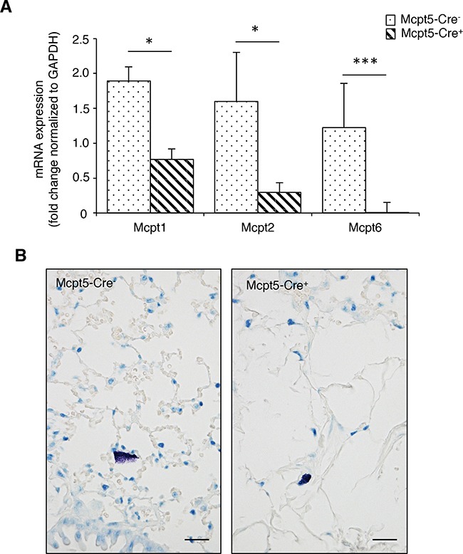
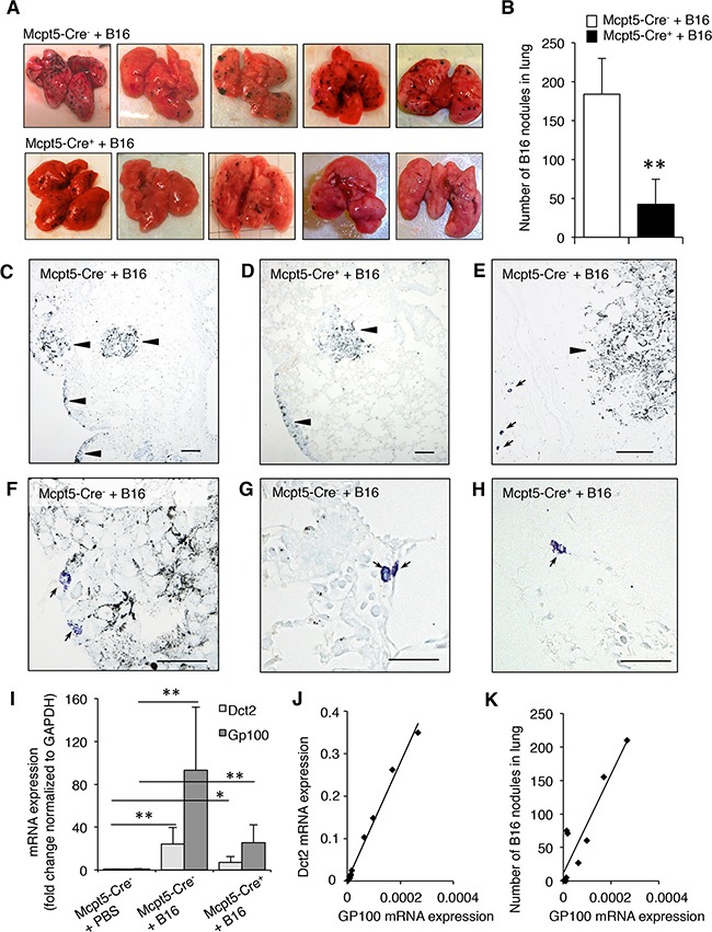
“Intriguingly however, B16 metastases are drastically reduced in the lung, 232 where only connective‐type mast cells are absent, but the mucosal‐type population marked by expression of integrin α4β7 is retained. 21”
From Mast cell ontogeny: From fetal development to life‐long health and disease (2023) · cites this paper as [232]
In the citing paper
“Finally, several mouse studies have found that the absence of mast cells has no effect on skin carcinogenesis or the growth of subcutaneously inoculated melanoma. 231 , 232 , 233”
From Mast cell ontogeny: From fetal development to life‐long health and disease (2023) · cites this paper as [232]
Self-check
Percentages, sums and reported differences re-computed from the paper’s own numbers.
The self-check needs the paper’s full text.
Citations
Cites
Inflammatory mast cells up-regulate angiogenesis during squamous epithelial carcinogenesis
Coussens, L. M., Raymond, W. W., Bergers, G., Laig-Webster, M., Behrendtsen, O., Werb, Z., Caughey, G. H., & Hanahan, D. (1999) · Genes & Development · doi:10.1101/gad.13.11.1382 · Cited by 946 · cited 2×
“To experimentally address the role of mast cells in malignancies, investigators have assessed various mast celldeficient mouse models, mainly such in which mast celldeficiency is a result of defective c-kit signaling [26-31].”
Background
Research works printed but not linked: 47
- Tsai, 2008, Immunomodulatory mast cells: negative, as well as positive, regulators of immunity, Nat Rev Immunol, 8, 478, 10.1038/nri2327 · DOI
- Voehringer, 2013, Protective and pathological roles of mast cells and basophils, Nat Rev Immunol, 13, 362, 10.1038/nri3427 · DOI · Paper page
- Shi, 2007, Mast cells promote atherosclerosis by releasing proinflammatory cytokines, Nat Med, 13, 719, 10.1038/nm1601 · DOI
- Dudeck, 2014, Mast cells promote T cell driven antigen-induced arthritis despite being dispensable in T cell bypassing antibody-induced arthritis, Arthritis Rheumatol
- Liu, 2001, Mast cells play a key role in neutrophil recruitment in experimental bullous pemphigoid, J Clin Invest, 108, 1151, 10.1172/JCI11494 · DOI
- Blank, 2015, Mast cells in renal inflammation and fibrosis: lessons learnt from animal studies, Mol Immunol, 63, 86, 10.1016/j.molimm.2014.03.002 · DOI
- Marshall, 2015, Mast cells as targets for immunotherapy of solid tumors, Mol Immunol, 63, 113, 10.1016/j.molimm.2014.02.020 · DOI
- Galli, 2013, Mast cells: potential positive and negative roles in tumor biology, Cancer Immunol Res, 1, 269, 10.1158/2326-6066.CIR-13-0119 · DOI
- Crivellato, 2009, The controversial role of mast cells in tumor growth, International review of cell and molecular biology, 275, 89, 10.1016/S1937-6448(09)75004-X · DOI
- Marone, 2016, Controversial role of mast cells in skin cancers, Exp Dermatol
- Noelle, 2009, Mast cells as regulators of adaptive immunity to tumours, Clin Exp Immunol, 155, 140, 10.1111/j.1365-2249.2008.03840.x · DOI
- Wanachantararak, 2003, Increase of mast cells and tumor angiogenesis in oral squamous cell carcinoma, J Oral Pathol Med, 32, 195, 10.1034/j.1600-0714.2003.00128.x · DOI
- Rudolph, 2005, Characterization of mast cell subpopulations in lip cancer, J Oral Pathol Med, 34, 268, 10.1111/j.1600-0714.2004.00297.x · DOI
- Enblad, 2002, Mast cell infiltration correlates with poor prognosis in Hodgkin's lymphoma, Br J Haematol, 119, 122, 10.1046/j.1365-2141.2002.03768.x · DOI
- Treon, 2006, Mast cells in Waldenstrom's macroglobulinemia support lymphoplasmacytic cell growth through CD154/CD40 signaling, Ann Oncol, 17, 1275, 10.1093/annonc/mdl109 · DOI
- Okuyama, 2007, Decreased number of mast cells infiltrating into needle biopsy specimens leads to a better prognosis of prostate cancer, British journal of cancer, 97, 952, 10.1038/sj.bjc.6603962 · DOI
- Hayes, 2004, The presence of stromal mast cells identifies a subset of invasive breast cancers with a favorable prognosis, Mod Pathol, 17, 690, 10.1038/modpathol.3800094 · DOI
- Huntsman, 2007, Stromal mast cells in invasive breast cancer are a marker of favourable prognosis: a study of 4,444 cases, Breast Cancer Res Treat
- Bradding, 2005, Macrophage and mast-cell invasion of tumor cell islets confers a marked survival advantage in non-small-cell lung cancer, J Clin Oncol, 23, 8959, 10.1200/JCO.2005.01.4910 · DOI
- Enblad, 2007, Mast cell infiltration is a favourable prognostic factor in diffuse large B-cell lymphoma, Br J Haematol, 138, 68, 10.1111/j.1365-2141.2007.06612.x · DOI
- Samoszuk, 2005, Mast cell density, angiogenesis, blood clotting, and prognosis in women with advanced ovarian cancer, Gynecologic oncology, 99, 20, 10.1016/j.ygyno.2005.05.042 · DOI
- Sirigu, 2003, Tumor vascularity and tryptase-positive mast cells correlate with a poor prognosis in melanoma, European journal of clinical investigation, 33, 420, 10.1046/j.1365-2362.2003.01152.x · DOI
- Kawamoto, 2000, Cutaneous malignant melanoma: correlation between neovascularization and peritumor accumulation of mast cells overexpressing vascular endothelial growth factor, Hum Pathol, 31, 955, 10.1053/hupa.2000.16658 · DOI
- Dammacco, 2003, Neovascularisation, expression of fibroblast growth factor-2, and mast cells with tryptase activity increase simultaneously with pathological progression in human malignant melanoma, Eur J Cancer, 39, 666, 10.1016/S0959-8049(02)00150-8 · DOI
- Harvima, 2015, Low numbers of tryptase+ and chymase+ mast cells associated with reduced survival and advanced tumor stage in melanoma, Melanoma Res, 25, 479, 10.1097/CMR.0000000000000192 · DOI
- Taubenberger, 1988, Mast-cell-deficient W/Wv mice exhibit a decreased rate of tumor angiogenesis, Int J Cancer, 42, 48, 10.1002/ijc.2910420110 · DOI
- Evan, 2007, Mast cells are required for angiogenesis and macroscopic expansion of Myc-induced pancreatic islet tumors, Nat Med, 13, 1211, 10.1038/nm1649 · DOI
- Zetter, 1994, Mast cell accumulation at sites of murine tumor implantation: implications for angiogenesis and tumor metastasis, Invasion Metastasis, 14, 395
- Paupe, 1985, Inverse correlation between tumor incidence and tissue histamine levels in W/WV, WV/+, and +/+ mice, J Natl Cancer Inst, 74, 671
- James, 1985, Growth of pulmonary metastases of B16 melanoma in mast cell-free mice, J Surg Res, 38, 24, 10.1016/0022-4804(85)90006-X · DOI
- Pejler, 2015, Approaches for Analyzing the Roles of Mast Cells and Their Proteases In Vivo, Adv Immunol, 126, 45, 10.1016/bs.ai.2014.11.002 · DOI · Paper page
- Rodewald, 2011, Cre-mediated cell ablation contests mast cell contribution in models of antibody- and T cell-mediated autoimmunity, Immunity, 35, 832, 10.1016/j.immuni.2011.09.015 · DOI
- Galli, 2011, Reduced mast cell and basophil numbers and function in Cpa3-Cre; Mcl-1fl/fl mice, Blood, 118, 6930, 10.1182/blood-2011-03-343962 · DOI · Paper page
- Roers, 2011, Mast cells are key promoters of contact allergy that mediate the adjuvant effects of haptens, Immunity, 34, 973, 10.1016/j.immuni.2011.03.028 · DOI
- Roers, 2008, Mast cell-specific Cre/loxP-mediated recombination in vivo, Transgenic research, 17, 307, 10.1007/s11248-007-9153-4 · DOI
- Wernersson, 2007, Mast cell proteases, Adv Immunol, 95, 167, 10.1016/S0065-2776(07)95006-3 · DOI
- Nieto, 2009, Epithelial-mesenchymal transitions in development and disease, Cell, 139, 871, 10.1016/j.cell.2009.11.007 · DOI · Paper page
- Wu, 2015, A twist tale of cancer metastasis and tumor angiogenesis, Histology and histopathology, 30, 1283
- Ronnstrand, 2006, The stem cell factor receptor/c-Kit as a drug target in cancer, Curr Cancer Drug Targets, 6, 65, 10.2174/156800906775471725 · DOI
- Jones, 2011, Protease phenotype of constitutive connective tissue and of induced mucosal mast cells in mice is regulated by the tissue, Proc Natl Acad Sci U S A, 108, 14210, 10.1073/pnas.1111048108 · DOI
- Papadopoulou, 2015, Dicer is indispensable for the development of murine mast cells, J Allergy Clin Immunol, 135, 1077, 10.1016/j.jaci.2014.10.005 · DOI
- Hartmann, 2012, Mast cells play a protumorigenic role in primary cutaneous lymphoma, Blood, 120, 2042, 10.1182/blood-2012-03-415638 · DOI
- Frossard, 2014, Targeting mast cells in inflammatory diseases, Pharmacology & therapeutics, 142, 416, 10.1016/j.pharmthera.2014.01.004 · DOI
- Pejler, 2012, Mast cell apoptosis induced by siramesine, a sigma-2 receptor agonist, Biochem Pharmacol, 84, 1671, 10.1016/j.bcp.2012.09.028 · DOI
- Olsson, 2015, Neutrophil Extracellular Traps Accumulate in Peripheral Blood Vessels and Compromise Organ Function in Tumor-Bearing Animals, Cancer Res, 75, 2653, 10.1158/0008-5472.CAN-14-3299 · DOI
- Melillo, 2015, Mast cells induce epithelial-to-mesenchymal transition and stem cell features in human thyroid cancer cells through an IL-8-Akt-Slug pathway, Oncogene, 34, 5175, 10.1038/onc.2014.441 · DOI
- Pejler, 2012, Serglycin: the master of the mast cell, Methods Mol Biol, 836, 201, 10.1007/978-1-61779-498-8_14 · DOI
Cited by
Sorted by citations
Mast cell ontogeny: From fetal development to life‐long health and disease
Chia, S. L., Kapoor, S., Carvalho, C., Bajénoff, M., & Gentek, R. (2023) · Immunological Reviews · doi:10.1111/imr.13191 · Cited by 60
Metadata
- Authors
- Öhrvik, H., Grujic, M., Waern, I., Gustafson, A.-M., Ernst, N., Roers, A., Hartmann, K., & Pejler, G.
- Affiliations
- Department of Medical Biochemistry and Microbiology, Uppsala University, Uppsala, Sweden
- Department of Anatomy, Physiology and Biochemistry, Swedish University of Agricultural Sciences, Uppsala, Sweden
- Department of Dermatology, University of Luebeck, Luebeck, Germany
- Institute for Immunology, University of Technology Dresden, Dresden, Germany
- Venue
- Oncotarget
- Publisher
- Impact Journals, LLC
- Published
- 2016-09-02
- Volume
- 7
- Issue
- 42
- Pages
- 68990-69001
- ISSN
- 1949-2553
- PMC
- PMC5356606
- Cited by
- 28
- References
- 1
- Language
- en
About these checks
Registry, retraction, Cites and statistics results are computed, not judged, with one exception: a printed reference that no index lists is read once by a model before it counts as a citation error. Claims are extracted by a model and kept only when their quote appears verbatim in the text; each cited source is then checked, with a model’s help, for whether it says what the claim relies on. Those verdicts can be wrong — read the claim’s quote and the cited source before relying on one.
Statistics and arithmetic are recomputed, not judged.
Confidence bands are shown only for fields calibrated on our annotated test set. None is calibrated yet, so no band is shown.
What counts as a claim ›How we measure confidence ›How we check this ›
Are you an author, or do you see a problem with this page? Request a review
How to cite
Cite CoChat for the checks on this page, and the paper for its own findings.
- This page
- CoChat. (2026). Citation check: Mast cells promote melanoma colonization of lungs. https://app.cochat.ai/papers/10.18632/oncotarget.11837
- The paper
- Öhrvik, H., Grujic, M., Waern, I., Gustafson, A.-M., Ernst, N., Roers, A., Hartmann, K., & Pejler, G. (2016). Mast cells promote melanoma colonization of lungs. Oncotarget. https://doi.org/10.18632/oncotarget.11837
