

The DOI matches its Crossref record; 46 of the 46 references it cites were found in a registry or an index.
Multimodality imaging in nanomedicine and nanotheranostics
Open accessAbstract
Accurate diagnosis of tumors needs much detailed information. However, available single imaging modality cannot provide complete or comprehensive data. Nanomedicine is the application of nanotechnology to medicine, and multimodality imaging based on nanoparticles has been receiving extensive attention. This new hybrid imaging technology could provide complementary information from different imaging modalities using only a single injection of contrast agent. In this review, we introduce recent developments in multifunctional nanoparticles and their biomedical applications to multimodal imaging and theragnosis as nanomedicine. Most of the reviewed studies are based on the intrinsic properties of nanoparticles and their application in clinical imaging technology. The imaging techniques include positron emission tomography, single-photon emission computed tomography, computerized tomography, magnetic resonance imaging, optical imaging, and ultrasound imaging.
Overview
The claims and statistics of this paper haven’t been checked yet.
Run the check — free, no account needed. It reads the open-access text and checks the sources it cites.
Integrity signals
Registry
Cites
Statistics
Figures
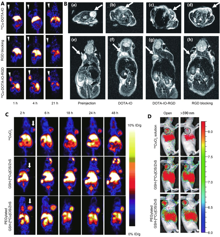
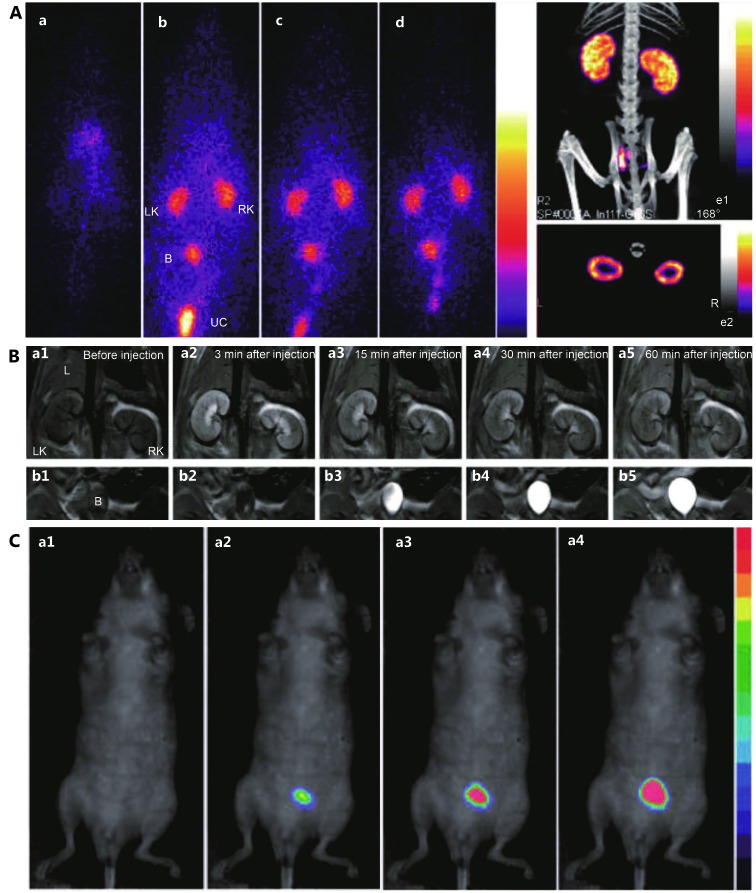
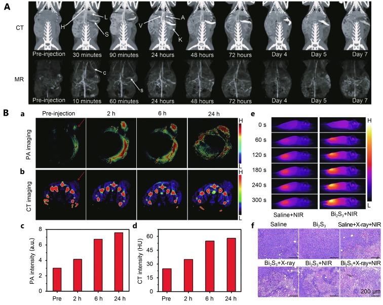
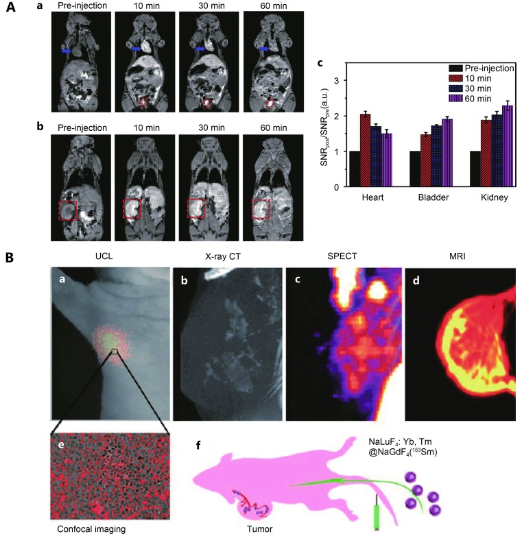
Verification
Claims the paper makes
The claims and statistics of this paper haven’t been checked yet.
Run the check — free, no account needed. It reads the open-access text and checks the sources it cites.
Claims other papers make about this one
Each is checked against this paper’s own text: does it say what the citing paper says it does?
The cited source says what the claim attributes to it.
In the citing paper
“Different types of NPs for multimodal imaging have been established such as modified Gd2O3 NPs, gold NPs, 64CuInS/ZnS QDs, Bi2S3 NPs, and liposomal nanocarrier conjugated 99mTc [137,138].”
From Approaches to Improve EPR-Based Drug Delivery for Cancer Therapy and Diagnosis (2023) · cites this paper as [137]
Self-check
Percentages, sums and reported differences re-computed from the paper’s own numbers.
The self-check needs the paper’s full text.
Citations
Cites
Chemistry and Properties of Nanocrystals of Different Shapes
Burda, C., Chen, X., Narayanan, R., & El-Sayed, M. A. (2005) · Chemical Reviews · doi:10.1021/cr030063a · Cited by 7,230
Wu, X., Liu, H., Liu, J., Haley, K. N., Treadway, J. A., Larson, J., Ge, N., Peale, F., & Bruchez, M. P. (2002) · Nature Biotechnology · doi:10.1038/nbt764 · Cited by 2,506
Nanochemistry and Nanomedicine for Nanoparticle-based Diagnostics and Therapy
Chen, G., Roy, I., Yang, C., & Prasad, P. N. (2016) · Chemical Reviews · doi:10.1021/acs.chemrev.5b00148 · Cited by 1,528
Recent advances in iron oxide nanocrystal technology for medical imaging☆
Corot, C., Robert, P., Idée, J., & Port, M. (2006) · Advanced Drug Delivery Reviews · doi:10.1016/j.addr.2006.09.013 · Cited by 1,498
Nanomedicine—Challenge and Perspectives
Riehemann, K., Schneider, S. W., Luger, T. A., Godin, B., Mauro Ferrari, B., & Fuchs, H. (2009) · Angewandte Chemie International Edition · doi:10.1002/anie.200802585 · Cited by 1,310
Molecular imaging in drug development
Willmann, J. K., van Bruggen, N., Dinkelborg, L. M., & Gambhir, S. S. (2008) · Nature Reviews Drug Discovery · doi:10.1038/nrd2290 · Cited by 1,175
Multimodality Imaging Probes: Design and Challenges
Louie, A. (2010) · Chemical Reviews · doi:10.1021/cr9003538 · Cited by 1,044
Fluorescence-guided surgery with live molecular navigation — a new cutting edge
Nguyen, Q. T., & Tsien, R. Y. (2013) · Nature reviews. Cancer · doi:10.1038/nrc3566 · Cited by 566
Molecular Imaging with Theranostic Nanoparticles
Jokerst, J. V., & Gambhir, S. S. (2011) · Accounts of Chemical Research · doi:10.1021/ar200106e · Cited by 565
Lee, H., Li, Z., Chen, K., Hsu, A. R., Xu, C., Xie, J., Sun, S., & Chen, X. S. (2008) · Journal of Nuclear Medicine · doi:10.2967/jnumed.108.051243 · Cited by 563
Research works printed but not linked: 1
- [53]Niu C Wang Z Lu G Krupka TM Sun Y You Y Gold–nanoshelled microcapsules: a theranostic agent for ultrasound contrast imaging and photothermal therapy Angew Chem Int Ed Engl 2011 50 3017 21 21404389
Cited by
Sorted by citations
Israel, L. L., Galstyan, A., Holler, E., & Ljubimova, J. Y. (2020) · Journal of Controlled Release · doi:10.1016/j.jconrel.2020.01.009 · Cited by 343
“Other than focusing on only the two main techniques of MRI and MPI, many NPs with dual or multimodality [22,153,154] imaging methods were developed, combining MRI and/or MPI with optical imaging [155–157], sensitive and high resolution positron emission tomography (PET) [129,158], computed tomography (CT) [159] and ot…”
Luminescent metal nanoclusters: Biosensing strategies and bioimaging applications
Xiao, Y. C., Wu, Z., Yao, Q., & Xie, J. (2021) · Aggregate · doi:10.1002/agt2.11 · Cited by 274
Magnetic Resonance Imaging and Iron-oxide Nanoparticles in the era of Personalized Medicine
Rahman, M. (2023) · Nanotheranostics · doi:10.7150/ntno.86467 · Cited by 244
“Their size has the great advantage to supply a large amount of material to the targeted site (15).”
Shang, L., Xu, J., & Nienhaus, G. U. (2019) · Nano Today · doi:10.1016/j.nantod.2019.100767 · Cited by 237
Two‐Dimensional Nanomaterials for Cancer Nanotheranostics
Chen, Y., Wu, Y., Sun, B., Liu, S., & Liu, H. (2017) · Small · doi:10.1002/smll.201603446 · Cited by 174
Multiplexed imaging for diagnosis and therapy
Heinzmann, K., Carter, L. M., Lewis, J. S., & Aboagye, E. O. (2017) · Nature Biomedical Engineering · doi:10.1038/s41551-017-0131-8 · Cited by 169
Conjugated Photosensitizers for Imaging and PDT in Cancer Research
Simões, J. C. S., Sarpaki, S., Papadimitroulas, P., Therrien, B., & Loudos, G. K. (2020) · Journal of Medicinal Chemistry · doi:10.1021/acs.jmedchem.0c00047 · Cited by 164
Magnetic Fe3O4 nanoparticles coated by natural rubber latex as MRI contrast agent
Arsalani, S., Guidelli, É. J., da Silveira, M. A., Salmón, C. E. G., Araújo, J. F. D. F., Bruno, A. C., & Baffa, O. (2018) · Journal of Magnetism and Magnetic Materials · doi:10.1016/j.jmmm.2018.11.132 · Cited by 116
Farzin, L., Sheibani, S., Moassesi, M. E., & Shamsipur, M. (2018) · Journal of Biomedical Materials Research Part A · doi:10.1002/jbm.a.36550 · Cited by 101
Approaches to Improve EPR-Based Drug Delivery for Cancer Therapy and Diagnosis
Subhan, M. A., Parveen, F., Filipczak, N., Yalamarty, S. S. K., & Torchilin, V. P. (2023) · Journal of Personalized Medicine · doi:10.3390/jpm13030389 · Cited by 98
“Different types of NPs for multimodal imaging have been established such as modified Gd2O3 NPs, gold NPs, 64CuInS/ZnS QDs, Bi2S3 NPs, and liposomal nanocarrier conjugated 99mTc [137,138].”
BackgroundMultiomics Research: Principles and Challenges in Integrated Analysis
Luo, Y., Zhao, C., & Chen, F. (2024) · BioDesign Research · doi:10.34133/bdr.0059 · Cited by 108
“One future direction is the integration of various imaging technologies, such as MRI, positron emission tomography (PET), CT, and high-resolution optical imaging technologies (e.g., fluorescence imaging), to achieve multimodal imaging of animals and plants [ 16 ].”
Aspects of high-performance and bio-acceptable magnetic nanoparticles for biomedical application
Kush, P., Kumar, P., Singh, R., & Kaushik, A. K. (2021) · Asian Journal of Pharmaceutical Sciences · doi:10.1016/j.ajps.2021.05.005 · Cited by 96
Camptothecin-based nanodrug delivery systems
Wen, Y. F., Yingze, W., Xiaoli, L., Wei, Z., Xinhe, X., Zhongxiao, H., & Xing-jie, L. (2017) · Cancer Biology and Medicine · doi:10.20892/j.issn.2095-3941.2017.0099 · Cited by 85
Covalent nano delivery systems for selective imaging and treatment of brain tumors
Ljubimova, J. Y., Sun, T., Mashouf, L. A., Ljubimov, A. V., Israel, L. L., Ljubimov, V. A., Falahatian, V., & Holler, E. (2017) · Advanced Drug Delivery Reviews · doi:10.1016/j.addr.2017.06.002 · Cited by 82
Rahme, K., & Dagher, N. (2019) · Pharmaceutics · doi:10.3390/pharmaceutics11070327 · Cited by 71
“Traditional clinical imaging techniques such as computed X-ray tomography (CT), magnetic resonance imaging (MRI), and ultrasound (US) do not always meet the need of personalized cancer diagnosis because of the shift toward the development of precise diagnosis of diseases, particularly cancer [57].”
MethodDendrimers based cancer nanotheranostics: An overview
Saluja, V., Mishra, Y., Mishra, V. K., Giri, N., & Nayak, P. (2021) · International Journal of Pharmaceutics · doi:10.1016/j.ijpharm.2021.120485 · Cited by 68
Ultrasound and nanomaterial: an efficient pair to fight cancer
Alphandéry, E. (2022) · Journal of Nanobiotechnology · doi:10.1186/s12951-022-01243-w · Cited by 67
Lipid-Encapsulated Fe3O4 Nanoparticles for Multimodal Magnetic Resonance/Fluorescence Imaging
Zhang, L., Tong, S., Zhang, Q., & Bao, G. (2020) · ACS Applied Nano Materials · doi:10.1021/acsanm.0c01193 · Cited by 58
Biocompatibility in Regenerative Nanomedicine
Zor, F., Selek, F. N., Orlando, G., & Williams, D. F. (2019) · Nanomedicine · doi:10.2217/nnm-2019-0140 · Cited by 57
Nanomedicine in Non-Small Cell Lung Cancer: From Conventional Treatments to Immunotherapy
García-Fernández, C., Fornaguera, C., & Borrós, S. (2020) · Cancers · doi:10.3390/cancers12061609 · Cited by 44
“The systemic administration of chemotherapy drugs involves their internalization by healthy cells [31,34,68].”
BackgroundMethodRare Earth Luminescence: Electronic Spectroscopy and Applications
Khan, L. U., & Khan, Z. U. (2018) · Springer eBooks · doi:10.1007/978-3-319-92955-2_10 · Cited by 43
Osteotropic Radiolabeled Nanophotosensitizer for Imaging and Treating Multiple Myeloma
Tang, R., Zheleznyak, A., Mixdorf, M., Ghai, A., Prior, J. L., Black, K. C. L., Shokeen, M., Reed, N., Biswas, P., & Achilefu, S. A. (2020) · ACS Nano · doi:10.1021/acsnano.9b09618 · Cited by 41
19 F MRI Probes for Multimodal Imaging**
Janasik, D., & Krawczyk, T. (2021) · Chemistry - A European Journal · doi:10.1002/chem.202102556 · Cited by 41
Magnetic nanoparticles: an emerging nano-based tool to fight against viral infections
Jat, S. K., Gandhi, H. A., Bhattacharya, J., & Sharma, M. K. (2021) · Materials Advances · doi:10.1039/d1ma00240f · Cited by 37
Nano-mediated strategy for targeting and treatment of non-small cell lung cancer (NSCLC)
Ashique, S., Garg, A., Mishra, N., Raina, N., Ming, L. C., Tuli, H. S., Behl, T., Rani, R., & Gupta, M. (2023) · Naunyn-Schmiedeberg s Archives of Pharmacology · doi:10.1007/s00210-023-02522-5 · Cited by 36
“Additionally, by changing these properties, it is feasible to employ them as contrast agents for CT/PET imaging methods, consequently establishing multifunctional theranostic platforms (Li et al. 2016b).”
BackgroundThao, T., Yoo, S. W., Park, S., Kim, J. Y., Choi, K., Kim, C., Kwon, S. Y., Min, J., & Lee, C. (2021) · Sensors · doi:10.3390/s21051776 · Cited by 36
“Optical imaging enables the obtaining of a high-resolution image at high speed, but the shallow imaging depth is an issue [9].”
BackgroundMagneto-silver core–shell nanohybrids for theragnosis
Haribabu, V., Girigoswami, K., & Girigoswami, A. (2020) · Nano-Structures & Nano-Objects · doi:10.1016/j.nanoso.2020.100636 · Cited by 35
Progress and Challenges in the Diagnosis and Treatment of Brain Cancer Using Nanotechnology
Ale, Y., & Nainwal, N. (2023) · Molecular Pharmaceutics · doi:10.1021/acs.molpharmaceut.3c00554 · Cited by 36
Iodinated Echogenic Glycol Chitosan Nanoparticles for X-ray CT/US Dual Imaging of Tumor
Choi, D., Jeon, S., You, D. G., Um, W., Kim, J. Y., Yoon, H. Y., Chang, H., Kim, D., Park, J. H., Kim, H., et al. (2017) · Nanotheranostics · doi:10.7150/ntno.18643 · Cited by 33
“[5] Among the diagnosis approaches, Ultrasound (US) imaging is widely utilized in clinical diagnosis due to high safety, low-cost and portability.[6] As an US imaging contrast agents, gas precursor-contained microbubbles have developed to enhance contrast effect during the US imaging.”
BackgroundAdvancements in Nanoporous Materials for Biomedical Imaging and Diagnostics
Parvin, N., Kumar, V., Mandal, T. K., & Joo, S. W. (2024) · Journal of Functional Biomaterials · doi:10.3390/jfb15080226 · Cited by 31
“These materials hold great potential for the early diagnosis of infectious diseases, cancer, and neurological disorders, offering improved sensitivity, specificity, and multiplexing capabilities compared to csonventional diagnostic methods.”
BackgroundIodinated Metallacrowns: Toward Combined Bimodal Near‐Infrared and X‐Ray Contrast Imaging Agents
Lutter, J. C., Eliseeva, S. V., Collet, G., Martinić, I., Kampf, J. W., Schneider, B. L., Carichner, A., Sobilo, J., Lerondel, S., Pètoud, S., et al. (2019) · Chemistry - A European Journal · doi:10.1002/chem.201905241 · Cited by 31
Tammaro, O., di Polidoro, A. C., Romano, E., Netti, P. A., & Torino, E. (2020) · Scientific Reports · doi:10.1038/s41598-020-63234-x · Cited by 31
Akakuru, O. U., Zhang, Z., Iqbal, M. Z., Zhu, C., Zhang, Y., & Wu, A. (2022) · Acta Pharmaceutica Sinica B · doi:10.1016/j.apsb.2022.02.016 · Cited by 26
Perry, W., Weiss, A., de Plas, R. V., Spraggins, J. M., Caprioli, R. M., & Skaar, E. P. (2020) · Current Opinion in Chemical Biology · doi:10.1016/j.cbpa.2020.01.008 · Cited by 24
“Excellent recent reviews on diagnostic imaging have been published [11, 12].”
BackgroundNanotechnology in interventional cardiology: A state-of-the-art review
Almas, T., Haider, R., Malik, J., Mehmood, A., Alvi, A., Naz, H., Satti, D. I., Zaidi, S. M. J., Alsubai, A. K., AlNajdi, S., et al. (2022) · IJC Heart & Vasculature · doi:10.1016/j.ijcha.2022.101149 · Cited by 25
Functionalization of theranostic AGuIX® nanoparticles for PET/MRI/optical imaging
Thakare, V., Tran, V., Natuzzi, M., Thomas, E., Moreau, M., Romieu, A., Collin, B., Courteau, A., Vrigneaud, J., Louis, C., et al. (2019) · RSC Advances · doi:10.1039/c9ra00365g · Cited by 25
Motiei, M., Dreifuss, T., Sadan, T., Omer, N., Blumenfeld‐Katzir, T., Fragogeorgi, E. A., Loudos, G. K., Popovtzer, R., & Ben‐Eliezer, N. (2019) · Chemistry Letters · doi:10.1246/cl.180780 · Cited by 23
Following nanomedicine activation with magnetic resonance imaging: why, how, and what’s next?
Reeβing, F., & Szymański, W. (2018) · Current Opinion in Biotechnology · doi:10.1016/j.copbio.2018.10.008 · Cited by 22
Shapoval, O., Oleksa, V., Šlouf, M., Lobaz, V., Trhlíková, O., Filipová, M., Janoušková, O., Engstová, H., Pankrác, J., Modrý, A., et al. (2021) · Nanomaterials · doi:10.3390/nano11010230 · Cited by 22
“Examples of nanosized particles used in imaging technologies include dendrimers, liposomes, gold, silver, quantum dots, iron oxides, silica, as well as lanthanides [2,4].”
BackgroundNanoengineered photoactive theranostic agents for cancer
Jain, N. K., Chathoth, B. M., Bhaskar, V. S., Meena, H., Prasad, R., & Srivastava, R. (2021) · Nanophotonics · doi:10.1515/nanoph-2021-0205 · Cited by 23
PET‐Based Dual‐Modal Probes for In Vivo Imaging
Zhao, R., Lan, D., Xia, B., Dong, M., Mu, J., & Zhao, Y. (2025) · Small · doi:10.1002/smll.202409713 · Cited by 23
Recent Advancements in Ln‐Ion‐Based Upconverting Nanomaterials and Their Biological Applications
Chhetri, B. P., Karmakar, A., & Ghosh, A. (2019) · Particle & Particle Systems Characterization · doi:10.1002/ppsc.201900153 · Cited by 22
Emerging nanoparticle-based strategies for advanced cancer imaging and diagnosis
Kamyab, H., Khalili, E., Yuzir, A., Taheri, M. M., Zambrano, A. K., & Rajendran, S. (2025) · International Journal of Pharmaceutics · doi:10.1016/j.ijpharm.2025.126046 · Cited by 24
Kiran, P., Debnath, S. K., Neekhra, S., Pawar, V. S., Khan, A., Dias, F., Pallod, S., & Srivastava, R. (2021) · Wiley Interdisciplinary Reviews Nanomedicine and Nanobiotechnology · doi:10.1002/wnan.1768 · Cited by 26
Najdian, A., Amanlou, M., Beiki, D., Bitarafan‐Rajabi, A., Mirzaei, M., & Ardestani, M. S. (2022) · Bioorganic Chemistry · doi:10.1016/j.bioorg.2022.105827 · Cited by 21
Morita, M., Tachikawa, T., Seino, S., Tanaka, K., & Majima, T. (2017) · ACS Applied Nano Materials · doi:10.1021/acsanm.7b00213 · Cited by 21
Iron–Gold Nanoflowers: A Promising Tool for Multimodal Imaging and Hyperthermia Therapy
Christou, E., Pearson, J. R., Beltrán, A. M., Fernández‐Afonso, Y., Gutiérrez, L., de la Fuente, J. M., Gámez, F., García‐Martín, M. L., & Caro, C. (2022) · Pharmaceutics · doi:10.3390/pharmaceutics14030636 · Cited by 21
“In fact, improved diagnostic accuracy has been demonstrated using MCNP-mediated multimodal imaging [38,39], as well as increased patient safety by reducing the amount of CAs injected.”
MethodBhattacharjee, S., & Brayden, D. J. (2020) · Expert Opinion on Drug Discovery · doi:10.1080/17460441.2021.1826434 · Cited by 20
Jun, E., Kim, S. C., Lee, C. M., Oh, J., Lee, S., & Shim, I. K. (2017) · Scientific Reports · doi:10.1038/s41598-017-12670-3 · Cited by 19
Triple-modal imaging of stem-cells labeled with multimodal nanoparticles, applied in a stroke model
da Silva, H. R., Mamani, J. B., Nucci, M. P., Nucci, L. P., Kondo, A. T., Fantacini, D. M. C., de Souza, L. E. B., Picanço‐Castro, V., Covas, D. T., Kutner, J. M., et al. (2019) · World Journal of Stem Cells · doi:10.4252/wjsc.v11.i2.100 · Cited by 19
Gosée, C., Cadiou, C., Moreau, J., Callewaert, M., Kowandy, C., Henoumont, C., Larbanoix, L., Laurent, S., & Chuburu, F. (2025) · Coordination Chemistry Reviews · doi:10.1016/j.ccr.2025.216523 · Cited by 18
“To avoid any biodistribution problems, it is also necessary to combine these different modalities within a single object [4].”
Roffo, F., Ponsiglione, A. M., Netti, P. A., & Torino, E. (2022) · Biomedicines · doi:10.3390/biomedicines10020438 · Cited by 18
“Among different diagnostic techniques, we addressed Gadolinium-based Contrast agents (CAs) for Magnetic Resonance Imaging (MRI) because, even though MRI represents the most clinically applied modality [27], Gadolinium-free CAs still suffer from transmetalation, nephrotoxic effects and abnormal brain deposition [28–30].”
MethodAron, A., & Zavaleta, C. L. (2023) · Molecular Imaging and Biology · doi:10.1007/s11307-023-01827-4 · Cited by 18
“…stars, spheres) under abundant construction paradigms (homogenous throughout, varying core-shell constructs, and various combinations and permutations of these concepts) and can be functionalized for use with various imaging modalities, such as PET, CT, MRI, ultrasound, and optical imaging [85].”
BackgroundpH and redox dual-sensitive drug delivery system constructed based on fluorescent carbon dots
Zhang, B., Duan, Q., Li, Y., Wang, J., Zhang, W., & Sang, S. (2021) · RSC Advances · doi:10.1039/d0ra09164b · Cited by 17
Advancing Cancer-Targeted Nanotherapies with Tumor Homing Peptides
Rocchi, A., Teesalu, T., & Celia, C. (2025) · ACS Pharmacology & Translational Science · doi:10.1021/acsptsci.5c00241 · Cited by 16
Modular Synthesis of Peptide-Based Single- and Multimodal Targeted Molecular Imaging Agents
Schmitthenner, H. F., Barrett, T. M., Beach, S. A., Heese, L. E., Weidman, C., Dobson, D. E., Mahoney, E. R., Schug, N. C., Jones, K. G., Durmaz, C., et al. (2021) · ACS Applied Bio Materials · doi:10.1021/acsabm.1c00157 · Cited by 16
Monaco, I., Armanetti, P., Locatelli, E., Flori, A., Maturi, M., Turco, S. D., Menichetti, L., & Franchini, M. C. (2018) · Journal of Materials Chemistry B · doi:10.1039/c8tb00453f · Cited by 16
Li, G., Wu, S., Chen, W., Duan, X., Sun, X., Li, S., Mai, Z., Wu, W., Zeng, G., Liu, H., et al. (2023) · Small Methods · doi:10.1002/smtd.202201313 · Cited by 18
Day, A. H., Domarkas, J., Nigam, S., Renard, I., Cawthorne, C., Burke, B. P., Bahra, G. S., Oyston, P. C. F., Fallis, I. A., Archibald, S. J., et al. (2019) · Dalton Transactions · doi:10.1039/c9dt04024b · Cited by 14
Dung, D. T. K., Umezawa, M., Ohnuki, K., Nigoghossian, K., Okubo, K., Kamimura, M., Yamaguchi, M., Fujii, H., & Soga, K. (2022) · Biomaterials Science · doi:10.1039/d1bm01574e · Cited by 14
XFCT-MRI hybrid multimodal contrast agents for complementary imaging
Saladino, G. M., Vogt, C., Brodin, B. A., Shaker, K., Kilic, N. I., Andersson, K., Arsenian‐Henriksson, M., Toprak, M. S., & Hertz, H. M. (2022) · Nanoscale · doi:10.1039/d2nr05829d · Cited by 13
Sabu, A., Lin, J.-Y., Doong, R., Huang, Y.-F., & Chiu, H.-C. (2021) · Materials Advances · doi:10.1039/d1ma00563d · Cited by 4
Genevois, C., Hocquelet, A., Mazzocco, C., Rustique, E., Couillaud, F., & Grenier, N. (2017) · International Journal of Molecular Sciences · doi:10.3390/ijms18122584 · Cited by 11
Farzin, L., Shamsipur, M., Moassesi, M. E., & Sheibani, S. (2018) · Bioorganic & Medicinal Chemistry · doi:10.1016/j.bmc.2018.11.031 · Cited by 11
Sieno, L. D., Contini, D., Presti, G. L., Cortese, L., Matéo, T., Rosinski, B., Venturini, E., Panizza, P., Mora, M., Aranda, G. B., et al. (2019) · Biomedical Optics Express · doi:10.1364/boe.10.003899 · Cited by 11
Hassan, S. M. U., Siddique, M. T., Fakhar-e-alam, M., Atif, M. M., Saifullah, A., Marwat, N., Khurshid, A., Noor, O., Hossain, N., Ahmad, S., et al. (2022) · Journal of King Saud University - Science · doi:10.1016/j.jksus.2022.102080 · Cited by 9
CT‐based radiomics prediction of CXCL13 expression in ovarian cancer
Xu, W., Zhu, C., Dan, J. I., Qian, H., Shi, L., Mao, X., Zhou, H., & Wang, L. (2023) · Medical Physics · doi:10.1002/mp.16730 · Cited by 9
Combining gene therapy with other therapeutic strategies and imaging agents for cancer theranostics
Azevedo, A., Farinha, D., Geraldes, C. F. G. C., & Faneca, H. (2021) · International Journal of Pharmaceutics · doi:10.1016/j.ijpharm.2021.120905 · Cited by 9
Subcellular Imaging and Diagnosis of Cancer using Engineered Nanoparticles
Nannuri, S. H., Nikam, A. N., Pandey, A. P., Mutalik, S., & George, S. D. (2021) · Current Pharmaceutical Design · doi:10.2174/1381612827666210525154131 · Cited by 8
Core/Shell Iron Oxide@Gold nanoparticles for dual-modal CT/MRI imaging
Betzer, O., Motiei, M., Dreifuss, T., Sadan, T., Omer, N., Blumenfeld‐Katzir, T., Liu, Z., Ben‐Eliezer, N., & Popovtzer, R. (2020) · Nanoscale Imaging, Sensing, and Actuation for Biomedical Applications · doi:10.1117/12.2548430 · Cited by 9
Emerging trends in NanoTheranostics: Integrating imaging and therapy for precision health care
Fahmy, H. M., Bayoumi, L., Helal, N. F., Mohamed, N. R. A., Emarh, Y., & Ahmed, A. M. (2025) · International Journal of Pharmaceutics · doi:10.1016/j.ijpharm.2025.126057 · Cited by 11
Carbon-coated (Eu/Dy-Enabled) erbium hydroxide nanostructures for biomedical imaging
Saifullah, A., Hassan, S. M. U., & Khurshid, A. (2024) · Materialia · doi:10.1016/j.mtla.2024.102108 · Cited by 8
Polymeric nanoparticles as tumor-targeting theranostic platform
Mukherjee, B., Paul, B., Hoque, A. A., Sen, R., Chakraborty, S., & Chakraborty, A. (2023) · Elsevier eBooks · doi:10.1016/b978-0-323-89953-6.00009-x · Cited by 7
Process analytics for the manufacturing of nanomedicines: Challenges and opportunities
Aguiam, N., Moura, L. I. F., Oliveira, M. I. L. L., Florindo, H. F., & Lopes, J. A. (2024) · Elsevier eBooks · doi:10.1016/b978-0-323-91819-0.00007-5 · Cited by 7
Hybrid Multifunctional Nanomaterials for Diagnostic and Therapeutic Applications
Lagopati, N., Efstathopoulos, E. P., Veroutis, D., Katifelis, H., Theocharous, G., Pantelis, P., Evangelou, K., Gorgoulis, V. G., & Gazouli, M. (2022) · Nanotechnology in the Life Sciences · doi:10.1007/978-3-031-12658-1_17 · Cited by 6
Gobernanza de la nanomedicina: una revisión sistemática
Soto-Vázquez, R., Lau, É. Z., & López, L. A. M. (2021) · Mundo Nano Revista Interdisciplinaria en Nanociencia y Nanotecnología · doi:10.22201/ceiich.24485691e.2022.28.69682 · Cited by 7
Multimodal Molecular Imaging in the Second Near-Infrared Window
Wu, M., Li, X., Mu, X., Zhang, X., Wang, H., & Zhang, X. (2022) · Nanomedicine · doi:10.2217/nnm-2022-0124 · Cited by 6
Nanoprobiotics: When Technology Meets Gut Health
Machado, D., Almeida, D., Seabra, C. L., Andrade, J. C., Gomes, A., & Freitas, A. C. (2020) · Nanotechnology in the life sciences · doi:10.1007/978-3-030-41464-1_17 · Cited by 6
Theranostic Nanomaterials and Its Use in Biomedicine
Mukhopadhyay, A. (2022) · Wiley eBooks · doi:10.1002/9781119857747.ch17 · Cited by 6
Bright Photoluminescent Gd-MXene Quantum Dots for Cell Imaging with Synergistic MRI/CT Capability
Noor, A., Hassan, S. M. U., Shahzad, F., Zhang, X., Khurshid, A., Iqbal, Z., Siddique, M. T., & Marwat, N. (2025) · Journal of Fluorescence · doi:10.1007/s10895-025-04621-3 · Cited by 5
Liu, S., Tegafaw, T., Ho, S. L., Yue, H., Zhao, D., Liu, Y., Mulugeta, E., Chen, X., Lee, H. D., Ahn, D., et al. (2025) · Molecules · doi:10.3390/molecules30122519 · Cited by 6
Biocompatible Fluorescent Nanomaterials for Molecular Imaging Applications
Walia, S., Sharma, C., & Acharya, A. (2020) · Springer eBooks · doi:10.1007/978-981-15-4280-0_3 · Cited by 4
Polymer–Lipid Hybrid Nanosystems: An Emerging Advanced Therapeutic Tool
Okafor, N. I., Igbokwe, N. N., Nnolum-Orji, N. F., & Choonara, Y. E. (2025) · Advances in Polymer Technology · doi:10.1155/adv/4707146 · Cited by 3
“Another challenge involves the immunogenicity of the surface modi fi cations, such as PEGy-lation, which, although bene fi cial for prolonging circulation time, may trigger the production of anti-PEG antibodies in some patients and reduce the stealth capability of the nanoparticles over time [101].”
InfluentialSriramvenugopal, M., & Pacak, C. A. (2020) · Methods in molecular biology · doi:10.1007/978-1-0716-0364-2_15 · Cited by 3
Alles, E. J., Dwyer, G., Colchester, R. J., Maneas, E., Stoyanov, D. V., & Desjardins, A. E. (2020) · IEEE International Ultrasonics Symposium · doi:10.1109/ius46767.2020.9251641 · Cited by 3
“Accurate visualisation is key for successful interventions; however, no single imaging modality offers sufficient resolution, penetration, and soft-tissue contrast to adequately monitor interventional treatment [2].”
BackgroundMultimodal Imaging with NIR Light
Doan, T. K. D. (2020) · Springer eBooks · doi:10.1007/978-981-15-9627-8_11 · Cited by 3
Luminescent Nanocomposites for Biomedical Applications
Popovich, K., Klepárnı́k, K., Ledvina, V., Neužilová, B., Fleismann, J., Škodová, M., Kobera, L., Mihóková, E., Múčka, V., & Čuba, V. (2020) · IEEE Transactions on Nuclear Science · doi:10.1109/tns.2020.2974316 · Cited by 3
Molecular imaging in tracking cancer stem cells: A review.
Ghaffari, H., Atashzar, M. R., & Abdollahi, H. (2020) · PubMed Central · doi:10.34171/mjiri.34.90 · Cited by 3
Magnetic resonance imaging in personalized medicine
Rahman, M. (2023) · Elsevier eBooks · doi:10.1016/b978-0-323-99924-3.00001-7 · Cited by 2
An overview of theranostic nanomedicine
González‐Méndez, I., Sorroza-Martínez, K., Cuétara‐Guadarrama, F., Vonlanthen, M., & Rivera, E. (2024) · Elsevier eBooks · doi:10.1016/b978-0-443-22044-9.00005-x · Cited by 2
Nanotechnology-based theranostics for bone tumors
Zhan, W., Zeng, S., Yang, L., Zhang, Y.-Z., & Zhang, J. (2025) · Giant · doi:10.1016/j.giant.2025.100365 · Cited by 2
Modified carbon nanomaterials for diagnosis, drug delivery and stem cell therapy
Chawla, S., Bansal, I., & Rai, P. (2023) · Elsevier eBooks · doi:10.1016/b978-0-12-824366-4.00001-7 · Cited by 2
Innovations in liposomal drug delivery: paving the way for personalized cancer therapy
Aljabali, A. A. A., Obeid, M. A., El‐Tanani, M., & Tambuwala, M. M. (2024) · Elsevier eBooks · doi:10.1016/b978-0-443-14044-0.00004-1 · Cited by 2
Nucleic Acid Guided Molecular Tool for In-Vivo Theranostic Applications
Baba, S. A., Mutreja, R., Beniwal, A., Jain, S., Yadav, E., Ghosh, T., Navani, N. K., & Kumar, P. (2019) · Springer eBooks · doi:10.1007/978-981-13-8836-1_7 · Cited by 2
Choice of Nanomedicine in Cancer Imaging and Therapeutics
Jadhav, K., Kole, E. B., Naik, J., Verma, R. K., Gholap, A. D., Rawat, S., Meena, S., & Rojekar, S. V. (2025) · Springer eBooks · doi:10.1007/978-981-95-0938-6_6 · Cited by 1
Cancer Imaging and Scope of Early-Stage Diagnosis: Molecular Imaging Modalities
Setia, A., Rani, K., Randhave, N. V., Verma, N., Malik, A. K., Kumar, V., Vaishali, V., & Muthu, M. S. (2025) · Springer eBooks · doi:10.1007/978-981-95-0938-6_11 · Cited by 1
Gallium‐Containing Agents for Tumor Diagnosis and Therapy: Current Status and Future Prospects
Han, Y., Li, Q., Zheng, Y., Xia, D., & Zhou, Y. (2026) · Small · doi:10.1002/smll.73698 · Cited by 2
2D nanomaterials: pioneering platforms for cancer theranostics
Mishra, A., & Tripathy, J. (2026) · RSC Applied Interfaces · doi:10.1039/d6lf00017g · Cited by 1
Applications of Colloidal Nanocrystals
(2021) · Royal Society of Chemistry eBooks · doi:10.1039/9781788016568-00209 · Cited by 1
Applications of Nanotechnology in Pulmonary Disease Diagnosis
Sharma, A. K., Kaurav, H., Sharma, A. K., Nagraik, R., & Kapoor, D. N. (2022) · Springer eBooks · doi:10.1007/978-981-16-4392-7_9 · Cited by 1
Metadata
- Authors
- Li, X., Li, X., Zhang, X.-N., Li, X.-D., Chang, J., Zhang, X.-N., Li, X.-D., & Chang, J.
- Venue
- Cancer Biology & Medicine
- Publisher
- China Anti-cancer Association
- Published
- 2016
- Volume
- 13
- Issue
- 3
- Pages
- 339-348
- ISSN
- 2095-3941 (ISSN-L)
- PMC
- PMC5069837
- OpenAlex
- W2527625794
- Cited by
- 126
- References
- 46
- Citations by year
- 2026: 5 · 2025: 16 · 2024: 12 · 2023: 13 · 2022: 13 · 2021: 19 · 2020: 20 · 2019: 12 · 2018: 8 · 2017: 8
- FWCI
- 4.50
- Type
- journal article
- Language
- en
- Open access
- gold
- doi.org
- Licence
- cc-by
- Locations
- Cancer Biology and Medicine (publishedVersion, cc-by)
- PubMed Central (publishedVersion, cc-by-nc-sa)
- PubMed (publishedVersion)
- DOAJ (DOAJ: Directory of Open Access Journals) (submittedVersion, cc-by-sa)
- Topics
- Physical Sciences › Engineering › Biomedical Engineering › Nanoplatforms for cancer theranostics
- Health Sciences › Medicine › Radiology, Nuclear Medicine and Imaging › Medical Imaging Techniques and Applications
- Physical Sciences › Engineering › Biomedical Engineering › Photoacoustic and Ultrasonic Imaging
- Keywords
- multimodal imaging, nanomedicine, theranostics, multifunctional nanoparticles, tumor diagnosis, hybrid imaging, positron emission tomography, single photon emission computed tomography, magnetic resonance imaging, optical imaging, ultrasonography, contrast media
About these checks
Registry, retraction, Cites and statistics results are computed, not judged, with one exception: a printed reference that no index lists is read once by a model before it counts as a citation error. Claims are extracted by a model and kept only when their quote appears verbatim in the text; each cited source is then checked, with a model’s help, for whether it says what the claim relies on. Those verdicts can be wrong — read the claim’s quote and the cited source before relying on one.
Statistics and arithmetic are recomputed, not judged.
Confidence bands are shown only for fields calibrated on our annotated test set. None is calibrated yet, so no band is shown.
What counts as a claim ›How we measure confidence ›How we check this ›
Are you an author, or do you see a problem with this page? Request a review
