

The DOI matches its Crossref record; 1 of the 1 references it cites were found in a registry or an index.
eEF2 improves dense connective tissue repair and healing outcome by regulating cellular death, autophagy, apoptosis, proliferation and migration
Open accessPreprint — published as 10.1007/s00018-023-04776-x
Abstract
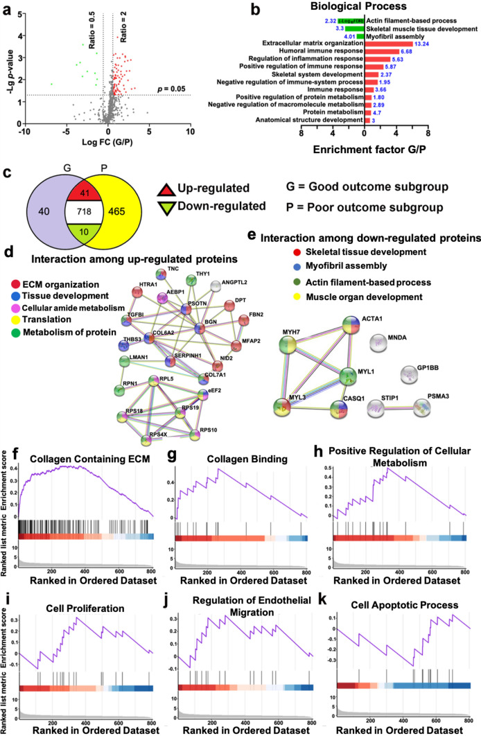
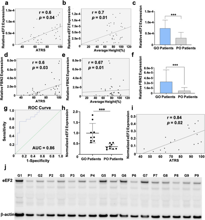
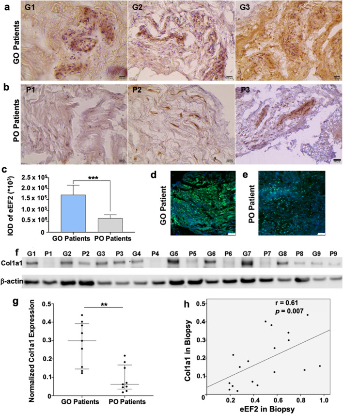
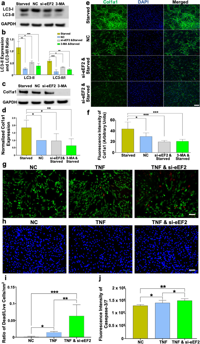
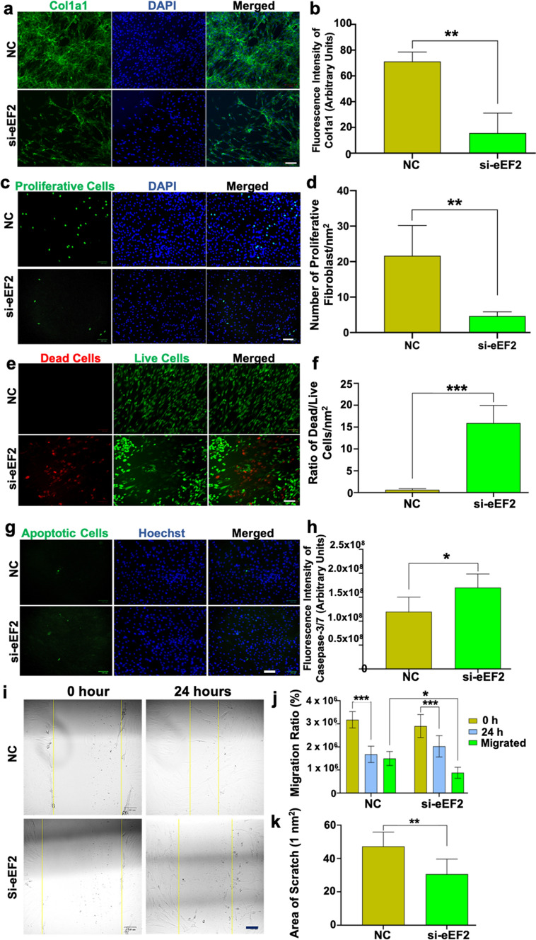
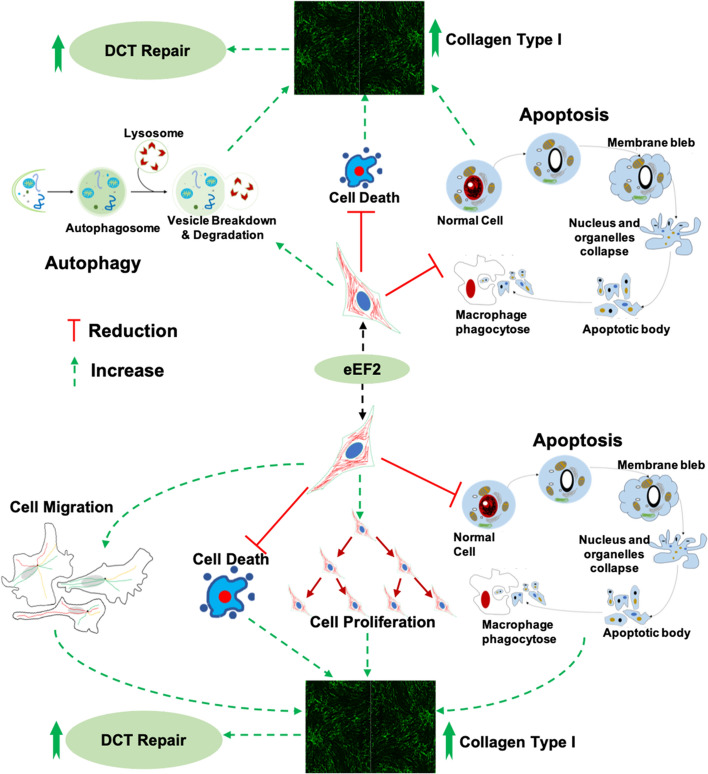
Outcomes following human dense connective tissue (DCT) repair are often variable and suboptimal, resulting in compromised function and development of chronic painful degenerative diseases. Moreover, biomarkers and mechanisms that guide good clinical outcomes after DCT injuries are mostly unknown. Here, we characterize the proteomic landscape of DCT repair following human tendon rupture and its association with long-term patient-reported outcome. Moreover, the regulatory mechanisms of relevant biomarkers were assessed partly by gene silencing experiments. A Mass-Spectrometry based proteomic approach quantified a large number (769) of proteins, including 51 differentially expressed proteins among 20 good versus 20 poor outcome patients. A novel biomarker, elongation factor-2 (eEF2) was identified as being strongly prognostic of the 1-year clinical outcome. Further bioinformatic and experimental investigation revealed that eEF2 positively regulated autophagy, cell proliferation and migration, as well as reduced cell death and apoptosis, leading to improved DCT repair and outcomes. Findings of eEF2 as novel prognostic biomarker could pave the way for new targeted treatments to improve healing outcomes after DCT injuries.
Overview
The claims and statistics of this paper haven’t been checked yet.
Run the check — free, no account needed. It reads the open-access text and checks the sources it cites.
Integrity signals
Registry
Cites
Statistics
Figures
Verification
Claims the paper makes
The claims and statistics of this paper haven’t been checked yet.
Run the check — free, no account needed. It reads the open-access text and checks the sources it cites.
Self-check
Percentages, sums and reported differences re-computed from the paper’s own numbers.
The self-check needs the paper’s full text.
Citations
Cites
Global quantification of mammalian gene expression control
Schwanhäusser, B., Busse, D., Li, N., Dittmar, G., Schuchhardt, J., Wolf, J., Chen, W., & Selbach, M. (2011) · Nature · doi:10.1038/nature10098 · Cited by 6,566
Research works printed but not linked: 50
- Abdul Alim M, Domeij-Arverud E, Nilsson G, Edman G, Ackermann PW (2018) Achilles tendon rupture healing is enhanced by intermittent pneumatic compression upregulating collagen type I synthesis. Knee Surg Sports Traumatol Arthrosc 26:2021–2029 · DOI
- Baranzini N, Pulze L, Tettamanti G, Acquati F, Grimaldi A (2021) HvRNASET2 Regulate Connective Tissue and Collagen I Remodeling During Wound Healing Process. Front Physiol 12:632506 · DOI
- Chen J, Svensson J, Sundberg CJ, Ahmed AS, Ackermann PW (2021) FGF gene expression in injured tendons as a prognostic biomarker of 1-year patient outcome after Achilles tendon repair. J Exp Orthop 8:20 · DOI
- Anderson-Baucum E, Pineros AR, Kulkarni A, Webb-Robertson BJ, Maier B, Anderson RM et al (2021) Deoxyhypusine synthase promotes a pro-inflammatory macrophage phenotype. Cell Metab 33:1883–1893e7 · DOI
- Hussein AI, Mancini C, Lybrand KE, Cooke ME, Matheny HE, Hogue BL et al (2018) Serum proteomic assessment of the progression of fracture healing. J Orthop Res 36:1153–1163 · DOI
- Fischer A, Wannemacher J, Christ S, Koopmans T, Kadri S, Zhao J et al (2022) Neutrophils direct preexisting matrix to initiate repair in damaged tissues. Nat Immunol 23:518–531 · DOI
- Tchoukalova YD, Zacharias SRC, Mitchell N, Madsen C, Myers CE, Gadalla D et al (2022) Human amniotic epithelial cell transplantation improves scar remodeling in a rabbit model of acute vocal fold injury: a pilot study. Stem Cell Res Ther 13:31 · DOI
- Ong CT, Khoo YT, Mukhopadhyay A, Masilamani J, Do DV, Lim IJ et al (2010) Comparative proteomic analysis between normal skin and keloid scar. Brit J Dermatol 162:1302–1315 · DOI
- Kim MS, Pinto SM, Getnet D, Nirujogi RS, Manda SS, Chaerkady R et al (2014) A draft map of the human proteome. Nature 509:575–581 · DOI · Paper page
- Wilhelm M, Schlegl J, Hahne H, Gholami AM, Lieberenz M, Savitski MM et al (2014) Mass-spectrometry-based draft of the human proteome. Nature 509:582–587 · DOI · Paper page
- Chen J, Wang J, Hart DA, Ahmed AS, Ackermann PW (2022) Complement factor D as a predictor of Achilles tendon healing and long-term patient outcomes. FASEB J 36:e22365 · DOI
- Mazzone MF, McCue T (2002) Common conditions of the achilles tendon. Am Fam Physician 65:1805–1810
- Myhrvold SB, Brouwer EF, Andresen TKM, Rydevik K, Amundsen M, Grun W et al (2022) Nonoperative or Surgical Treatment of Acute Achilles' Tendon Rupture. N Engl J Med 386:1409–1420 · DOI
- Maffulli N, Moller HD, Evans CH (2002) Tendon healing: can it be optimised? Br J Sports Med 36:315–316 · DOI
- Alim MA, Svedman S, Edman G, Ackermann PW (2016) Procollagen markers in microdialysate can predict patient outcome after Achilles tendon rupture. BMJ Open Sport Exerc Med 2:e000114 · DOI
- Li B, Wang JH (2011) Fibroblasts and myofibroblasts in wound healing: force generation and measurement. J Tissue Viability 20:108–120 · DOI · Paper page
- Liu JY, Luo CQ, Yin ZQ, Li P, Wang SH, Chen J et al (2016) Downregulation of let-7b promotes COL1A1 and COL1A2 expression in dermis and skin fibroblasts during heat wound repair. Mol Med Rep 13:2683–2688 · DOI
- Svedman S, Juthberg R, Edman G, Ackermann PW (2018) Reduced Time to Surgery Improves Patient-Reported Outcome After Achilles Tendon Rupture. Am J Sports Med 46:2929–2934 · DOI
- Kong AT, Leprevost FV, Avtonomov DM, Mellacheruvu D, Nesvizhskii AI (2017) MSFragger: ultrafast and comprehensive peptide identification in mass spectrometry-based proteomics. Nat Methods 14:513–520 · DOI
- Chu J, Lu M, Pfeifer CG, Alt V, Docheva D (2020) Rebuilding Tendons: A Concise Review on the Potential of Dermal Fibroblasts.Cells. ; 9
- Yao Q, Liu BQ, Li H, McGarrigle D, Xing BW, Zhou MT et al (2014) C-terminal Src kinase (Csk)-mediated phosphorylation of eukaryotic elongation factor 2 (eEF2) promotes proteolytic cleavage and nuclear translocation of eEF2. J Biol Chem 289:12666–12678 · DOI
- Sagnol S, Yang Y, Bessin Y, Allemand F, Hapkova I, Notarnicola C et al (2014) Homodimerization of RBPMS2 through a new RRM-interaction motif is necessary to control smooth muscle plasticity. Nucleic Acids Res 42:10173–10184 · DOI
- An Y, Liu WJ, Xue P, Ma Y, Zhang LQ, Zhu B et al (2018) Autophagy promotes MSC-mediated vascularization in cutaneous wound healing via regulation of VEGF secretion. Cell Death Dis 9:58 · DOI
- Han YF, Sun TJ, Han YQ, Xu G, Liu J, Tao R (2015) Clinical perspectives on mesenchymal stem cells promoting wound healing in diabetes mellitus patients by inducing autophagy. Eur Rev Med Pharmacol Sci 19:2666–2670
- Vescarelli E, Pilloni A, Dominici F, Pontecorvi P, Angeloni A, Polimeni A et al (2017) Autophagy activation is required for myofibroblast differentiation during healing of oral mucosa. J Clin Periodontol 44:1039–1050 · DOI
- Ren H, Zhao F, Zhang Q, Huang X, Wang Z (2022) Autophagy and skin wound healing. Burns Trauma 10:tkac003 · DOI
- Arulselvan P, Fard MT, Tan WS, Gothai S, Fakurazi S, Norhaizan ME et al (2016) Role of Antioxidants and Natural Products in Inflammation. Oxid Med Cell Longev 2016:5276130 · DOI
- Litwiniuk M, Krejner A, Speyrer MS, Gauto AR, Grzela T (2016) Hyaluronic Acid in Inflammation and Tissue Regeneration. Wounds 28:78–88
- Guo M, Hay BA (1999) Cell proliferation and apoptosis. Curr Opin Cell Biol 11:745–752 · DOI
- Oh SJ, Erb HH, Hobisch A, Santer FR, Culig Z (2012) Sorafenib decreases proliferation and induces apoptosis of prostate cancer cells by inhibition of the androgen receptor and Akt signaling pathways. Endocr Relat Cancer 19:305–319 · DOI
- Fronza M, Heinzmann B, Hamburger M, Laufer S, Merfort I (2009) Determination of the wound healing effect of Calendula extracts using the scratch assay with 3T3 fibroblasts. J Ethnopharmacol 126:463–467 · DOI
- Qu F, Pintauro MP, Haughan JE, Henning EA, Esterhai JL, Schaer TP et al (2015) Repair of dense connective tissues via biomaterial-mediated matrix reprogramming of the wound interface. Biomaterials 39:85–94 · DOI
- Yang Y, Lin Z, Cheng J, Ding S, Mao WW, Shi S et al (2020) The roles of autophagy in osteogenic differentiation in rat ligamentum fibroblasts: Evidence and possible implications. FASEB J 34:8876–8886 · DOI
- Xie ZY, Xiao ZH, Wang FF (2018) Inhibition of autophagy reverses alcohol-induced hepatic stellate cells activation through activation of Nrf2-Keap1-ARE signaling pathway. Biochimie 147:55–62 · DOI
- Deng Z, Luo P, Lai W, Song T, Peng J, Wei HK (2017) Myostatin inhibits eEF2K-eEF2 by regulating AMPK to suppress protein synthesis. Biochem Biophys Res Commun 494:278–284 · DOI
- Koskinen SOA, Heinemeier KM, Olesen JL, Langberg H, Kjaer M (2004) Physical exercise can influence local levels of matrix metalloproteinases and their inhibitors in tendon-related connective tissue. J Appl Physiol 96:861–864 · DOI
- Saraswati S, Marrow SMW, Watch LA, Young PP (2019) Identification of a pro-angiogenic functional role for FSP1-positive fibroblast subtype in wound healing. Nat Commun 10:3027 · DOI
- Koos JA, Bassett A (2018) Genetics Home Reference: A Review. Med Ref Serv Q 37:292–299 · DOI
- Zhang L, Wang K, Lei Y, Li Q, Nice EC, Huang C (2015) Redox signaling: Potential arbitrator of autophagy and apoptosis in therapeutic response. Free Radic Biol Med 89:452–465 · DOI
- Rose AJ, Alsted TJ, Jensen TE, Kobbero JB, Maarbjerg SJ, Jensen J et al (2009) A Ca(2+)-calmodulin-eEF2K-eEF2 signalling cascade, but not AMPK, contributes to the suppression of skeletal muscle protein synthesis during contractions. J Physiol 587:1547–1563 · DOI
- Shah JM, Omar E, Pai DR, Sood S (2012) Cellular events and biomarkers of wound healing. Indian J Plast Surg 45:220–228 · DOI · Paper page
- Landen NX, Li D, Stahle M (2016) Transition from inflammation to proliferation: a critical step during wound healing. Cell Mol Life Sci 73:3861–3885 · DOI · Paper page
- Saxena S, Vekaria H, Sullivan PG, Seifert AW (2019) Connective tissue fibroblasts from highly regenerative mammals are refractory to ROS-induced cellular senescence. Nat Commun 10:4400 · DOI
- Kaleci B, Koyuturk M (2020) Efficacy of resveratrol in the wound healing process by reducing oxidative stress and promoting fibroblast cell proliferation and migration. Dermatol Ther 33:e14357 · DOI
- Shi N, Chen X, Liu R, Wang D, Su M, Wang Q et al (2018) Eukaryotic elongation factors 2 promotes tumor cell proliferation and correlates with poor prognosis in ovarian cancer. Tissue Cell 53:53–60 · DOI
- Ackermann PW, Domeij-Arverud E, Leclerc P, Amoudrouz P, Nader GA (2013) Anti-inflammatory cytokine profile in early human tendon repair. Knee Surg Sports Traumatol Arthrosc 21:1801–1806 · DOI
- Bainbridge P (2013) Wound healing and the role of fibroblasts. J Wound Care 22:407–408 · DOI
- Tracy LE, Minasian RA, Caterson EJ (2016) Extracellular Matrix and Dermal Fibroblast Function in the Healing Wound. Adv Wound Care (New Rochelle) 5:119–136 · DOI · Paper page
- Wong T, McGrath JA, Navsaria H (2007) The role of fibroblasts in tissue engineering and regeneration. Br J Dermatol 156:1149–1155 · DOI
- Addis R, Cruciani S, Santaniello S, Bellu E, Sarais G, Ventura C et al (2020) Fibroblast Proliferation and Migration in Wound Healing by Phytochemicals: Evidence for a Novel Synergic Outcome. Int J Med Sci 17:1030–1042 · DOI
Metadata
- Authors
- Chen, J., Wang, J., Wu, X., Simon, N., Svensson, C. I., Yuan, J., Hart, D. A., Ahmed, A. S., & Ackermann, P. W.
- Affiliations
- Karolinska Institutet (SE)
- Zhejiang Provincial People's Hospital (CN)
- Peking University (CN)
- University of Calgary (CA)
- University of Helsinki (FI)
- Corresponding author
- Chen, Junyu
- Ahmed, Aisha S
- ORCID
- Venue
- Cellular and Molecular Life Sciences
- Published
- 2023-01-11
- Volume
- 80
- Issue
- 5
- Pages
- 128-128
- ISSN
- 1420-682X (ISSN-L)
- PMC
- PMC10121543
- OpenAlex
- W4366603743
- Cited by
- 19
- References
- 1
- Citations by year
- 2026: 3 · 2025: 8 · 2024: 5 · 2023: 3
- FWCI
- 5.63
- Type
- journal article
- Language
- en
- Open access
- hybrid
- doi.org
- Locations
- Cellular and Molecular Life Sciences (publishedVersion, cc-by)
- bioRxiv (Cold Spring Harbor Laboratory) (acceptedVersion)
- Research Square (acceptedVersion, cc-by)
- PubMed Central (publishedVersion, cc-by)
- PubMed (publishedVersion)
- Työväentutkimus Vuosikirja (submittedVersion)
- Funders
- European Commission (814244, HORIZON2020)
- Karolinska Institutet
- Knut och Alice Wallenbergs Stiftelse (2018.0161, 2019.0437)
- Vetenskapsrådet (2017-00202, 2018-)
- Centrum för idrottsforskning (2020-0019, 2022-0066)
- Stiftelsen Konung Gustaf V:s 80-årsfond (FAI-2019-0611, FAI-2018-0524)
- Reumatikerförbundet (R‐940565, R-968148)
- Health and Medical Care Committee of the Regional Executive Board, Region Västra Götaland (SLL20180348)
- H2020 Marie Skłodowska-Curie Actions (814244)
- Topics
- Health Sciences › Medicine › Orthopedics and Sports Medicine › Tendon Structure and Treatment
- Health Sciences › Medicine › Surgery › Wound Healing and Treatments
- Health Sciences › Dentistry › Periodontics › Periodontal Regeneration and Treatments
- Keywords
- elongation factor 2, Achilles tendon rupture, tissue repair, prognostic factors, proteomics, autophagy, apoptosis, cell proliferation, cell migration, patient-reported outcomes
- MeSH terms
- Achilles Tendon, Autophagy, Connective Tissue, Humans, Biomarkers, Cell Death, Apoptosis, Peptide Elongation Factor 2, Proteomics
About these checks
Registry, retraction, Cites and statistics results are computed, not judged, with one exception: a printed reference that no index lists is read once by a model before it counts as a citation error. Claims are extracted by a model and kept only when their quote appears verbatim in the text; each cited source is then checked, with a model’s help, for whether it says what the claim relies on. Those verdicts can be wrong — read the claim’s quote and the cited source before relying on one.
Statistics and arithmetic are recomputed, not judged.
Confidence bands are shown only for fields calibrated on our annotated test set. None is calibrated yet, so no band is shown.
What counts as a claim ›How we measure confidence ›How we check this ›
Are you an author, or do you see a problem with this page? Request a review
