PreprintCited by 102Bacteriophages and microbial interactionsBacterial biofilms and quorum sensingEvolution and Genetic Dynamics

The DOI matches its Crossref record; 1 of the 1 references it cites were found in a registry or an index.

Targeting MDR Pseudomonas aeruginosa biofilm with an evolutionary trained bacteriophage cocktail exploiting phage resistance trade-offs

Open access
Kunisch, Fabian, Wagemans, Jeroen, Yildirim, Selma, Chan, Benjamin, Schaudinn, Christoph, Lavigne, Rob, Turner, Paul, Raschke, Michael, Trampuz, Andrej, Moreno, Mercedes González·2023·Nature Communications

Preprint — published as 10.1038/s41467-024-52595-w · Paper page

Abstract

Spread of multidrug-resistant Pseudomonas aeruginosa strains threatens to render currently available antibiotics obsolete, with limited prospects for the development of new antibiotics. Lytic bacteriophages (phages), the natural predators of bacteria, represent a path to combat this threat. In vitro directed evolution is traditionally applied to expand the phage host range or increase bacterial suppression in planktonic cultures. However, while 80% of human microbial infections are biofilm-associated, research towards targeted improvement of phages’ ability to combat biofilms remains scarce. Here, we describe an in vitro evolution assay that improves multiple phage parameters in parallel, while optimizing phage cocktail design by exploiting a bacterial phage resistance trade-off. The obtained evolved phages show an expanded host spectrum, improved antimicrobial efficacy under isothermal microcalorimetric monitoring and enhanced antibiofilm performance, as determined by RT-qPCR. Our two-phage cocktail revealed further improved antimicrobial efficacy without incurring dual-phage-resistance in treated bacteria. We anticipate this assay will allow a better understanding of phenotypic-genomic relationships in phages and enable the training of phages against other desired pathogens. This, in turn, will strengthen phage therapy as a treatment adjunct to improve clinical outcomes of multidrug-resistant bacterial infections.

Overview

The claims and statistics of this paper haven’t been checked yet.

Run the check — free, no account needed. It reads the open-access text and checks the sources it cites.

A few free checks a day · papers already verified open instantly · results are public

Integrity signals

Registry

Crossref · 10.21203/rs.3.rs-2545888/v1

Cites

1 of 1 references verified0 citation errors · 0 not checkable

Statistics

Figures

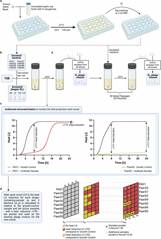
Fig. 1 Experimental design of the antibiofilm evolution assay. Experimental setup of the evolution assay to extend the host range and improve the antimicrobial and antibiofilm activity of bacteriophages (phages). a Biofilm formation on porous glass beads by incubation with P. aeruginosa in tryptic soy broth (TSB) under agitation and 37 °C. Dip-washing of beads with 24 h pre-established biofilm in sterile phosphate-buffered saline (PBS) before transferal into the calorimetric ampules. b Representation of one calorimetric ampule containing a 24 h pre-stablished biofilm bead of one P. aeruginosa strain in TSB and the phage mixture at round 0 (R0) containing the four ancestral phages. A total of 43 ampules (one growth control and four phage mixture dilutions per strain and three sterility controls) were used during each round of evolution. c Heat production (J) was recorded for each individual ampule by isothermal calorimetry during 24 h. d At the end of each round, the percentage heat reduction of phage-containing ampules relative to growth control ampules was determined at a 24 h (rounds 1–15) or 8 h (rounds 16–30) time point. Samples showing a heat reduction equal or above 75% (referred to as active samples) were selected and pooled together with the undiluted phage samples (always included) into the new phage mixture. e Across all eight bacterial strains, the undiluted and the active samples after each round were pooled together, creating the phage mixture that served as starting point for the next round, and hence was added to pre-stablished biofilm beads anew. In total, 30 rounds of evolution were performed. Figure 1 was created with BioRender.com and released under a Creative Commons Attribution-NonCommercial-NoDerivs 4.0 International license (https://creativecommons.org/licences/by-nc-nd/4.0/deed.en).
Fig. 1 Experimental design of the antibiofilm evolution assay. Experimental setup of the evolution assay to extend the host range and improve the antimicrobial and antibiofilm activity of bacteriophages (phages). a Biofilm formation on porous glass beads by incubation with P. aeruginosa in tryptic soy broth (TSB) under agitation and 37 °C. Dip-washing of beads with 24 h pre-established biofilm in sterile phosphate-buffered saline (PBS) before transferal into the calorimetric ampules. b Representation of one calorimetric ampule containing a 24 h pre-stablished biofilm bead of one P. aeruginosa strain in TSB and the phage mixture at round 0 (R0) containing the four ancestral phages. A total of 43 ampules (one growth control and four phage mixture dilutions per strain and three sterility controls) were used during each round of evolution. c Heat production (J) was recorded for each individual ampule by isothermal calorimetry during 24 h. d At the end of each round, the percentage heat reduction of phage-containing ampules relative to growth control ampules was determined at a 24 h (rounds 1–15) or 8 h (rounds 16–30) time point. Samples showing a heat reduction equal or above 75% (referred to as active samples) were selected and pooled together with the undiluted phage samples (always included) into the new phage mixture. e Across all eight bacterial strains, the undiluted and the active samples after each round were pooled together, creating the phage mixture that served as starting point for the next round, and hence was added to pre-stablished biofilm beads anew. In total, 30 rounds of evolution were performed. Figure 1 was created with BioRender.com and released under a Creative Commons Attribution-NonCommercial-NoDerivs 4.0 International license (https://creativecommons.org/licences/by-nc-nd/4.0/deed.en).CC BY · PubMed Central
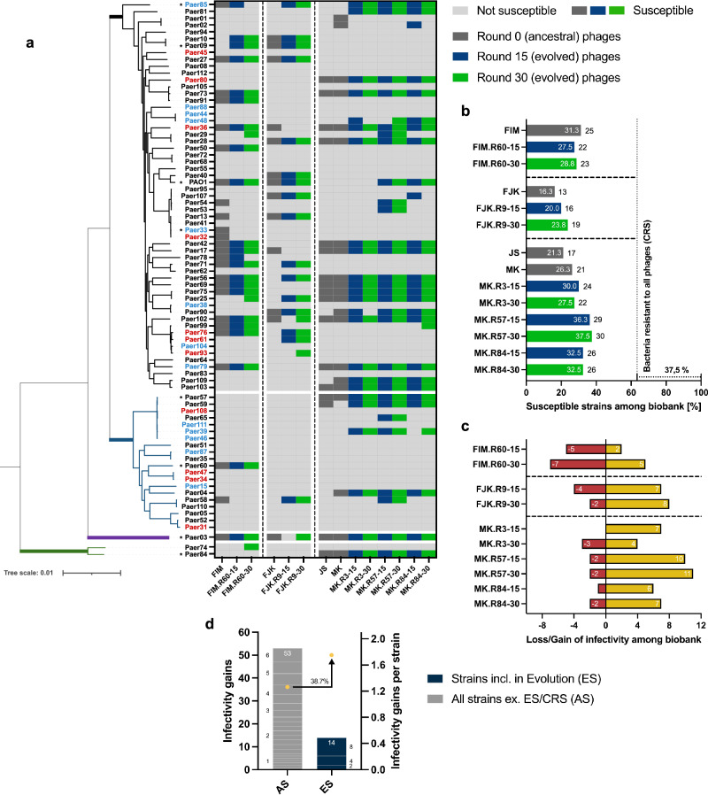
Fig. 2 Host range analysis of unevolved and evolved phages. a Phylogenetic tree based on the core genes (3,667 of 19,556 genes) of the P. aeruginosa strain collection (80 strains) encompassing four genomic clusters (Cluster 1, black branch, 71.3%; Cluster 2, blue branch, 25.0%; Cluster 3, violet branch, 1.3%; Cluster 4, green branch, 2.5%). Classification of each strain according to its resistance profile (4MRGN, red lettering; 3MRGN, blue lettering; MRGN, multi-resistance Gram-negative). Strains marked with an asterisk were included in the experimental evolution. Activity determination of the unevolved (dark grey) and evolved (R15, blue; R30, green) phages against the bacterial collection by spot assay overlay (heat map). b Percentual (white lettering) and absolute (black lettering) infectivity of each phage among the bacterial collection. Vertical dotted line represents the percentage of bacterial strains susceptible to at least one phage (62.5%). 37.5% (30 strains) of bacterial strains were not susceptible to any phage (CRS, completely resistant strains). c Representation of the infectivity gains and losses of the evolved phages. An infectivity gain (yellow bar) is defined as the capability of an evolved phage to infect a bacterial strain that was initially not susceptible to the phage’s genomically closest unevolved progenitor phage. A loss of infectivity (red bar) occurred when a bacterial strain is susceptible to the unevolved ancestral phage but resistant to the corresponding evolved phage. d Representation of the cumulated infectivity gains of the evolved phages among the evolution strains (ES) or all strains (AS, excluding the ES and CRS). Among the ES (n = 8), three strains (stacked blue boxes, numbers at the side of the boxes indicate the infectivity gains on the according strain) resulted in 14 infectivity gains (Paer85, 8 gains; PAO1, 4 gains; Paer09, 2 gains). The 53 infectivity gains among AS (n = 42) correspond to gains on 22 strains (stacked grey boxes, numbers at the side of the boxes indicate the infectivity gains on the according strain). For direct comparability of the two strain populations (ES and AS), the cumulated values were divided by the number of strains in each group, resulting in averaged gains per strain (yellow circles).
Fig. 2 Host range analysis of unevolved and evolved phages. a Phylogenetic tree based on the core genes (3,667 of 19,556 genes) of the P. aeruginosa strain collection (80 strains) encompassing four genomic clusters (Cluster 1, black branch, 71.3%; Cluster 2, blue branch, 25.0%; Cluster 3, violet branch, 1.3%; Cluster 4, green branch, 2.5%). Classification of each strain according to its resistance profile (4MRGN, red lettering; 3MRGN, blue lettering; MRGN, multi-resistance Gram-negative). Strains marked with an asterisk were included in the experimental evolution. Activity determination of the unevolved (dark grey) and evolved (R15, blue; R30, green) phages against the bacterial collection by spot assay overlay (heat map). b Percentual (white lettering) and absolute (black lettering) infectivity of each phage among the bacterial collection. Vertical dotted line represents the percentage of bacterial strains susceptible to at least one phage (62.5%). 37.5% (30 strains) of bacterial strains were not susceptible to any phage (CRS, completely resistant strains). c Representation of the infectivity gains and losses of the evolved phages. An infectivity gain (yellow bar) is defined as the capability of an evolved phage to infect a bacterial strain that was initially not susceptible to the phage’s genomically closest unevolved progenitor phage. A loss of infectivity (red bar) occurred when a bacterial strain is susceptible to the unevolved ancestral phage but resistant to the corresponding evolved phage. d Representation of the cumulated infectivity gains of the evolved phages among the evolution strains (ES) or all strains (AS, excluding the ES and CRS). Among the ES (n = 8), three strains (stacked blue boxes, numbers at the side of the boxes indicate the infectivity gains on the according strain) resulted in 14 infectivity gains (Paer85, 8 gains; PAO1, 4 gains; Paer09, 2 gains). The 53 infectivity gains among AS (n = 42) correspond to gains on 22 strains (stacked grey boxes, numbers at the side of the boxes indicate the infectivity gains on the according strain). For direct comparability of the two strain populations (ES and AS), the cumulated values were divided by the number of strains in each group, resulting in averaged gains per strain (yellow circles).CC BY · PubMed Central
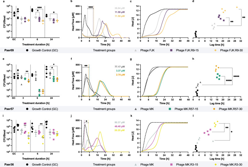
Fig. 3 Antibiofilm and antimicrobial activity of unevolved and evolved phages. a qPCR-determined bacterial load (CFU/bead) of 24 h pre-established Paer09 (included in the evolution) biofilm untreated (GC) and after co-incubation (3, 6 and 24 h) with phage FJK (unevolved), FJK.R9-15 (15 rounds of evolution) and FJK.R9-30 (30 rounds of evolution). The p-values are 0.0365 (6 h, FJK vs. FJK.R9-15), 0.0010 (6 h, FJK vs. FJK.R9-30) and 0.0242 (24 h, FJK vs. FJK.R9-30). b Heat flow (µW) curves measured for 48 h of Paer09 biofilm co-incubated with phage FJK, FJK.R9-15, FJK.R9-30, or untreated (GC, dashed line). Vertical dotted lines indicate the minimum heat flow (µW values in the right upper corner) detected in each treated sample, related to the highest suppressive effect of the phage on bacterial cells. The p-value is 0.0004 (FJK vs. FJK.R9-15/30). c, d Respective heat (J) curves of phage-treated Paer09 biofilm and the calculated suppression time for each tested condition. The p-values are 0.0014 (FJK vs. FJK.R9-15) and 0.0085 (FJK vs. FJK.R9-30). Identical setup for Paer57 (included in the evolution) treated with phage MK, MK.R57-15, MK.R57-30 or untreated (GC). The p-values are 0.0032 (e, 3 h, MK.R57-15 vs. MK.R57-30), 0.0493 (e, 24 h, MK vs. MK.R57-15), 0.0431 (e, 24 h, MK vs. MK.R57-30), 0.0056 (f, MK vs. MK.R57-15), 0.0053 (f, MK vs. MK.R57-30) and <0.0001 (h, MK vs. MK.R57-15/30). Identical setup for Paer36 (not included in the evolution) treated with phage MK, MK.R3-15, MK.R3-30 or untreated (GC). The p-values are 0.0067 (i, 6 h, MK.R3-15 vs. MK.R3-30), 0.0182 (j, MK vs. MK.R3-30), 0.0369 (j, MK.R3-15 vs. MK.R3-30), 0.0105 (l, MK vs. MK.R3-30) and 0.0012 (l, MK.R3-15 vs. MK.R3-30). All experiments were conducted in fourfold and the curves (b, c, f, g, j, k) display the mean. The error bars represent the standard error of the mean. Statistical analysis was conducted by unpaired two-tailed Student’s t test and p-values indicated with asterisks (*, p = <0.05; **, p = <0.01; ***, p = <0.001; ****, p = <0.0001). Source data are provided as a source data file.
Fig. 3 Antibiofilm and antimicrobial activity of unevolved and evolved phages. a qPCR-determined bacterial load (CFU/bead) of 24 h pre-established Paer09 (included in the evolution) biofilm untreated (GC) and after co-incubation (3, 6 and 24 h) with phage FJK (unevolved), FJK.R9-15 (15 rounds of evolution) and FJK.R9-30 (30 rounds of evolution). The p-values are 0.0365 (6 h, FJK vs. FJK.R9-15), 0.0010 (6 h, FJK vs. FJK.R9-30) and 0.0242 (24 h, FJK vs. FJK.R9-30). b Heat flow (µW) curves measured for 48 h of Paer09 biofilm co-incubated with phage FJK, FJK.R9-15, FJK.R9-30, or untreated (GC, dashed line). Vertical dotted lines indicate the minimum heat flow (µW values in the right upper corner) detected in each treated sample, related to the highest suppressive effect of the phage on bacterial cells. The p-value is 0.0004 (FJK vs. FJK.R9-15/30). c, d Respective heat (J) curves of phage-treated Paer09 biofilm and the calculated suppression time for each tested condition. The p-values are 0.0014 (FJK vs. FJK.R9-15) and 0.0085 (FJK vs. FJK.R9-30). Identical setup for Paer57 (included in the evolution) treated with phage MK, MK.R57-15, MK.R57-30 or untreated (GC). The p-values are 0.0032 (e, 3 h, MK.R57-15 vs. MK.R57-30), 0.0493 (e, 24 h, MK vs. MK.R57-15), 0.0431 (e, 24 h, MK vs. MK.R57-30), 0.0056 (f, MK vs. MK.R57-15), 0.0053 (f, MK vs. MK.R57-30) and <0.0001 (h, MK vs. MK.R57-15/30). Identical setup for Paer36 (not included in the evolution) treated with phage MK, MK.R3-15, MK.R3-30 or untreated (GC). The p-values are 0.0067 (i, 6 h, MK.R3-15 vs. MK.R3-30), 0.0182 (j, MK vs. MK.R3-30), 0.0369 (j, MK.R3-15 vs. MK.R3-30), 0.0105 (l, MK vs. MK.R3-30) and 0.0012 (l, MK.R3-15 vs. MK.R3-30). All experiments were conducted in fourfold and the curves (b, c, f, g, j, k) display the mean. The error bars represent the standard error of the mean. Statistical analysis was conducted by unpaired two-tailed Student’s t test and p-values indicated with asterisks (*, p = <0.05; **, p = <0.01; ***, p = <0.001; ****, p = <0.0001). Source data are provided as a source data file.CC BY · PubMed Central
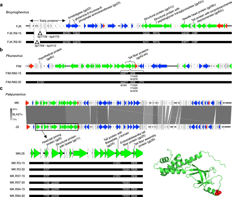
Fig. 4 Schematic genome representation of the evolved phages. a–c For each bacteriophage (phage) genus, a genome map is provided for the ancestral phage (FJK, FIM, or MK/JS) with the relevant functional annotations highlighted on top. Each coloured arrow represents a coding sequence with white encoding hypothetical proteins, blue (DNA) metabolism-associated proteins, green virion proteins, and red genome packaging or cell lysis proteins. The genomic changes in the evolved phages after round 15 and round 30 are displayed below each ancestral phage genome, with deletions being shown as a delta symbol and the individual SNPs annotated. For the Pakpunavirus members, a BLAST comparison between both ancestral phages is also shown. d Tertiary structure prediction of EPS depolymerase FJK_gp62. In red, the cysteine residue on position 98 is highlighted, which is mutated to the aromatic amino acid phenylalanine in the evolved phage.
Fig. 4 Schematic genome representation of the evolved phages. a–c For each bacteriophage (phage) genus, a genome map is provided for the ancestral phage (FJK, FIM, or MK/JS) with the relevant functional annotations highlighted on top. Each coloured arrow represents a coding sequence with white encoding hypothetical proteins, blue (DNA) metabolism-associated proteins, green virion proteins, and red genome packaging or cell lysis proteins. The genomic changes in the evolved phages after round 15 and round 30 are displayed below each ancestral phage genome, with deletions being shown as a delta symbol and the individual SNPs annotated. For the Pakpunavirus members, a BLAST comparison between both ancestral phages is also shown. d Tertiary structure prediction of EPS depolymerase FJK_gp62. In red, the cysteine residue on position 98 is highlighted, which is mutated to the aromatic amino acid phenylalanine in the evolved phage.CC BY · PubMed Central
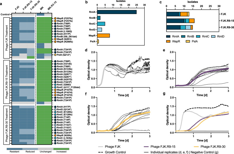
Fig. 5 Phenotypic and genomic characterisation of phage-treated Paer09 isolates. a Efficiency of plating (EOP) of five bacteriophages (phages) (FJK, FJK.R9-15, FJK.R9-30, MK, and MK.R3-15) for 48 Paer09 strains treated with individual phages (FJK, FJK.R9-15, FJK.R9-30). The EOP is defined as the concentration ratio of a phage on the phage-treated isolate (numerator) and the naive Paer09 strain (denominator; Control). An EOP above 10 was considered as increased efficiency (green), while an EOP of 0.1 to 10 was ranked as unchanged efficiency (grey). A reduced efficiency was defined as an EOP between 0.001 and 0.1 (light blue). When no individual plaques were visible or the EOP was equal to or under 0.001 the isolate was determined resistant to the phage (blue). Mutation-associated protein of each strain with amino acid change in parentheses. A lack of labelling refers to isolates in which no apparent mutation could be identified. b Illustration of the cumulative number of isolates by mutant protein. c Illustration of the number of isolates by mutant protein according to the treatment group (phage FJK; phage FJK.R9-15; phage FJK.R9-30). Optical density (OD600) measurements of a three-day co-incubation of planktonic naive Paer09 strain with phage FJK (d), phage FJK.R9-15 (e) and phage FJK.R9-30 (f) at an MOI of 0.001. The thin black lines represent each individual replicate (n = 8), and the thick coloured line represents the average. For phages FJK.R9-15/30 the completely suppressed replicate was excluded from the average calculation. g Illustration of the averaged co-incubation curves for phages FJK (grey line; panel d), FJK.R9-15 (violet line; e) and FJK.R9-30 (yellow line; f) over three days in comparison to a growth control (dotted black line) and negative control (black line). Source data are provided as a source data file.
Fig. 5 Phenotypic and genomic characterisation of phage-treated Paer09 isolates. a Efficiency of plating (EOP) of five bacteriophages (phages) (FJK, FJK.R9-15, FJK.R9-30, MK, and MK.R3-15) for 48 Paer09 strains treated with individual phages (FJK, FJK.R9-15, FJK.R9-30). The EOP is defined as the concentration ratio of a phage on the phage-treated isolate (numerator) and the naive Paer09 strain (denominator; Control). An EOP above 10 was considered as increased efficiency (green), while an EOP of 0.1 to 10 was ranked as unchanged efficiency (grey). A reduced efficiency was defined as an EOP between 0.001 and 0.1 (light blue). When no individual plaques were visible or the EOP was equal to or under 0.001 the isolate was determined resistant to the phage (blue). Mutation-associated protein of each strain with amino acid change in parentheses. A lack of labelling refers to isolates in which no apparent mutation could be identified. b Illustration of the cumulative number of isolates by mutant protein. c Illustration of the number of isolates by mutant protein according to the treatment group (phage FJK; phage FJK.R9-15; phage FJK.R9-30). Optical density (OD600) measurements of a three-day co-incubation of planktonic naive Paer09 strain with phage FJK (d), phage FJK.R9-15 (e) and phage FJK.R9-30 (f) at an MOI of 0.001. The thin black lines represent each individual replicate (n = 8), and the thick coloured line represents the average. For phages FJK.R9-15/30 the completely suppressed replicate was excluded from the average calculation. g Illustration of the averaged co-incubation curves for phages FJK (grey line; panel d), FJK.R9-15 (violet line; e) and FJK.R9-30 (yellow line; f) over three days in comparison to a growth control (dotted black line) and negative control (black line). Source data are provided as a source data file.CC BY · PubMed Central
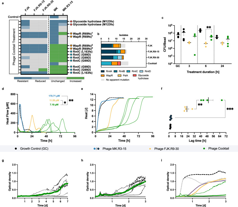
Fig. 6 Antibiofilm and antimicrobial activity of the phage cocktail. a Efficiency of plating (EOP) of five phages (FJK, FJK.R9-15, FJK.R9-30, MK, and MK.R3-15) on 18 Paer09 strains treated with the phage cocktail (FJK.R9-30 + MK.R3-15). EOP is defined within the methods. Mutation-associated protein of each strain with amino acid change in parentheses (1c.203_204dupTC p.R69fs and 2c.205delA p.R69fs). Lack of labelling refers to isolates with no apparent mutation. b Illustration of the number of isolates by mutant protein and treatment group (FJK; FJK.R9-15; FJK.R9-30; Cocktail). c qPCR-determined bacterial load (CFU/bead) of 24 h pre-established Paer09 biofilm untreated (GC) and after co-incubation (3, 6 and 24 h) with FJK.R9-30 or the cocktail. The p-value is 0.0077 (6 h, Cocktail vs. FJK.R9-30). d, e Heat flow (µW) and heat (J) curves measured for 48, 72 and 96 h of Paer09 biofilm co-incubated with MK.R3-15, FJK.R9-30, cocktail (individual replicates) or untreated (GC, dashed black line). Minimum heat flow values in the right upper corner (d). The p-values are 0.0404 (FJK.R9-30 vs. Cocktail) and 0.0024 (MK.R3-15 vs. Cocktail). f Calculated lag time (h) from the heat curves for each tested condition. The p-values are 0.0051 (FJK.R9-30 vs. Cocktail) and 0.0002 (MK.R3-15 vs. Cocktail). OD600 measurement of a seven-day (g) and three-day (h) co-incubation of planktonic naive Paer09 strain with the cocktail (MOI: 0.001, g; MOI: 0.0001, h). Thin lines represent the individual replicates (n = 8), and the thick coloured line the average, excluding completely suppressed replicates (n = 4, g; n = 2, h). i Illustration of averaged co-incubation curves (including grey, violet, and yellow curves from Fig. 5d) over three days compared to a growth control (dotted black line) and negative control (black line). The cocktail (MOI: 0.0001) is shown as a dotted green curve. All experiments were performed in fourfold and unless specified otherwise, the calorimetric curves (d, e) display the mean. The error bars represent the standard error of the mean. Statistical analysis was conducted by unpaired two-tailed Student’s t test and p-values indicated with asterisks (*p = <0.05; **p = <0.01; ***p = <0.001). Source data are provided as a source data file.
Fig. 6 Antibiofilm and antimicrobial activity of the phage cocktail. a Efficiency of plating (EOP) of five phages (FJK, FJK.R9-15, FJK.R9-30, MK, and MK.R3-15) on 18 Paer09 strains treated with the phage cocktail (FJK.R9-30 + MK.R3-15). EOP is defined within the methods. Mutation-associated protein of each strain with amino acid change in parentheses (1c.203_204dupTC p.R69fs and 2c.205delA p.R69fs). Lack of labelling refers to isolates with no apparent mutation. b Illustration of the number of isolates by mutant protein and treatment group (FJK; FJK.R9-15; FJK.R9-30; Cocktail). c qPCR-determined bacterial load (CFU/bead) of 24 h pre-established Paer09 biofilm untreated (GC) and after co-incubation (3, 6 and 24 h) with FJK.R9-30 or the cocktail. The p-value is 0.0077 (6 h, Cocktail vs. FJK.R9-30). d, e Heat flow (µW) and heat (J) curves measured for 48, 72 and 96 h of Paer09 biofilm co-incubated with MK.R3-15, FJK.R9-30, cocktail (individual replicates) or untreated (GC, dashed black line). Minimum heat flow values in the right upper corner (d). The p-values are 0.0404 (FJK.R9-30 vs. Cocktail) and 0.0024 (MK.R3-15 vs. Cocktail). f Calculated lag time (h) from the heat curves for each tested condition. The p-values are 0.0051 (FJK.R9-30 vs. Cocktail) and 0.0002 (MK.R3-15 vs. Cocktail). OD600 measurement of a seven-day (g) and three-day (h) co-incubation of planktonic naive Paer09 strain with the cocktail (MOI: 0.001, g; MOI: 0.0001, h). Thin lines represent the individual replicates (n = 8), and the thick coloured line the average, excluding completely suppressed replicates (n = 4, g; n = 2, h). i Illustration of averaged co-incubation curves (including grey, violet, and yellow curves from Fig. 5d) over three days compared to a growth control (dotted black line) and negative control (black line). The cocktail (MOI: 0.0001) is shown as a dotted green curve. All experiments were performed in fourfold and unless specified otherwise, the calorimetric curves (d, e) display the mean. The error bars represent the standard error of the mean. Statistical analysis was conducted by unpaired two-tailed Student’s t test and p-values indicated with asterisks (*p = <0.05; **p = <0.01; ***p = <0.001). Source data are provided as a source data file.CC BY · PubMed Central

Verification

Claims the paper makes

The claims and statistics of this paper haven’t been checked yet.

Run the check — free, no account needed. It reads the open-access text and checks the sources it cites.

A few free checks a day · papers already verified open instantly · results are public

Self-check

Percentages, sums and reported differences re-computed from the paper’s own numbers.

The self-check needs the paper’s full text.

Citations

Cites

Show all 1 references

Metadata

Authors
Kunisch, F., Wagemans, J., Yildirim, S., Chan, B., Schaudinn, C., Lavigne, R., Turner, P., Raschke, M., Trampuz, A., & Moreno, M. G.
Affiliations
  • University of Münster (DE)
  • KU Leuven (BE)
  • Berlin Institute of Health at Charité – Universitätsmedizin Berlin
  • Yale University (US)
  • Robert Koch Institute (DE)
  • Westfälische Wilhelms-Universität Münster
  • Charité – Universitätsmedizin Berlin
  • Humboldt-Universität zu Berlin (DE)
Venue
Nature Communications
Published
2023-06-19
Volume
15
Issue
1
Pages
8572-8572
ISSN
2041-1723 (ISSN-L)
OpenAlex
W4403098193
Cited by
102
References
1
Citations by year
2026: 61 · 2025: 39 · 2024: 2
FWCI
27.92
Type
journal article
Language
en
Open access
Locations
  • Nature Communications (publishedVersion, cc-by-nc-nd)
  • Research Square (acceptedVersion, cc-by)
  • PubMed Central (publishedVersion, cc-by-nc-nd)
  • PubMed (publishedVersion)
  • Publication server of the Robert Koch Institute (Robert Kock Institute) (submittedVersion, cc-by)
  • Lirias (KU Leuven) (submittedVersion, cc-by-nc-nd)
  • RePEc: Research Papers in Economics (submittedVersion)
  • DOAJ (DOAJ: Directory of Open Access Journals) (submittedVersion, cc-by-sa)
Funders
  • Yale University
  • Sonnenfeld Stiftung
  • Joint Programming Initiative on Antimicrobial Resistance
  • Bundesministerium für Bildung und Forschung (01KI1823)
  • KU Leuven (IDN/20/024)
  • Studienstiftung des Deutschen Volkes
  • Università di Pisa
Topics
  • Life Sciences › Immunology and Microbiology › Virology › Bacteriophages and microbial interactions
  • Life Sciences › Biochemistry, Genetics and Molecular Biology › Molecular Biology › Bacterial biofilms and quorum sensing
  • Life Sciences › Biochemistry, Genetics and Molecular Biology › Genetics › Evolution and Genetic Dynamics
Keywords
Pseudomonas aeruginosa, biofilm, phage therapy, multidrug resistance, host range expansion, antibiofilm activity, isothermal microcalorimetry, real-time PCR
MeSH terms
Phage Therapy, Anti-Bacterial Agents, Bacteriophages, Humans, Microbial Sensitivity Tests, Pseudomonas aeruginosa, Pseudomonas Infections, Pseudomonas Phages, Biofilms, Drug Resistance, Multiple, Bacterial, Host Specificity

About these checks

Registry, retraction, Cites and statistics results are computed, not judged, with one exception: a printed reference that no index lists is read once by a model before it counts as a citation error. Claims are extracted by a model and kept only when their quote appears verbatim in the text; each cited source is then checked, with a model’s help, for whether it says what the claim relies on. Those verdicts can be wrong — read the claim’s quote and the cited source before relying on one.

Statistics and arithmetic are recomputed, not judged.

Confidence bands are shown only for fields calibrated on our annotated test set. None is calibrated yet, so no band is shown.

What counts as a claim ›How we measure confidence ›How we check this ›

Are you an author, or do you see a problem with this page? Request a review

How to cite

Cite CoChat for the checks on this page, and the paper for its own findings.

This page
CoChat. (2026). Citation check: Targeting MDR Pseudomonas aeruginosa biofilm with an evolutionary trained bacteriophage cocktail exploiting phage resistance trade-offs. https://app.cochat.ai/papers/10.21203/rs.3.rs-2545888/v1
The paper
Kunisch, F., Wagemans, J., Yildirim, S., Chan, B., Schaudinn, C., Lavigne, R., Turner, P., Raschke, M., Trampuz, A., & Moreno, M. G. (2023). Targeting MDR Pseudomonas aeruginosa biofilm with an evolutionary trained bacteriophage cocktail exploiting phage resistance trade-offs. Nature Communications. https://doi.org/10.21203/rs.3.rs-2545888/v1