

The DOI matches its Crossref record; 1 of the 1 references it cites were found in a registry or an index.
Anti-viral chimeric protein RetroMAD1™ potently block SARS-CoV-2 viral entry and propagation
Open accessPreprint — published as 10.1038/s41598-023-47511-z · Paper page
Abstract
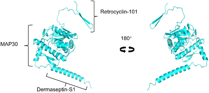
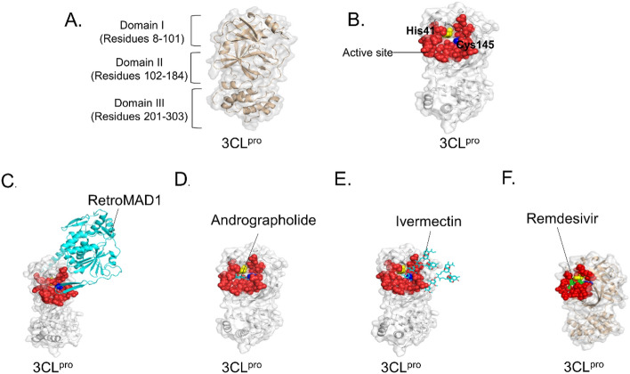
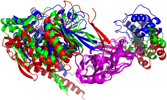
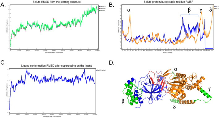
COVID-19 is a disease caused by the highly transmissible and pathogenic SARS-CoV-2 virus. Since its first case was documented in 2019, it has rapidly widespread and has caused millions of deaths worldwide. Many intervention strategies targeting these proteins have been developed. However, frequently mutation of SARS-CoV-2 poses a challenge to the effectiveness of current treatments. Therefore, it is critical to develop new therapeutic drugs against this disease. In this present study, in silico approach was used to study the interaction between RetroMAD1™and SARS-CoV-2 proteins including Spike proteins (S), 3-chymotrypsin-like protease (3CL pro) and papain-like protease (PL pro). The interaction of these viral proteins and RetroMAD1™ was performed through HDOCK server and visualised using PyMOL. Docking results revealed that all the complexes of SARS-CoV-2 proteins binding with RetroMAD1™ have relatively high docking scores. The binding energy of RetroMAD1™ complexes with SARS-CoV-2 S, 3CL pro, PL pro were − 15, -12.3 and − 15.4, respectively. RetroMAD1™antiviral efficiency and cytotoxicity was also evaluated using EpiAirway™ Model. In vitro validation of viral inhibitory effect of RetroMAD1™was performed with 3CL pro Inhibition Assay. The outcome showed that RetroMAD1™ represents a potential drug candidate against SARS-CoV-2 for its promising viral inhibitory effect.
Overview
The claims and statistics of this paper haven’t been checked yet.
Run the check — free, no account needed. It reads the open-access text and checks the sources it cites.
Integrity signals
Registry
Cites
Statistics
Figures
Verification
Claims the paper makes
The claims and statistics of this paper haven’t been checked yet.
Run the check — free, no account needed. It reads the open-access text and checks the sources it cites.
Self-check
Percentages, sums and reported differences re-computed from the paper’s own numbers.
The self-check needs the paper’s full text.
Citations
Cites
Highly accurate protein structure prediction with AlphaFold
Jumper, J., Evans, R., Pritzel, A., Green, T., Figurnov, M., Ronneberger, O., Tunyasuvunakool, K., Bates, R., Žídek, A., Potapenko, A., et al. (2021) · Nature · doi:10.1038/s41586-021-03819-2 · Cited by 43,764
Research works printed but not linked: 61
- Kozlov, M. Why scientists are racing to develop more COVID antivirals. Nature. 601, 496 (2002). · DOI
- Dolgin, E. The race for antiviral drugs to beat COVID - and the next pandemic. Nature. 592, 340–343 (2021). · DOI
- Huan, U. E., Da Silva, P. M. F. & Floriano, A. P. Case studies on RetroMAD1™, an orally administered recombinant chimeric protein to treat naturally infected Feline Leukemia Virus (FeLV) cats. Archives of Veterinary Science and Medicine. 2, 41–57 (2019).
- Bakar, A. M. S. A. & Ung, E. H. Antimicrobial fusion compounds and uses thereof. U.S. Patent No. 9,155,800. 13 Oct. (2015).
- Cole, A. M. et al. The formulated microbicide RC-101 was safe and antivirally active following intravaginal application in pigtailed macaques. PLoS One. 5, e15111 (2010). · DOI
- Cole, A. M. et al. Retrocyclin: a primate peptide that protects cells from infection by T- and M-tropic strains of HIV-1. Proc Natl Acad Sci USA. 99, 1813–8 (2002). · DOI
- Lehrer, R. I., Cole, A. M. & Selsted, M. E. θ-Defensins: cyclic peptides with endless potential. J Biol Chem. 287, 27014–9 (2012). · DOI
- Owen, S. M. et al. RC-101, a retrocyclin-1 analogue with enhanced activity against primary HIV type 1 isolates. AIDS Res Hum Retroviruses. 20, 1157–65 (2004). · DOI
- Cole, A. L. et al. The retrocyclin analogue RC-101 prevents human immunodeficiency virus type 1 infection of a model human cervicovaginal tissue construct. Immunology. 121, 140–145 (2007). · DOI
- Jia, X. et al. Mechanism through Which Retrocyclin Targets Flavivirus Multiplication. J Virol. 95, e0056021 (2021). · DOI
- Prantner, D. et al. The θ-defensin retrocyclin 101 inhibits TLR4- and TLR2-dependent signaling and protects mice against influenza infection. J Leukoc Biol. 102, 1103–1113 (2017). · DOI
- Hooven, T. A., Randis, T. M., Hymes, S. R., Rampersaud, R. & Ratner, A. J. Retrocyclin inhibits Gardnerella vaginalis biofilm formation and toxin activity. J Antimicrob Chemother. 67, 2870–2 (2012). · DOI
- Lamers, R. P., Eade, C. R., Waring, A. J., Cole, A. L. & Cole, A. M. Characterization of the retrocyclin analogue RC-101 as a preventative of Staphylococcus aureus nasal colonization. Antimicrob Agents Chemother. 55, 5338–46 (2011). · DOI
- Yasin, B. et al. Ɵ Defensins Protect Cells from Infection by Herpes Simplex Virus by Inhibiting Viral Adhesion and Entry. Journal of Virology. 78, 5147–5156 (2004). · DOI
- Kudryashova, E. et al. Inhibition of SARS-CoV-2 Infection by Human Defensin HNP1 and Retrocyclin RC-101. Journal of molecular biology. 434, 167225–167225 (2022). · DOI
- Lee-Huang, S. et al. MAP 30: a new inhibitor of HIV-1 infection and replication. FEBS Lett. 272, 12–8 (1990). · DOI
- Lee-Huang, S. et al. Anti-HIV and anti-tumor activities of recombinant MAP30 from bitter melon. Gene. 161, 151–156 (1995). · DOI
- Lee-Huang, S. et al. Inhibition of MDA-MB-231 human breast tumor xenografts and HER2 expression by anti-tumor agents GAP31 and MAP30. Anticancer Research. 20, 653–659 (2000).
- Jiang, Y. et al. MAP30 promotes apoptosis of U251 and U87 cells by suppressing the LGR5 and Wnt/β-catenin signaling pathway, and enhancing Smac expression. Oncol Lett 15, 5833–5840 (2018).
- Bourinbaiar, A. S. & Lee-Huang, S. The Activity of Plant-Derived Antiretroviral Proteins MAP30 and GAP31 against Herpes Simplex Virus Infection in Vitro. Biochemical and Biophysical Research Communications. 219, 923–929 (1996). · DOI
- Chang, C. D., Lin, P. Y., Chen, Y. C., Huang, H. H. & Shih, W. L. Novel purification method and antibiotic activity of recombinant Momordica charantia MAP30. 3 Biotech. 7, 3 (2017). · DOI
- Sun, Y. et al. Anti-HIV Agent MAP30 Modulates the Expression Profile of Viral and Cellular Genes for Proliferation and Apoptosis in AIDS-Related Lymphoma Cells Infected with Kaposi's Sarcoma-Associated Virus. Biochemical and Biophysical Research Communications. 287, 983–994 (2001). · DOI
- Fan, J. M. et al. Inhibition on Hepatitis B virus in vitro of recombinant MAP30 from bitter melon. Mol Biol Rep. 36, 381–8 (2009). · DOI
- Barbieri, L., Battelli, M. G. & Stirpe, F. Ribosome-inactivating proteins from plants. Biochimica et Biophysica Acta (BBA)-Reviews on Biomembranes. 1154, 237–282 (1993). · DOI
- Fan, X. et al. αMMC and MAP30, two ribosomeinactivating proteins extracted from Momordica charantia, induce cell cycle arrest and apoptosis in A549 human lung carcinoma cells. Molecular Medicine Reports. 11, 3553–3558 (2015). · DOI
- Puri, M. et al. Ribosome inactivating proteins (RIPs) from Momordica charantia for anti viral therapy. Current Molecular Medicine. 9, 1080–1094 (2009). · DOI
- Hartley, M. R., Chaddock, J. A. & Bonness, M. S. The structure and function of ribosome-inactivating proteins. Trends in Plant Science. 1, 252 (1996). · DOI
- Wang, Yun-Xing. et al. Solution Structure of Anti-HIV-1 and Anti-Tumor Protein MAP30: Structural Insights into Its Multiple Functions. Cell. 99, 433–442 (1999). · DOI
- Schreiber, C. A. et al. The antiviral agents, MAP30 and GAP31, are not toxic to human spermatozoa and may be useful in preventing the sexual transmission of human immunodeficiency virus type 1. Fertility and sterility. 72, 686–690 (1999). · DOI
- Mor, A., Van Huong, N., Delfour, A., Migliore-Samour, D. & Nicolas, P. Isolation, amino acid sequence and synthesis of dermaseptin, a novel antimicrobial peptide of amphibian skin. Biochemistry, 30, 8824–8830 (1991). · DOI · Paper page
- Savoia, D., Guerrini, R., Marzola, E. & Salvadori, S. Synthesis and antimicrobial activity of dermaseptin S1 analogues. Bioorg Med Chem. 16, 8205–9 (2008). · DOI
- Yang, Z. et al. Targeted delivery of antimicrobial peptide by Cry protein crystal to treat intramacrophage infection. Biomaterials. 217, 119286 (2019). · DOI
- Savoia, D., Donalisio, M., Civra, A., Salvadori, S. & Guerrini, R. In vitro activity of dermaseptin S1 derivatives against genital pathogens. Apmis. 118, 674–80 (2010). · DOI
- Mirdita, M., Schütze, K., Moriwaki, Y., Ovchinnikov, S. & Steinegger, M. ColabFold: Making protein folding accessible to all. Nature Methods. 19, 679–692 (2021). · DOI · Paper page
- Steinegger M, Söding J. MMseqs2 enables sensitive protein sequence searching for the analysis of massive data sets. Nat Biotechnol. 35, 1026–1028 (2017). · DOI · Paper page
- Mirdita, M., Steinegger, M. & Söding, J. MMseqs2 desktop and local web server app for fast, interactive sequence searches. Bioinformatics. 35, 2856–2858 (2019). · DOI · Paper page
- Krieger, E., & Vriend, G. YASARA View—molecular graphics for all devices—from smartphones to workstations. Bioinformatics. 30, 2981–2982 (2014). · DOI
- Waterhouse, A. et al. SWISS-MODEL: homology modelling of protein structures and complexes. Nucleic acids research. 46, W296-W303 (2018). · DOI · Paper page
- Kim, S. et al. PubChem 2023 update. Nucleic Acids Res. 51, D1373–D1380 (2023). · DOI · Paper page
- Reed, L. J. & Muench, H. A simple method of estimating fifty per cent endpoints. American journal of epidemiology. 27, 493–497 (1938). · DOI · Paper page
- Jawad, B., Adhikari, P., Podgornik, R., & Ching, W. Y. Key interacting residues between RBD of SARS-CoV-2 and ACE2 receptor: combination of molecular dynamics simulation and density functional calculation. Journal of chemical information and modeling, 61, 4425–4441 (2021). · DOI
- Razali, R., Asis, H., & Budiman, C. Structure-function characteristics of SARS-CoV-2 proteases and their potential inhibitors from microbial sources. Microorganisms. 9, 2481 (2021). · DOI
- Zhu, J. et al. Progress on SARS-CoV-2 3CLpro inhibitors: inspiration from SARS-CoV 3CLpro peptidomimetics and small-molecule anti-inflammatory compounds. Drug Design, Development and Therapy. 1067–1082 (2022). · DOI
- Ferreira, J. C., Fadl, S., Villanueva, A. J. & Rabeh, W. M. Catalytic dyad residues His41 and Cys145 impact the catalytic activity and overall conformational fold of the main SARS-CoV-2 protease 3-chymotrypsin-like protease. Frontiers in Chemistry. 9, 692168 (2021). · DOI
- Novak, J. et al. Proposition of a new allosteric binding site for potential SARS-CoV-2 3CL protease inhibitors by utilizing molecular dynamics simulations and ensemble docking. Journal of Biomolecular Structure and Dynamics. 40, 9347–9360 (2022). · DOI
- Maiti, B. K. Can papain-like protease inhibitors halt SARS-CoV-2 replication? ACS pharmacology & translational science. 3, 1017–1019 (2020). · DOI
- Ke, Z. et al. Structures and distributions of SARS-CoV-2 spike proteins on intact virions. Nature. 588, 498–502 (2020). · DOI · Paper page
- Tai, W. et al. Characterization of the receptor-binding domain (RBD) of 2019 novel coronavirus: implication for development of RBD protein as a viral attachment inhibitor and vaccine. Cellular & molecular immunology. 17, 613–620 (2020). · DOI · Paper page
- Durai, P., Batool, M., Shah, M. & Choi, S. Middle East respiratory syndrome coronavirus: transmission, virology and therapeutic targeting to aid in outbreak control. Experimental & molecular medicine. 47, e181-e181 (2015). · DOI
- Arya, R. et al. Structural insights into SARS-CoV-2 proteins. Journal of molecular biology. 433, 166725 (2021). · DOI · Paper page
- Shin, D. et al. Papain-like protease regulates SARS-CoV-2 viral spread and innate immunity. Nature. 587, 657–662 (2020). · DOI · Paper page
- Moustaqil, M. et al. SARS-CoV-2 proteases PLpro and 3CLpro cleave IRF3 and critical modulators of inflammatory pathways (NLRP12 and TAB1): implications for disease presentation across species. Emerging microbes & infections. 10, 178–195 (2021). · DOI · Paper page
- Sekar, P. C. & Rajasekaran, R. Could Dermaseptin Analogue be a Competitive Inhibitor for ACE2 Towards Binding with Viral Spike Protein Causing COVID19?: Computational Investigation. Int J Pept Res Ther. 27, 1043–1056 (2021). · DOI
- Jia, Z. & Gong, W. Will Mutations in the Spike Protein of SARS-CoV-2 Lead to the Failure of COVID-19 Vaccines? Journal of Korean medical science. 36, e124-e124 (2021).
- Alenquer, M. et al. Signatures in SARS-CoV-2 spike protein conferring escape to neutralizing antibodies. PLoS pathogens. 17, e1009772-e1009772 (2021). · DOI
- Cao, Y. et al. Omicron escapes the majority of existing SARS-CoV-2 neutralizing antibodies. Nature. 602, 657–663 (2022). · DOI · Paper page
- Mahase, E. Covid-19: What do we know about XBB. 1.5 and should we be worried? British Medical Journal Publishing Group. 380, p153 (2023). · DOI
- Sreekanth Reddy, O. & Lai, W.-F. Tackling COVID-19 Using Remdesivir and Favipiravir as Therapeutic Options. Chembiochem: a European journal of chemical biology. 22, 939–948 (2021). · DOI
- Vitiello, A. & Ferrara, F. Association and pharmacological synergism of the triple drug therapy baricitinib/remdesivir/rhACE2 for the management of COVID-19 infection. Naunyn Schmiedebergs Arch Pharmacol. 395, 99–104 (2022). · DOI
- Agostini, M. L. et al. Coronavirus Susceptibility to the Antiviral Remdesivir (GS-5734) Is Mediated by the Viral Polymerase and the Proofreading Exoribonuclease. mBio. 9, 2, e00221-18 (2018). · DOI · Paper page
- Jeong, G. U., Song, H., Yoon, G. Y., Kim, D. & Kwon, Y.-C. Therapeutic Strategies Against COVID-19 and Structural Characterization of SARS-CoV-2: A Review. Frontiers in Microbiology. 11, 1723 (2020). · DOI
Metadata
- Authors
- Chan, L.-C., Yassim, A. S. M., Leow, T. C., Sabri, S., Yahaya, R. S. R., & Bakar, A. M. S. A.
- Affiliations
- Biovalence Sdn. Bhd.
- Universiti Sains Malaysia (MY)
- Universiti Putra Malaysia (MY)
- Jabatan Perkhidmatan Veterinar (MY)
- Corresponding author
- Yassim, Aini Syahida Mat
- Bakar, Awang Muhammad Sagaf Abu
- Venue
- Scientific Reports
- Publisher
- Research Square Platform LLC
- Published
- 2023-04-05
- Volume
- 13
- Issue
- 1
- Pages
- 20178-20178
- ISSN
- 2045-2322 (ISSN-L)
- PMC
- PMC10656507
- OpenAlex
- W4388751591
- Cited by
- 5
- References
- 1
- Citations by year
- 2026: 1 · 2025: 1 · 2024: 3
- FWCI
- 1.03
- Type
- journal article
- Language
- en
- Open access
- gold
- doi.org
- Locations
- Scientific Reports (publishedVersion, cc-by)
- Research Square (acceptedVersion, cc-by)
- PubMed Central (publishedVersion, cc-by)
- PubMed (publishedVersion)
- DOAJ (DOAJ: Directory of Open Access Journals) (submittedVersion, cc-by-sa)
- Universiti Putra Malaysia Institutional Repository (Universiti Putra Malaysia) (submittedVersion, cc-by)
- Greater South Information System
- Funders
- Utah State University
- Topics
- Health Sciences › Medicine › Infectious Diseases › SARS-CoV-2 and COVID-19 Research
- Life Sciences › Pharmacology, Toxicology and Pharmaceutics › Drug Discovery › Computational Drug Discovery Methods
- Life Sciences › Biochemistry, Genetics and Molecular Biology › Molecular Biology › vaccines and immunoinformatics approaches
- Keywords
- SARS-CoV-2, COVID-19 pandemic, main protease, main protease inhibition, fusion protein, antiviral drug candidates, protein-protein complex stability, molecular dynamics simulation, human bronchial epithelial cells, cytotoxicity
- MeSH terms
- COVID-19, SARS-CoV-2, Antiviral Agents, Cysteine Endopeptidases, Humans, Peptide Hydrolases, Protease Inhibitors, Recombinant Fusion Proteins, Viral Nonstructural Proteins
About these checks
Registry, retraction, Cites and statistics results are computed, not judged, with one exception: a printed reference that no index lists is read once by a model before it counts as a citation error. Claims are extracted by a model and kept only when their quote appears verbatim in the text; each cited source is then checked, with a model’s help, for whether it says what the claim relies on. Those verdicts can be wrong — read the claim’s quote and the cited source before relying on one.
Statistics and arithmetic are recomputed, not judged.
Confidence bands are shown only for fields calibrated on our annotated test set. None is calibrated yet, so no band is shown.
What counts as a claim ›How we measure confidence ›How we check this ›
Are you an author, or do you see a problem with this page? Request a review
How to cite
Cite CoChat for the checks on this page, and the paper for its own findings.
- This page
- CoChat. (2026). Citation check: Anti-viral chimeric protein RetroMAD1™ potently block SARS-CoV-2 viral entry and propagation. https://app.cochat.ai/papers/10.21203/rs.3.rs-2712307/v1
- The paper
- Chan, L.-C., Yassim, A. S. M., Leow, T. C., Sabri, S., Yahaya, R. S. R., & Bakar, A. M. S. A. (2023). Anti-viral chimeric protein RetroMAD1™ potently block SARS-CoV-2 viral entry and propagation. Scientific Reports. https://doi.org/10.21203/rs.3.rs-2712307/v1
Data sources
Bibliographic data: Semantic Scholar, OpenAlex, Crossref.
Contains information from the Semantic Scholar Academic Graph Datasets, made available under the ODC Attribution License (ODC-BY 1.0).
Semantic Scholar’s data is described in: Kinney, R., et al. (2023). The Semantic Scholar Open Data Platform. arXiv:2301.10140.
