

The DOI matches its Crossref record; 1 of the 1 references it cites were found in a registry or an index.
Unveiling the Crucial Role of Ferroptosis in Host Resistance to Streptococcus agalactiae Infection
Open accessPreprint — published as 10.1038/s41420-024-02189-8
Abstract
IL-1β represents an important inflammatory factor involved in the host response against GBS infection. Prior research has suggested a potential involvement of IL-1β in the process of ferroptosis. However, the relationship between IL-1β and ferroptosis in the context of anti-GBS infection remains uncertain. This research demonstrates that the significance of ferroptosis in the host's immune defense against GBS infection in a mouse model of abdominal infection, with peritoneal macrophages identified as the primary cells undergoing ferroptosis. Subsequent studies have shown that IL-1β promotes lipid oxidation in macrophages by upregulating pathways associated with lipid oxidation. Concurrently, IL-1β is not only involved in the initiation of ferroptosis in macrophages, but its production is intricately linked to the onset of ferroptosis. Ultimately, we posit that ferroptosis acts as a crucial initiating factor in the host response to GBS infection, with IL-1β playing a significant role in the resistance to infection by serving as a key inducer of ferroptosis.
Overview
The claims and statistics of this paper haven’t been checked yet.
Run the check — free, no account needed. It reads the open-access text and checks the sources it cites.
Integrity signals
Registry
Cites
Statistics
Figures
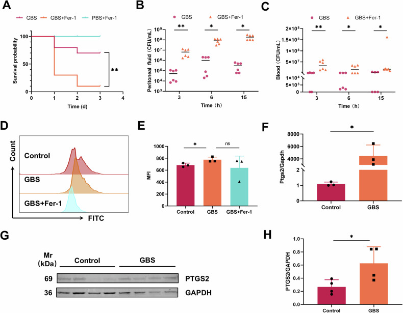
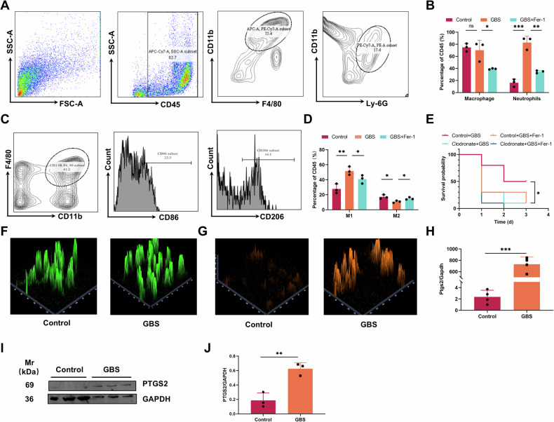
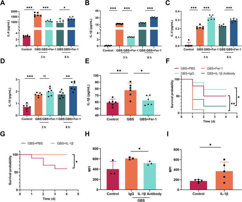
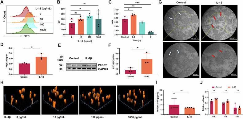
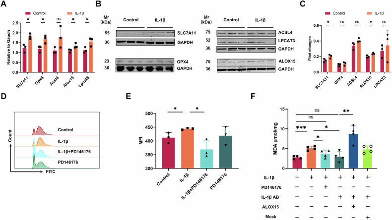
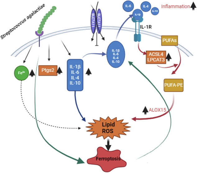
Verification
Claims the paper makes
The claims and statistics of this paper haven’t been checked yet.
Run the check — free, no account needed. It reads the open-access text and checks the sources it cites.
Self-check
Percentages, sums and reported differences re-computed from the paper’s own numbers.
The self-check needs the paper’s full text.
Citations
Cites
Iron in infection and immunity
Cassat, J. E., & Skaar, E. P. (2013) · Cell Host & Microbe · doi:10.1016/j.chom.2013.04.010 · Cited by 1,214
Research works printed but not linked: 42
- S.M. Hansen, N. Uldbjerg, M. Kilian, and U.B. Sorensen, Dynamics of Streptococcus agalactiae colonization in women during and after pregnancy and in their infants. J Clin Microbiol 42 (2004) 83–9. · DOI
- C. Biondo, G. Mancuso, A. Midiri, G. Signorino, M. Domina, V. Lanza Cariccio, N. Mohammadi, M. Venza, I. Venza, G. Teti, and C. Beninati, The interleukin-1beta/CXCL1/2/neutrophil axis mediates host protection against group B streptococcal infection. Infect Immun 82 (2014) 4508–17. · DOI
- T. Persichini, N. Maio, M.C. di Patti, G. Rizzo, S. Toscano, M. Colasanti, and G. Musci, Interleukin-1beta induces ceruloplasmin and ferroportin-1 gene expression via MAP kinases and C/EBPbeta, AP-1, and NF-kappaB activation. Neurosci Lett 484 (2010) 133–8. · DOI
- Z. Sun, W. Huang, Y. Zheng, P. Liu, W. Yang, Z. Guo, D. Kong, Q. Lv, X. Zhou, Z. Du, H. Jiang, and Y. Jiang, Fpr2/CXCL1/2 Controls Rapid Neutrophil Infiltration to Inhibit Streptococcus agalactiae Infection. Frontiers in Immunology 12 (2021).
- J. Li, F. Cao, H.L. Yin, Z.J. Huang, Z.T. Lin, N. Mao, B. Sun, and G. Wang, Ferroptosis: past, present and future. Cell Death Dis 11 (2020) 88. · DOI · Paper page
- Q. Liu, J. Wu, X. Zhang, X. Wu, Y. Zhao, and J. Ren, Iron homeostasis and disorders revisited in the sepsis. Free Radic Biol Med 165 (2021) 1–13. · DOI
- G. Corna, L. Campana, E. Pignatti, A. Castiglioni, E. Tagliafico, L. Bosurgi, A. Campanella, S. Brunelli, A.A. Manfredi, P. Apostoli, L. Silvestri, C. Camaschella, and P. Rovere-Querini, Polarization dictates iron handling by inflammatory and alternatively activated macrophages. Haematologica 95 (2010) 1814–22. · DOI · Paper page
- C.B. Billesbolle, C.M. Azumaya, R.C. Kretsch, A.S. Powers, S. Gonen, S. Schneider, T. Arvedson, R.O. Dror, Y. Cheng, and A. Manglik, Structure of hepcidin-bound ferroportin reveals iron homeostatic mechanisms. Nature 586 (2020) 807–811. · DOI · Paper page
- F. Du, Z.M. Qian, Q. Gong, Z.J. Zhu, L. Lu, and Y. Ke, The iron regulatory hormone hepcidin inhibits expression of iron release as well as iron uptake proteins in J774 cells. J Nutr Biochem 23 (2012) 1694–700. · DOI
- J.M. Muriuki, A.J. Mentzer, G. Band, J.J. Gilchrist, T. Carstensen, S.A. Lule, M.M. Goheen, F. Joof, W. Kimita, R. Mogire, C.L. Cutland, A. Diarra, A. Rautanen, C. Pomilla, D. Gurdasani, K. Rockett, N. Mturi, F.M. Ndungu, J.A.G. Scott, S.B. Sirima, A. Morovat, A.M. Prentice, S.A. Madhi, E.L. Webb, A.M. Elliott, P. Bejon, M.S. Sandhu, A.V.S. Hill, D.P. Kwiatkowski, T.N. Williams, C. Cerami, and S.H. Atkinson, The ferroportin Q248H mutation protects from anemia, but not malaria or bacteremia. Sci Adv 5 (2019) eaaw0109. · DOI · Paper page
- N.K. Shanmugam, K. Chen, and B.J. Cherayil, Commensal Bacteria-induced Interleukin 1beta (IL-1beta) Secreted by Macrophages Up-regulates Hepcidin Expression in Hepatocytes by Activating the Bone Morphogenetic Protein Signaling Pathway. J Biol Chem 290 (2015) 30637–47. · DOI
- P.J. Schmidt, Regulation of Iron Metabolism by Hepcidin under Conditions of Inflammation. J Biol Chem 290 (2015) 18975–83. · DOI
- Y. Kanamori, M. Murakami, M. Sugiyama, O. Hashimoto, T. Matsui, and M. Funaba, Interleukin-1beta (IL-1beta) transcriptionally activates hepcidin by inducing CCAAT enhancer-binding protein delta (C/EBPdelta) expression in hepatocytes. J Biol Chem 292 (2017) 10275–10287. · DOI
- X. Chen, R. Kang, G. Kroemer, and D. Tang, Ferroptosis in infection, inflammation, and immunity. J Exp Med 218 (2021). · DOI
- T. Hirschhorn, and B.R. Stockwell, The development of the concept of ferroptosis. Free Radic Biol Med 133 (2019) 130–143. · DOI · Paper page
- B. Zhou, J. Liu, R. Kang, D.J. Klionsky, G. Kroemer, and D. Tang, Ferroptosis is a type of autophagy-dependent cell death. Seminars in Cancer Biology 66 (2020) 89–100. · DOI
- E. Park, and S.W. Chung, ROS-mediated autophagy increases intracellular iron levels and ferroptosis by ferritin and transferrin receptor regulation. Cell Death & Disease 10 (2019). · DOI
- S. Dolma, S.L. Lessnick, W.C. Hahn, and B.R. Stockwell, Identification of genotype-selective antitumor agents using synthetic lethal chemical screening in engineered human tumor cells. Cancer Cell 3 (2003) 285–96. · DOI · Paper page
- H. Yuk, M. Abdullah, D.H. Kim, H. Lee, and S.J. Lee, Necrostatin-1 Prevents Ferroptosis in a RIPK1- and IDO-Independent Manner in Hepatocellular Carcinoma. Antioxidants (Basel) 10 (2021).
- A. Donovan, A. Brownlie, Y. Zhou, J. Shepard, S.J. Pratt, J. Moynihan, B.H. Paw, A. Drejer, B. Barut, A. Zapata, T.C. Law, C. Brugnara, S.E. Lux, G.S. Pinkus, J.L. Pinkus, P.D. Kingsley, J. Palis, M.D. Fleming, N.C. Andrews, and L.I. Zon, Positional cloning of zebrafish ferroportin1 identifies a conserved vertebrate iron exporter. Nature 403 (2000) 776–81. · DOI · Paper page
- H.H. Dar, Y.Y. Tyurina, K. Mikulska-Ruminska, I. Shrivastava, H.C. Ting, V.A. Tyurin, J. Krieger, C.M. St Croix, S. Watkins, E. Bayir, G. Mao, C.R. Armbruster, A. Kapralov, H. Wang, M.R. Parsek, T.S. Anthonymuthu, A.F. Ogunsola, B.A. Flitter, C.J. Freedman, J.R. Gaston, T.R. Holman, J.M. Pilewski, J.S. Greenberger, R.K. Mallampalli, Y. Doi, J.S. Lee, I. Bahar, J.M. Bomberger, H. Bayir, and V.E. Kagan, Pseudomonas aeruginosa utilizes host polyunsaturated phosphatidylethanolamines to trigger theft-ferroptosis in bronchial epithelium. J Clin Invest 128 (2018) 4639–4653. · DOI · Paper page
- J. Ousingsawat, R. Schreiber, E. Gulbins, M. Kamler, and K. Kunzelmann, P. aeruginosa Induced Lipid Peroxidation Causes Ferroptotic Cell Death in Airways. Cell Physiol Biochem 55 (2021) 590–604. · DOI
- E.P. Amaral, D.L. Costa, S. Namasivayam, N. Riteau, O. Kamenyeva, L. Mittereder, K.D. Mayer-Barber, B.B. Andrade, and A. Sher, A major role for ferroptosis in Mycobacterium tuberculosis-induced cell death and tissue necrosis. J Exp Med 216 (2019) 556–570. · DOI · Paper page
- C.A. Dinarello, Immunological and inflammatory functions of the interleukin-1 family. Annu Rev Immunol 27 (2009) 519–50. · DOI · Paper page
- E. Pandur, K. Tamasi, R. Pap, G. Janosa, and K. Sipos, Distinct Effects of Escherichia coli, Pseudomonas aeruginosa and Staphylococcus aureus Cell Wall Component-Induced Inflammation on the Iron Metabolism of THP-1 Cells. Int J Mol Sci 22 (2021). · DOI
- R. Ma, L. Fang, L. Chen, X. Wang, J. Jiang, and L. Gao, Ferroptotic stress promotes macrophages against intracellular bacteria. Theranostics 12 (2022) 2266–2289. · DOI
- K.P. Haley, and E.P. Skaar, A battle for iron: host sequestration and Staphylococcus aureus acquisition. Microbes Infect 14 (2012) 217–27. · DOI · Paper page
- B.R. Stockwell, Ferroptosis turns 10: Emerging mechanisms, physiological functions, and therapeutic applications. Cell 185 (2022) 2401–2421. · DOI
- Y. Ou, S.J. Wang, D. Li, B. Chu, and W. Gu, Activation of SAT1 engages polyamine metabolism with p53-mediated ferroptotic responses. Proc Natl Acad Sci U S A 113 (2016) E6806-E6812.
- A.J. Schwartz, N.K. Das, S.K. Ramakrishnan, C. Jain, M.T. Jurkovic, J. Wu, E. Nemeth, S. Lakhal-Littleton, J.A. Colacino, and Y.M. Shah, Hepatic hepcidin/intestinal HIF-2alpha axis maintains iron absorption during iron deficiency and overload. J Clin Invest 129 (2019) 336–348. · DOI · Paper page
- W.S. Yang, R. SriRamaratnam, M.E. Welsch, K. Shimada, R. Skouta, V.S. Viswanathan, J.H. Cheah, P.A. Clemons, A.F. Shamji, C.B. Clish, L.M. Brown, A.W. Girotti, V.W. Cornish, S.L. Schreiber, and B.R. Stockwell, Regulation of ferroptotic cancer cell death by GPX4. Cell 156 (2014) 317–331. · DOI · Paper page
- S.J. Dixon, K.M. Lemberg, M.R. Lamprecht, R. Skouta, E.M. Zaitsev, C.E. Gleason, D.N. Patel, A.J. Bauer, A.M. Cantley, W.S. Yang, B. Morrison, 3rd, and B.R. Stockwell, Ferroptosis: an iron-dependent form of nonapoptotic cell death. Cell 149 (2012) 1060–72. · DOI · Paper page
- L. Yang, H. Wang, X. Yang, Q. Wu, P. An, X. Jin, W. Liu, X. Huang, Y. Li, S. Yan, S. Shen, T. Liang, J. Min, and F. Wang, Auranofin mitigates systemic iron overload and induces ferroptosis via distinct mechanisms. Signal Transduction and Targeted Therapy 5 (2020). · DOI
- -. Yingying Yu, * Li Jiang,1,* Hao Wang,3 Zhe Shen,1 Qi Cheng,1 Pan Zhang,1 Jiaming Wang,1 Qian Wu,1 Xuexian Fang,1 Lingyan Duan,1, and K.W. Shufen Wang, 1 Peng An,2 Tuo Shao,4 Raymond T. Chung,4 Shusen Zheng,1 Junxia Min,1 and Fudi Wang1-3, <hepatic transferrin plays a role in systemic iron homeostasis and.pdf>. Blood 136(6):726–739) (2020).
- X. Fang, Z. Cai, H. Wang, D. Han, Q. Cheng, P. Zhang, F. Gao, Y. Yu, Z. Song, Q. Wu, P. An, S. Huang, J. Pan, H.-Z. Chen, J. Chen, A. Linkermann, J. Min, and F. Wang, Loss of Cardiac Ferritin H Facilitates Cardiomyopathy via Slc7a11-Mediated Ferroptosis. Circulation Research 127 (2020) 486–501. · DOI
- X. Fang, H. Ardehali, J. Min, and F. Wang, The molecular and metabolic landscape of iron and ferroptosis in cardiovascular disease. Nature Reviews Cardiology 20 (2022) 7–23. · DOI
- X. Fang, H. Wang, D. Han, E. Xie, X. Yang, J. Wei, S. Gu, F. Gao, N. Zhu, X. Yin, Q. Cheng, P. Zhang, W. Dai, J. Chen, F. Yang, H.T. Yang, A. Linkermann, W. Gu, J. Min, and F. Wang, Ferroptosis as a target for protection against cardiomyopathy. Proc Natl Acad Sci U S A 116 (2019) 2672–2680. · DOI
- S. Gao, Y. Gao, L. Cai, and R. Qin, Luteolin attenuates Staphylococcus aureus-induced endometritis through inhibiting ferroptosis and inflammation via activating the Nrf2/GPX4 signaling pathway. Microbiol Spectr (2024) e0327923. · DOI
- Y. Du, and Z. Guo, Recent progress in ferroptosis: inducers and inhibitors. Cell Death Discov 8 (2022) 501. · DOI
- Y. Huang, W. Xu, and R. Zhou, NLRP3 inflammasome activation and cell death. Cell Mol Immunol 18 (2021) 2114–2127. · DOI · Paper page
- L. Zhao, L. Jin, and B. Yang, Diosmetin alleviates S. aureus-induced mastitis by inhibiting SIRT1/GPX4 mediated ferroptosis. Life Sci 331 (2023) 122060. · DOI
- M. Qi, H. Geng, N. Geng, Y. Cui, C. Qi, G. Cheng, K. Song, L. Hu, Y. Liu, J. Liu, and B. Han, Streptococcus agalactiae-induced autophagy of bovine mammary epithelial cell via PI3K/AKT/mTOR pathway. Journal of Dairy Research 89 (2022) 178–184. · DOI
Metadata
- Authors
- jiang, H., yi, J., sun, Z., Li, J., Jiang, Y., Liu, P., Wang, Y., Liu, H., Lv, Q., Kong, D., Huang, W., Ren, Y., & Li, Q.
- Affiliations
- Academy of Military Medical Sciences (CN)
- Life Science College, Southwest Forestry University
- Southwest Forestry University (CN)
- Corresponding author
- jiang, Hua
- Li, Jing
- Jiang, Yongqiang
- ORCID
- Venue
- Cell Death Discovery
- Published
- 2024-04-18
- Volume
- 10
- Issue
- 1
- Pages
- 423-423
- ISSN
- 2058-7716 (ISSN-L)
- PMC
- PMC11445261
- OpenAlex
- W4403032921
- Cited by
- 6
- References
- 1
- Citations by year
- 2026: 3 · 2025: 3
- FWCI
- 1.73
- Type
- journal article
- Language
- en
- Open access
- gold
- doi.org
- Locations
- Cell Death Discovery (publishedVersion, cc-by)
- Research Square (acceptedVersion, cc-by)
- PubMed Central (publishedVersion, cc-by)
- PubMed (publishedVersion)
- DOAJ (DOAJ: Directory of Open Access Journals) (submittedVersion, cc-by-sa)
- Funders
- State Key Laboratory of Pathogen and Biosecurity
- Topics
- Life Sciences › Biochemistry, Genetics and Molecular Biology › Cancer Research › Ferroptosis and cancer prognosis
- Health Sciences › Medicine › Pediatrics, Perinatology and Child Health › Neonatal and Maternal Infections
- Health Sciences › Medicine › Hematology › Iron Metabolism and Disorders
- Keywords
- ferroptosis, Streptococcus agalactiae, IL-1β, peritoneal macrophages, lipid peroxidation, host resistance to infection, intra-abdominal infection, innate immunity
About these checks
Registry, retraction, Cites and statistics results are computed, not judged, with one exception: a printed reference that no index lists is read once by a model before it counts as a citation error. Claims are extracted by a model and kept only when their quote appears verbatim in the text; each cited source is then checked, with a model’s help, for whether it says what the claim relies on. Those verdicts can be wrong — read the claim’s quote and the cited source before relying on one.
Statistics and arithmetic are recomputed, not judged.
Confidence bands are shown only for fields calibrated on our annotated test set. None is calibrated yet, so no band is shown.
What counts as a claim ›How we measure confidence ›How we check this ›
Are you an author, or do you see a problem with this page? Request a review
