PreprintCited by 18Bacteriophages and microbial interactionsInvertebrate Immune Response MechanismsBacterial Genetics and Biotechnology

The DOI matches its Crossref record; 1 of the 1 references it cites were found in a registry or an index.

Filamentation activates bacterial NLR-like antiviral protein

Open access
Zheng, Jianting, Wang, Yiqun, Tian, Yuqing, Yang, Xu, Yu, Feng·2024·Nature Communications

Preprint — published as 10.1038/s41467-025-57732-7 · Paper page

Abstract

Bacterial antiviral STANDs (Avs) are evolutionarily related to the nucleotide-binding leucine-rich repeat containing receptors (NLRs) widely distributed in immune systems across animals and plants. Ef Avs5, an Avs type 5 protein from Escherichia fergusonii, contains an N-terminal SIR2 effector domain, a nucleotide-binding oligomerization domain (NOD) and a C-terminal sensor domain, conferring protection against diverse phage invasions. Despite the established roles of SIR2 and STAND in prokaryotic and eukaryotic immunity, the mechanism underlying their collaboration remains unclear. Here we present cryo-EM structures of Ef Avs5 filaments, elucidating the mechanisms of dimerization, filamentation, filament clustering, ATP binding and NAD+ hydrolysis, all of which are crucial for anti-phage defense. The SIR2 domains and NODs engage in the intra- and inter-dimer interaction to form an individual filament, while the outward C-terminal domains contribute to bundle formation. Filamentation potentially stabilizes the dimeric SIR2 configuration, thereby activating the NADase activity of Ef Avs5. Ef Avs5 is deficient in the ATPase activity, but elevated ATP concentrations can impede its NADase activity. Together, we uncover the filament assembly of Avs5 as a unique mechanism to switch enzyme activities and perform anti-phage defenses, emphasizing the conserved role of filamentation in immune signaling across diverse life forms.

Overview

The claims and statistics of this paper haven’t been checked yet.

Run the check — free, no account needed. It reads the open-access text and checks the sources it cites.

A few free checks a day · papers already verified open instantly · results are public

Integrity signals

Registry

Crossref · 10.21203/rs.3.rs-5156926/v1

Cites

1 of 1 references verified0 citation errors · 0 not checkable

Statistics

Figures

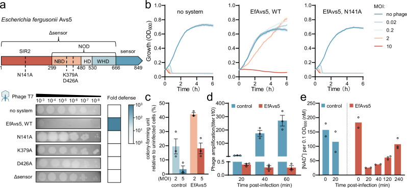
Fig. 1 EfAvs5 consumes NAD+ to confer defense. a Domain organization of the EfAvs5 protein from Escherichia fergusonii and efficiency of plaquing of phages infecting E. coli expressing EfAvs5 or an empty vector. Data are representative images of n = 3 biological replicates. The mutations used are indicated below the domain organization. NBD, nucleotide-binding domain; HD, helical domain; WHD, wing-helical domain; NOD, nucleotide-binding oligomerization domain. b Infection time courses for liquid cultures of E. coli, with EfAvs5, EfAvs5 N141A or empty vector, infected at different multiplicities of infection (MOI) of phage T7. Data represent the mean of three replicates, and shaded regions represent the standard error of the mean (SEM). c Colony-forming units of E. coli cells expressing either the EfAvs5 or empty vector as a control infected at an MOI of 2 and 5 with T7 for 20 minutes. Data represent the mean of three replicates, and error bars represent the SEM, with individual data points overlaid. d Titer of phage T7 propagated on E. coli cells expressing either the EfAvs5 or empty vector as a control infected at an MOI of 0.1 with T7. Data represent the titer of T7 measured in plaque-forming units (PFU)/mL after indicated time from initial infection, divided by the original phage titer prior to infection. Data represent the mean of three replicates, and error bars represent the SEM, with individual data points overlaid. e Measurement of NAD+ concentration in E. coli expressing EfAvs5 or empty vector as a control at the indicated time points after infection with phage T7 at an MOI of 2, normalized to an OD600 of 0.1. The control group underwent near-complete lysis after 20 minutes of infection, precluding the quantification of NAD+ content. Data with the avs5 system at 120 and 240 minutes represent three biological replicates; others represent two replicates; all individual data points are shown. Source data are provided as a Source Data file.
Fig. 1 EfAvs5 consumes NAD+ to confer defense. a Domain organization of the EfAvs5 protein from Escherichia fergusonii and efficiency of plaquing of phages infecting E. coli expressing EfAvs5 or an empty vector. Data are representative images of n = 3 biological replicates. The mutations used are indicated below the domain organization. NBD, nucleotide-binding domain; HD, helical domain; WHD, wing-helical domain; NOD, nucleotide-binding oligomerization domain. b Infection time courses for liquid cultures of E. coli, with EfAvs5, EfAvs5 N141A or empty vector, infected at different multiplicities of infection (MOI) of phage T7. Data represent the mean of three replicates, and shaded regions represent the standard error of the mean (SEM). c Colony-forming units of E. coli cells expressing either the EfAvs5 or empty vector as a control infected at an MOI of 2 and 5 with T7 for 20 minutes. Data represent the mean of three replicates, and error bars represent the SEM, with individual data points overlaid. d Titer of phage T7 propagated on E. coli cells expressing either the EfAvs5 or empty vector as a control infected at an MOI of 0.1 with T7. Data represent the titer of T7 measured in plaque-forming units (PFU)/mL after indicated time from initial infection, divided by the original phage titer prior to infection. Data represent the mean of three replicates, and error bars represent the SEM, with individual data points overlaid. e Measurement of NAD+ concentration in E. coli expressing EfAvs5 or empty vector as a control at the indicated time points after infection with phage T7 at an MOI of 2, normalized to an OD600 of 0.1. The control group underwent near-complete lysis after 20 minutes of infection, precluding the quantification of NAD+ content. Data with the avs5 system at 120 and 240 minutes represent three biological replicates; others represent two replicates; all individual data points are shown. Source data are provided as a Source Data file.CC BY-NC-ND · PubMed Central
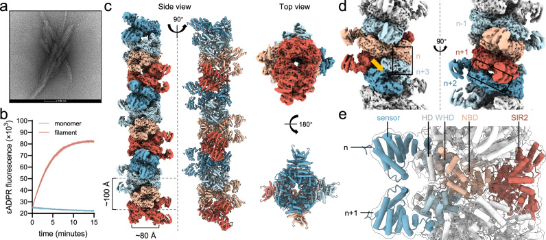
Fig. 2 Cryo-EM structure of the EfAvs5 filament. a Negative-stain micrograph images of EfAvs5, representative of >5 fields of view with similar results. The scale bar represents 100 nm. b EfAvs5’s NADase activity is activated by filamentation. After the addition of ε-NAD+, total fluorescence was measured over time. Data represent the mean of three replicates, and shaded regions represent the SEM. c Top and side views of the density map and the atomic model of EfAvs5 filament. d Detailed view of EfAvs5 filament to show the subunit interfaces. Two interfaces are indicated with boxes, and the NAD+ active site is indicated with an orange arrow. e The core of EfAvs5 filament is composed of the SIR2 domain and the NOD. Source data are provided as a Source Data file.
Fig. 2 Cryo-EM structure of the EfAvs5 filament. a Negative-stain micrograph images of EfAvs5, representative of >5 fields of view with similar results. The scale bar represents 100 nm. b EfAvs5’s NADase activity is activated by filamentation. After the addition of ε-NAD+, total fluorescence was measured over time. Data represent the mean of three replicates, and shaded regions represent the SEM. c Top and side views of the density map and the atomic model of EfAvs5 filament. d Detailed view of EfAvs5 filament to show the subunit interfaces. Two interfaces are indicated with boxes, and the NAD+ active site is indicated with an orange arrow. e The core of EfAvs5 filament is composed of the SIR2 domain and the NOD. Source data are provided as a Source Data file.CC BY-NC-ND · PubMed Central
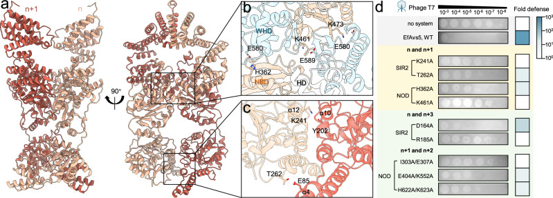
Fig. 3 Dimerization of EfAvs5. a The structure of the EfAvs5 dimer. b Details of the NOD-based dimeric interfaces. c Details of the SIR2-based dimeric interfaces. d Phage plaque assay of E. coli expressing EfAvs5 variants with mutations at the interfaces. The yellow background highlights the intra-dimer interactions, while the green background indicates the inter-dimer interactions. Data are representative images of n = 3 biological replicates. Source data are provided as a Source Data file.
Fig. 3 Dimerization of EfAvs5. a The structure of the EfAvs5 dimer. b Details of the NOD-based dimeric interfaces. c Details of the SIR2-based dimeric interfaces. d Phage plaque assay of E. coli expressing EfAvs5 variants with mutations at the interfaces. The yellow background highlights the intra-dimer interactions, while the green background indicates the inter-dimer interactions. Data are representative images of n = 3 biological replicates. Source data are provided as a Source Data file.CC BY-NC-ND · PubMed Central
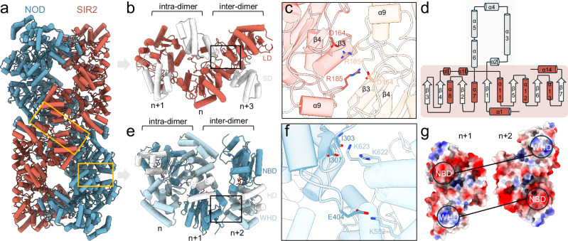
Fig. 4 SIR2- and NOD-dependent filamentation. a Two spirals dominated by NOD (blue) and SIR2 domains (red), respectively. The C-terminal domains of EfAvs5 are not displayed. The inter-dimer interactions are indicated by orange boxes. b The intra-dimer and inter-dimer interface of SIR2 domain of the n + 1, n, n + 3 subunits. LD, large domain; SD, small domain. c Detailed inter-dimer interface of SIR2 domain. The n, n + 3 subunits of EfAvs5 were colored red and orange, respectively. d SIR2 topology diagrams of EfAvs5. The small domain is shaded grey, and the large domain is shaded red. e The intra-dimer and inter-dimer interface of NOD domain of the n, n + 1, n + 2 subunits. f Detailed inter-dimer interface of NOD domain. The NBD of n + 1 subunit, and the WHD of n + 2 subunit of EfAvs5 were colored dark and light blue, respectively. g The electrostatic potential inter-dimer interface of two adjacent NOD domains.
Fig. 4 SIR2- and NOD-dependent filamentation. a Two spirals dominated by NOD (blue) and SIR2 domains (red), respectively. The C-terminal domains of EfAvs5 are not displayed. The inter-dimer interactions are indicated by orange boxes. b The intra-dimer and inter-dimer interface of SIR2 domain of the n + 1, n, n + 3 subunits. LD, large domain; SD, small domain. c Detailed inter-dimer interface of SIR2 domain. The n, n + 3 subunits of EfAvs5 were colored red and orange, respectively. d SIR2 topology diagrams of EfAvs5. The small domain is shaded grey, and the large domain is shaded red. e The intra-dimer and inter-dimer interface of NOD domain of the n, n + 1, n + 2 subunits. f Detailed inter-dimer interface of NOD domain. The NBD of n + 1 subunit, and the WHD of n + 2 subunit of EfAvs5 were colored dark and light blue, respectively. g The electrostatic potential inter-dimer interface of two adjacent NOD domains.CC BY-NC-ND · PubMed Central
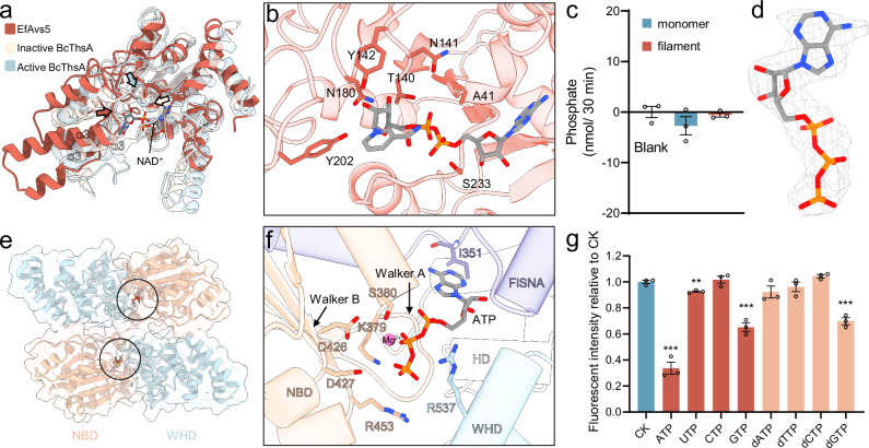
Fig. 5 The NAD+ hydrolyzing pocket and ATP binding pocket of EfAvs5. a Overlay of SIR2 domain (red) of EfAvs5 with the inactive (6LHX, light orange) and activated (8BTO, light blue) Bacillus cereus ThsA (BcThsA) SIR2 domains. NAD+ from the activated ThsA (1ICI) is shown. The loops above the NAD+ pocket of EfAvs5 and ThsA SIR2 are indicated by arrows colored as in the color scheme. b Residues of EfAvs5 interacting with NAD+ molecule highlighted in sticks in the predicted EfAvs5-NAD+ binding model. c ATPase activity assay of the monomer or the filament EfAvs5. Data represent the mean of three replicates, and error bars represent the SEM, with individual data points overlaid. P-values are compared to the blank group by two-sided Student’s t-test, and exact values are provided in the Source Data File. d Cryo-EM map of ATP. e The binding pocket of ATP in the NOD dimer. f Residues of EfAvs5 coordinating ATP molecule highlighted in sticks. The highly conserved Walker A motif and Walker B motif were indicated by arrows. FISNA, fish-specific NACHT-associated domain. g Effects of NTPs and dNTPs (5 mM) on the NADase activity of EfAvs5, showing the ratio of fluorescence changes observed within 15 minutes compared to the control group (CK), which had no NTPs/dNTPs added. Data represent the mean of three biological replicates, and error bars represent the SEM, with individual data points overlaid. P-values are compared to CK using a two-sided Student’s t-test. Asterisks (**, and ***) indicate statistical significance at p < 0.01 and p < 0.001, respectively. The exact P-values and source data are provided in the Source Data File.
Fig. 5 The NAD+ hydrolyzing pocket and ATP binding pocket of EfAvs5. a Overlay of SIR2 domain (red) of EfAvs5 with the inactive (6LHX, light orange) and activated (8BTO, light blue) Bacillus cereus ThsA (BcThsA) SIR2 domains. NAD+ from the activated ThsA (1ICI) is shown. The loops above the NAD+ pocket of EfAvs5 and ThsA SIR2 are indicated by arrows colored as in the color scheme. b Residues of EfAvs5 interacting with NAD+ molecule highlighted in sticks in the predicted EfAvs5-NAD+ binding model. c ATPase activity assay of the monomer or the filament EfAvs5. Data represent the mean of three replicates, and error bars represent the SEM, with individual data points overlaid. P-values are compared to the blank group by two-sided Student’s t-test, and exact values are provided in the Source Data File. d Cryo-EM map of ATP. e The binding pocket of ATP in the NOD dimer. f Residues of EfAvs5 coordinating ATP molecule highlighted in sticks. The highly conserved Walker A motif and Walker B motif were indicated by arrows. FISNA, fish-specific NACHT-associated domain. g Effects of NTPs and dNTPs (5 mM) on the NADase activity of EfAvs5, showing the ratio of fluorescence changes observed within 15 minutes compared to the control group (CK), which had no NTPs/dNTPs added. Data represent the mean of three biological replicates, and error bars represent the SEM, with individual data points overlaid. P-values are compared to CK using a two-sided Student’s t-test. Asterisks (**, and ***) indicate statistical significance at p < 0.01 and p < 0.001, respectively. The exact P-values and source data are provided in the Source Data File.CC BY-NC-ND · PubMed Central
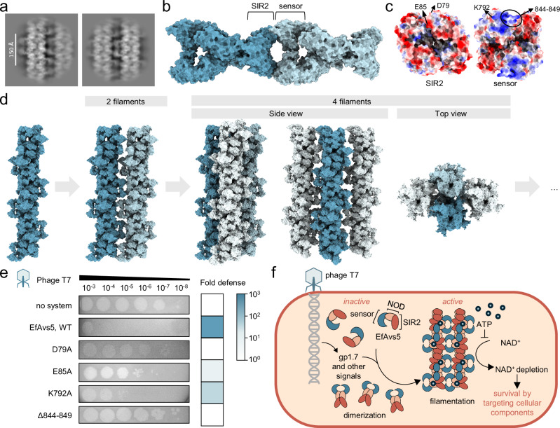
Fig. 6 Filament bundle formation mediates EfAvs5 anti-phage activity. a Representative 2D class averages showing the bundled filament assembly. The scale bar represents 150 Å. b Interacting EfAvs5 dimers in adjacent filaments. c The electrostatic potential interface of C-terminal sensor and N-terminal SIR2 domain. d A schematic diagram of the filament bundle assembly of EfAvs5. e Phage plaque assay of E. coli expressing EfAvs5 variants with mutations at the bundling interfaces. Data are representative images of n = 3 biological replicates. f Proposed model of EfAvs5 immune mechanism. Without phage infection, EfAvs5 remains inactive as a monomer. Phage infection activates EfAvs5, forming filament bundles where SIR2 enables survival by targeting cellular components NAD+. Source data are provided as a Source Data file.
Fig. 6 Filament bundle formation mediates EfAvs5 anti-phage activity. a Representative 2D class averages showing the bundled filament assembly. The scale bar represents 150 Å. b Interacting EfAvs5 dimers in adjacent filaments. c The electrostatic potential interface of C-terminal sensor and N-terminal SIR2 domain. d A schematic diagram of the filament bundle assembly of EfAvs5. e Phage plaque assay of E. coli expressing EfAvs5 variants with mutations at the bundling interfaces. Data are representative images of n = 3 biological replicates. f Proposed model of EfAvs5 immune mechanism. Without phage infection, EfAvs5 remains inactive as a monomer. Phage infection activates EfAvs5, forming filament bundles where SIR2 enables survival by targeting cellular components NAD+. Source data are provided as a Source Data file.CC BY-NC-ND · PubMed Central

Verification

Claims the paper makes

The claims and statistics of this paper haven’t been checked yet.

Run the check — free, no account needed. It reads the open-access text and checks the sources it cites.

A few free checks a day · papers already verified open instantly · results are public

Self-check

Percentages, sums and reported differences re-computed from the paper’s own numbers.

The self-check needs the paper’s full text.

Citations

Cites

Research works printed but not linked: 41

  1. Millman A et al (2022) An expanded arsenal of immune systems that protect bacteria from phages. Cell Host Microbe 30:1556–1569e1555. https://doi.org:10.1016/j.chom.2022.09.017 · DOI · Paper page
  2. Vassallo CN, Doering CR, Littlehale ML, Teodoro GIC, Laub MT (2022) A functional selection reveals previously undetected anti-phage defence systems in the E. coli pangenome. Nat Microbiol 7:1568–1579. https://doi.org:10.1038/s41564-022-01219-4 · DOI
  3. Gao L et al (2020) Diverse enzymatic activities mediate antiviral immunity in prokaryotes. Science 369:1077–1084. https://doi.org:10.1126/science.aba0372 · DOI
  4. Gao LA et al (2022) Prokaryotic innate immunity through pattern recognition of conserved viral proteins. Science 377. https://doi.org:10.1126/science.abm4096 · DOI
  5. Huang S et al (2023) NLR signaling in plants: from resistosomes to second messengers. Trends Biochem Sci 48:776–787. https://doi.org:10.1016/j.tibs.2023.06.002 · DOI
  6. Mitchell PS, Sandstrom A, Vance RE (2019) The NLRP1 inflammasome: new mechanistic insights and unresolved mysteries. Curr Opin Immunol 60:37–45. https://doi.org:10.1016/j.coi.2019.04.015 · DOI
  7. Koopal B et al (2022) Short prokaryotic Argonaute systems trigger cell death upon detection of invading DNA. Cell 185:1471–1486e1419. https://doi.org:10.1016/j.cell.2022.03.012 · DOI
  8. Ka D, Oh H, Park E, Kim J-H, Bae E (2020) Structural and functional evidence of bacterial antiphage protection by Thoeris defense system via NAD + degradation. Nat Commun 11. https://doi.org:10.1038/s41467-020-16703-w · DOI
  9. Garb J et al (2022) Multiple phage resistance systems inhibit infection via SIR2-dependent NAD + depletion. Nat Microbiol 7:1849–1856. https://doi.org:10.1038/s41564-022-01207-8 · DOI
  10. Tang D et al (2023) Multiple enzymatic activities of a Sir2-HerA system cooperate for anti-phage defense. Mol Cell 83:4600–4613e4606. https://doi.org:10.1016/j.molcel.2023.11.010 · DOI
  11. Tamulaitiene G et al (2024) Activation of Thoeris antiviral system via SIR2 effector filament assembly. Nature. https://doi.org:10.1038/s41586-024-07092-x · DOI
  12. Yang X, Wang Y, Zheng J (2024) Structural insights into autoinhibition and activation of defense-associated sirtuin protein. Int J Biol Macromol 277:134145. https://doi.org:10.1016/j.ijbiomac.2024.134145 · DOI
  13. Fillol-Salom A et al (2022) Bacteriophages benefit from mobilizing pathogenicity islands encoding immune systems against competitors. Cell 185:3248–3262e3220. https://doi.org:10.1016/j.cell.2022.07.014 · DOI
  14. Morehouse BR et al (2022) Cryo-EM structure of an active bacterial TIR–STING filament complex. Nature 608:803–807. https://doi.org:10.1038/s41586-022-04999-1 · DOI
  15. Hogrel G et al (2022) Cyclic nucleotide-induced helical structure activates a TIR immune effector. Nature 608:808–812. https://doi.org:10.1038/s41586-022-05070-9 · DOI · Paper page
  16. Du J et al (2011) Sirt5 is a NAD-dependent protein lysine demalonylase and desuccinylase. Science 334:806–809. https://doi.org:10.1126/science.1207861 · DOI · Paper page
  17. Yin H et al (2024) Insights into the modulation of bacterial NADase activity by phage proteins. Nat Commun 15. https://doi.org:10.1038/s41467-024-47030-z · DOI
  18. Kaur G, Iyer LM, Burroughs AM, Aravind L (2021) Bacterial death and TRADD-N domains help define novel apoptosis and immunity mechanisms shared by prokaryotes and metazoans. Elife 10. https://doi.org:10.7554/eLife.70394 · DOI
  19. Holm L (2022) Dali server: structural unification of protein families. Nucleic Acids Res 50:W210–w215. https://doi.org:10.1093/nar/gkac387 · DOI
  20. Wang J et al (2019) Reconstitution and structure of a plant NLR resistosome conferring immunity. Science 364. https://doi.org:10.1126/science.aav5870 · DOI
  21. Zhou M et al (2015) Atomic structure of the apoptosome: mechanism of cytochrome c- and dATP-mediated activation of Apaf-1. Genes Dev 29:2349–2361. https://doi.org:10.1101/gad.272278.115 · DOI
  22. Xiao L, Magupalli VG, Wu H (2023) Cryo-EM structures of the active NLRP3 inflammasome disc. Nature 613:595–600. https://doi.org:10.1038/s41586-022-05570-8 · DOI
  23. Ma S et al (2020) Direct pathogen-induced assembly of an NLR immune receptor complex to form a holoenzyme. Science 370. https://doi.org:10.1126/science.abe3069 · DOI
  24. Wang J et al (2019) Reconstitution and structure of a plant NLR resistosome conferring immunity. Science 364. https://doi.org:10.1126/science.aav5870 · DOI
  25. Pang Y et al (2015) Structure of the apoptosome: mechanistic insights into activation of an initiator caspase from Drosophila. Genes Dev 29:277–287. https://doi.org:10.1101/gad.255877.114 · DOI
  26. Ma S et al (2024) Oligomerization-mediated autoinhibition and cofactor binding of a plant NLR. Nature 632:869–876. https://doi.org:10.1038/s41586-024-07668-7 · DOI
  27. Zhang Y, Dorey S, Swiderski M, Jones JD (2004) Expression of RPS4 in tobacco induces an AvrRps4-independent HR that requires EDS1, SGT1 and HSP90. Plant J 40:213–224. https://doi.org:10.1111/j.1365-313X.2004.02201.x · DOI
  28. Horsefield S et al (2019) NAD(+) cleavage activity by animal and plant TIR domains in cell death pathways. Science 365:793–799. https://doi.org:10.1126/science.aax1911 · DOI
  29. Conte AN et al (2024) https://doi.org:10.1101/2024.06.04.597415 · DOI
  30. Song W et al (2024) Substrate-induced condensation activates plant TIR domain proteins. Nature 627:847–853. https://doi.org:10.1038/s41586-024-07183-9 · DOI
  31. Punjani A, Rubinstein JL, Fleet DJ, Brubaker MA (2017) cryoSPARC: algorithms for rapid unsupervised cryo-EM structure determination. Nat Methods 14:290–296. https://doi.org:10.1038/nmeth.4169 · DOI · Paper page
  32. Casanal A, Lohkamp B, Emsley P (2020) Current developments in Coot for macromolecular model building of Electron Cryo-microscopy and Crystallographic Data. Protein Sci 29:1069–1078. https://doi.org:10.1002/pro.3791 · DOI
  33. Pettersen EF et al (2021) UCSF ChimeraX: Structure visualization for researchers, educators, and developers. Protein Sci 30:70–82. https://doi.org:10.1002/pro.3943 · DOI
  34. Liebschner D et al (2019) Macromolecular structure determination using X-rays, neutrons and electrons: recent developments in Phenix. Acta Crystallogr D Struct Biol 75:861–877. https://doi.org:10.1107/S2059798319011471 · DOI · Paper page
  35. Tesson F et al (2022) Systematic and quantitative view of the antiviral arsenal of prokaryotes. Nat Commun 13:2561. https://doi.org:10.1038/s41467-022-30269-9 · DOI
  36. van Kempen M et al (2024) Fast and accurate protein structure search with Foldseek. Nat Biotechnol 42:243–246. https://doi.org:10.1038/s41587-023-01773-0 · DOI · Paper page
  37. Steinegger M, Söding J (2017) MMseqs2 enables sensitive protein sequence searching for the analysis of massive data sets. Nat Biotechnol 35:1026–1028. https://doi.org:10.1038/nbt.3988 · DOI · Paper page
  38. Katoh K, Rozewicki J, Yamada K (2019) D. MAFFT online service: multiple sequence alignment, interactive sequence choice and visualization. Brief Bioinform 20:1160–1166. https://doi.org:10.1093/bib/bbx108 · DOI · Paper page
  39. Capella-Gutiérrez S, Silla-Martínez JM, Gabaldón T (2009) trimAl: a tool for automated alignment trimming in large-scale phylogenetic analyses. Bioinformatics 25:1972–1973. https://doi.org:10.1093/bioinformatics/btp348 · DOI
  40. Nguyen LT, Schmidt HA, von Haeseler A, Minh BQ (2015) IQ-TREE: a fast and effective stochastic algorithm for estimating maximum-likelihood phylogenies. Mol Biol Evol 32:268–274. https://doi.org:10.1093/molbev/msu300 · DOI · Paper page
  41. Letunic I, Bork P (2021) Interactive Tree Of Life (iTOL) v5: an online tool for phylogenetic tree display and annotation. Nucleic Acids Res 49:W293–w296. https://doi.org:10.1093/nar/gkab301 · DOI

Metadata

Authors
Zheng, J., Wang, Y., Tian, Y., Yang, X., & Yu, F.
Affiliations
Shanghai Jiao Tong University (CN)
Corresponding author
Zheng, Jianting
Venue
Nature Communications
Published
2024-10-15
Volume
16
Issue
1
Pages
2408-2408
ISSN
2041-1723 (ISSN-L)
OpenAlex
W4408312757
Cited by
18
References
1
Citations by year
2026: 12 · 2025: 6
FWCI
9.52
Type
journal article
Language
en
Open access
Locations
  • Nature Communications (publishedVersion, cc-by-nc-nd)
  • Research Square (acceptedVersion, cc-by)
  • Europe PMC (PubMed Central) (publishedVersion, cc-by-nc-nd)
  • PubMed Central (publishedVersion, cc-by-nc-nd)
  • PubMed (publishedVersion)
  • RePEc: Research Papers in Economics (submittedVersion)
  • DOAJ (DOAJ: Directory of Open Access Journals) (submittedVersion, cc-by-sa)
Funders
  • National Natural Science Foundation of China (32370071, 32070040)
  • National Key Research and Development Program of China
Topics
  • Life Sciences › Immunology and Microbiology › Virology › Bacteriophages and microbial interactions
  • Life Sciences › Immunology and Microbiology › Immunology › Invertebrate Immune Response Mechanisms
  • Life Sciences › Biochemistry, Genetics and Molecular Biology › Genetics › Bacterial Genetics and Biotechnology
Keywords
filamentation, nucleotide-binding oligomerization domain, NOD-like receptors, Escherichia fergusonii, phage defense, NADase activity, filament bundling, ATP binding, protein dimerization, cryo-EM structure
MeSH terms
Protein Domains, Adenosine Triphosphate, Bacterial Proteins, Models, Molecular, NAD, Protein Binding, Bacteriophage T7, Cryoelectron Microscopy, Protein Multimerization

About these checks

Registry, retraction, Cites and statistics results are computed, not judged, with one exception: a printed reference that no index lists is read once by a model before it counts as a citation error. Claims are extracted by a model and kept only when their quote appears verbatim in the text; each cited source is then checked, with a model’s help, for whether it says what the claim relies on. Those verdicts can be wrong — read the claim’s quote and the cited source before relying on one.

Statistics and arithmetic are recomputed, not judged.

Confidence bands are shown only for fields calibrated on our annotated test set. None is calibrated yet, so no band is shown.

What counts as a claim ›How we measure confidence ›How we check this ›

Are you an author, or do you see a problem with this page? Request a review