

The DOI matches its Crossref record; 1 of the 1 references it cites were found in a registry or an index.
Recruitment of Mre11 to recombination sites during meiosis
Open accessPreprint — published as 10.1038/s41467-026-71310-5
Abstract
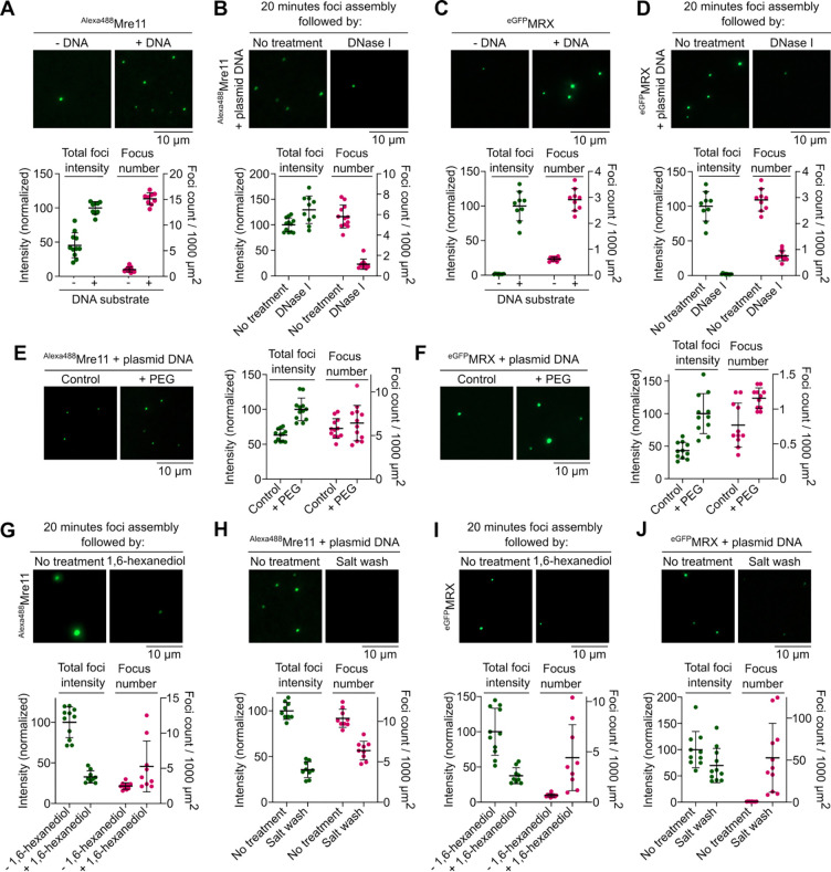
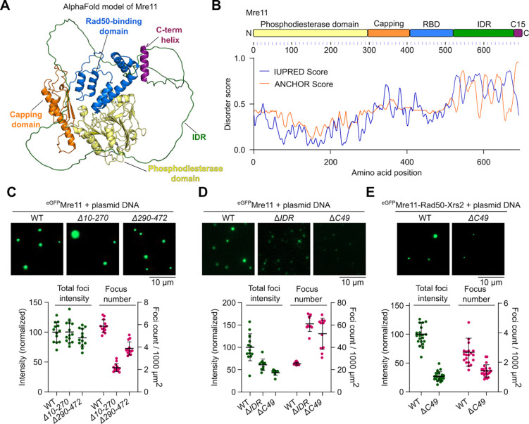
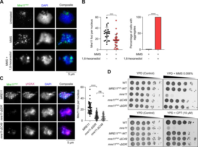
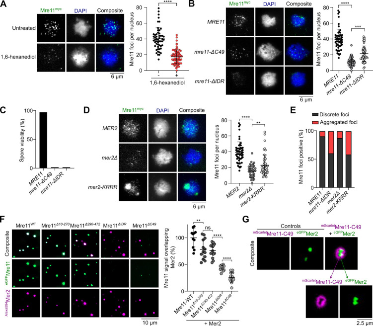
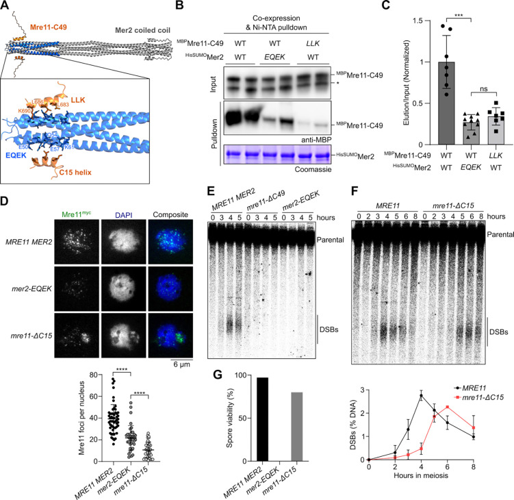
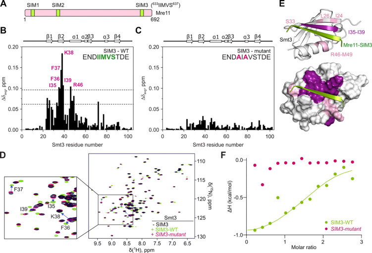
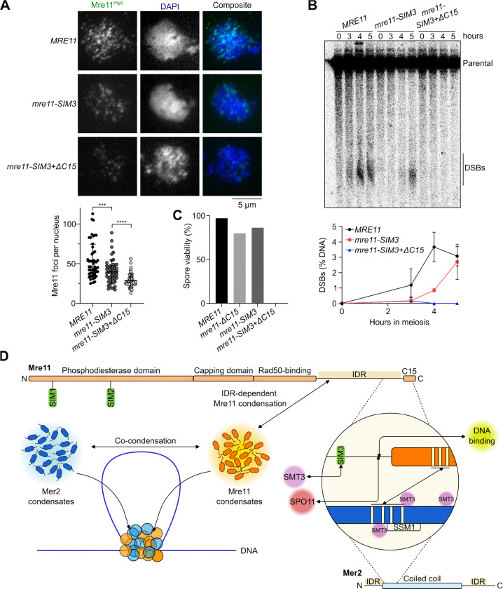
The Mre11 nuclease is part of the highly conserved MRX complex involved in the repair of DNA double-strand breaks (DSBs). During meiosis in budding yeast, MRX is also required for the programmed induction of DSBs by Spo11, thereby initiating homologous recombination to promote accurate chromosome segregation. Recruitment of Mre11 to meiotic DSB sites depends on Rec114-Mei4 and Mer2 (RMM), which are thought to organize the meiotic DSB machinery by a mechanism involving biomolecular condensation. Here, we explored the role of Mre11 during meiosis and its relationship to RMM condensation. We show that both Mre11 and MRX complexes form DNA-dependent, hexanediol sensitive condensates in vitro . In vivo , Mre11 assembles into DNA damage-dependent foci in vegetative cells and DSB-independent foci in meiotic cells. In vitro condensates and in vivo foci both depend on the C-terminal intrinsically-disordered region (IDR) of Mre11. Importantly, while the Mre11 IDR is dispensable for vegetative DNA repair it is essential during meiosis. The C-terminal region of Mre11 forms a short α-helix that binds a conserved region of Mer2, and mutating residues within this interface reduces Mre11 foci and DSB formation. Finally, we identified a SUMO-interacting motif within the Mre11 IDR that enhances recruitment of Mre11 during meiosis and facilitates DSB formation. Our results provide new insights into the biophysical properties of Mre11 and its role in initiating meiotic recombination.
Overview
The claims and statistics of this paper haven’t been checked yet.
Run the check — free, no account needed. It reads the open-access text and checks the sources it cites.
Integrity signals
Registry
Cites
Statistics
Figures
Verification
Claims the paper makes
The claims and statistics of this paper haven’t been checked yet.
Run the check — free, no account needed. It reads the open-access text and checks the sources it cites.
Self-check
Percentages, sums and reported differences re-computed from the paper’s own numbers.
The self-check needs the paper’s full text.
Citations
Cites
Highly accurate protein structure prediction with AlphaFold
Jumper, J., Evans, R., Pritzel, A., Green, T., Figurnov, M., Ronneberger, O., Tunyasuvunakool, K., Bates, R., Žídek, A., Potapenko, A., et al. (2021) · Nature · doi:10.1038/s41586-021-03819-2 · Cited by 43,764
Research works printed but not linked: 72
- Cooke MS, Evans MD, Dizdaroglu M, Lunec J, Oxidative (2003) DNA damage: mechanisms, mutation, and disease. FASEB j 17(10):1195–1214. 10.1096/fj.02-0752rev · DOI · Paper page
- De Almeida LC, Calil FA, Machado-Neto JA, Costa-Lotufo LV (2021) DNA damaging agents and DNA repair: From carcinogenesis to cancer therapy. Cancer Genet 252–253:6–24. 10.1016/j.cancergen.2020.12.002 · DOI
- Tubbs A, Nussenzweig A (2017) Endogenous DNA Damage as a Source of Genomic Instability in Cancer. Cell 168(4):644–656. 10.1016/j.cell.2017.01.002 · DOI · Paper page
- Oster S, Aqeilan RI, Programmed DNA (2020) Damage and Physiological DSBs: Mapping, Biological Significance and Perturbations in Disease States. Cells 9(8):1870. 10.3390/cells9081870 · DOI
- Börner GV, Hochwagen A, MacQueen AJ (2023) Meiosis in budding yeast. Genetics 225(2):iyad125. 10.1093/genetics/iyad125 · DOI
- Zickler D, Kleckner N, Meiosis (2023) Dances Between Homologs. Annu Rev Genet 57(1):1–63. 10.1146/annurev-genet-061323-044915 · DOI
- Lam I, Keeney S (2015) Mechanism and Regulation of Meiotic Recombination Initiation. Cold Spring Harb Perspect Biol 7(1):a016634. 10.1101/cshperspect.a016634 · DOI
- Yadav VK, Claeys Bouuaert C (2021) Mechanism and Control of Meiotic DNA Double-Strand Break Formation in S. cerevisiae. Front Cell Dev Biol 9:642737. 10.3389/fcell.2021.642737 · DOI
- Claeys Bouuaert C, Pu S, Wang J et al (2021) DNA-driven condensation assembles the meiotic DNA break machinery. Nature 592(7852):144–149. 10.1038/s41586-021-03374-w · DOI
- Claeys Bouuaert C, Tischfield SE, Pu S et al (2021) Structural and functional characterization of the Spo11 core complex. Nat Struct Mol Biol 28(1):92–102. 10.1038/s41594-020-00534-w · DOI · Paper page
- Johnson D, Crawford M, Cooper T et al (2021) Concerted cutting by Spo11 illuminates meiotic DNA break mechanics. Nature 594(7864):572–576. 10.1038/s41586-021-03389-3 · DOI
- Oger C, Claeys Bouuaert C (2025) SPO11 dimers are sufficient to catalyse DNA double-strand breaks in vitro. Nature 639(8055):792–799. 10.1038/s41586-024-08574-8 · DOI
- Kleckner N (2006) Chiasma formation: chromatin/axis interplay and the role(s) of the synaptonemal complex. Chromosoma 115(3):175–194. 10.1007/s00412-006-0055-7 · DOI
- Panizza S, Mendoza MA, Berlinger M et al (2011) Spo11-Accessory Proteins Link Double-Strand Break Sites to the Chromosome Axis in Early Meiotic Recombination. Cell 146(3):372–383. 10.1016/j.cell.2011.07.003 · DOI
- Alani E, Padmore R, Kleckner N (1990) Analysis of Wild-Type and rad50 mutants of Yeast Suggests an Intimate Relationship between Meiotic Chromosome Synapsis and Recombination. Cell 61(3):419–436. 10.1016/0092-8674(90)90524-I · DOI
- Ivanov EL, Korolevt VG, Fabre F (1992) Xrs2, a DNA Repair Gene of Saccharomyces Cerevisiae, Is Needed for Meiotic Recombination. Genetics 132(3):651–664. 10.1093/genetics/132.3.651 · DOI
- Nairz K, Klein F (1997) mre11S —a yeast mutation that blocks double-strand-break processing and permits nonhomologous synapsis in meiosis. Genes Dev. ;11(17):2272–2290. 10.1101/gad.11.17.2272 · DOI
- Keeney S (2001) Mechanism and control of meiotic recombination initiation. Curr Top Dev Biol 52:1–53. 10.1016/S0070-2153(01)52008-6 · DOI
- Moore JK, Haber JE (1996) Cell Cycle and Genetic Requirements of Two Pathways of Nonhomologous End-Joining Repair of Double-Strand Breaks in Saccharomyces cerevisiae. Mol Cell Biol 16(5):2164–2173. 10.1128/MCB.16.5.2164 · DOI · Paper page
- Paull TT, Gellert M (1998) The 3′ to 5′ Exonuclease Activity of Mre11 Facilitates Repair of DNA Double-Strand Breaks. Mol Cell 1(7):969–979. 10.1016/S1097-2765(00)80097-0 · DOI
- Garcia V, Phelps SEL, Gray S, Neale MJ (2011) Bidirectional resection of DNA double-strand breaks by Mre11 and Exo1. Nature 479(7372):241–244. 10.1038/nature10515 · DOI
- Casari E, Rinaldi C, Marsella A et al (2019) Processing of DNA Double-Strand Breaks by the MRX Complex in a Chromatin Context. Front Mol Biosci 6:43. 10.3389/fmolb.2019.00043 · DOI
- Gobbini E, Cassani C, Villa M, Bonetti D, Longhese M (2016) Functions and regulation of the MRX complex at DNA double-strand breaks. Microb Cell 3(8):329–337. 10.15698/mic2016.08.517 · DOI
- Paull TT (2018) 20 Years of Mre11 Biology: No End in Sight. Mol Cell 71(3):419–427. 10.1016/j.molcel.2018.06.033 · DOI
- Tisi R, Vertemara J, Zampella G, Longhese MP (2020) Functional and structural insights into the MRX/MRN complex, a key player in recognition and repair of DNA double-strand breaks. Comput Struct Biotechnol J 18:1137–1152. 10.1016/j.csbj.2020.05.013 · DOI
- Lamarche BJ, Orazio NI, Weitzman MD (2010) The MRN complex in double-strand break repair and telomere maintenance. FEBS Lett 584(17):3682–3695. 10.1016/j.febslet.2010.07.029 · DOI · Paper page
- Lu R, Zhang H, Jiang YN, Wang ZQ, Sun L, Zhou ZW (2021) Post-Translational Modification of MRE11: Its Implication in DDR and Diseases. Genes 12(8):1158. 10.3390/genes12081158 · DOI
- Oh J, Symington LS (2018) Role of the Mre11 Complex in Preserving Genome Integrity. Genes 9(12):589. 10.3390/genes9120589 · DOI
- Furuse M, Nagase Y, Tsubouchi H, Murakami-Murofushi K, Shibata T, Ohta K (1998) Distinct roles of two separable in vitro activities of yeast Mre11 in mitotic and meiotic recombination. EMBO J 17(21):6412–6425. 10.1093/emboj/17.21.6412 · DOI
- Usui T, Ohta T, Oshiumi H, Tomizawa J ichi, Ogawa H, Ogawa T (1998) Complex Formation and Functional Versatility of Mre11 of Budding Yeast in Recombination. Cell. ;95(5):705–716. 10.1016/S0092-8674(00)81640-2 · DOI
- Aithal R, Nangalia K, Spirek M, Chen D, Klein F, Krejci L (2024) Physical interaction with Spo11 mediates the localisation of Mre11 to chromatin in meiosis and promotes its nuclease activity. Nucleic Acids Res 52(8):4328–4343. 10.1093/nar/gkae111 · DOI
- Wen Q, Scorah J, Phear G, Rodgers G, Rodgers S, Meuth M (2008) A Mutant Allele of MRE11 Found in Mismatch Repair-deficient Tumor Cells Suppresses the Cellular Response to DNA Replication Fork Stress in a Dominant Negative Manner. Cohen-Fix O, ed. MBoC. ;19(4):1693–1705. 10.1091/mbc.e07-09-0975 · DOI
- Tamai T, Reginato G, Ojiri R et al (2024) Sae2 controls Mre11 endo- and exonuclease activities by different mechanisms. Nat Commun 15(1):7221. 10.1038/s41467-024-51493-5 · DOI
- Kissling VM, Reginato G, Bianco E et al (2022) Mre11-Rad50 oligomerization promotes DNA double-strand break repair. Nat Commun 13(1):2374. 10.1038/s41467-022-29841-0 · DOI
- Kroschwald S, Maharana S, Simon A (2017) Hexanediol: a chemical probe to investigate the material properties of membrane-less compartments. Matters Published online May 22. 10.19185/matters.201702000010 · DOI
- Lammens K, Bemeleit DJ, Möckel C et al (2011) The Mre11:Rad50 Structure Shows an ATP-Dependent Molecular Clamp in DNA Double-Strand Break Repair. Cell 145(1):54–66. 10.1016/j.cell.2011.02.038 · DOI
- Rotheneder M, Stakyte K, Van De Logt E et al (2023) Cryo-EM structure of the Mre11-Rad50-Nbs1 complex reveals the molecular mechanism of scaffolding functions. Mol Cell 83(2):167–185e9. 10.1016/j.molcel.2022.12.003 · DOI
- Petrini J, Hohl M, Yu Y, Kuryavyi V, Patel D (2024) Structure guided functional analysis of the S. cerevisiae Mre11 complex. Published online Dec 9. 10.21203/rs.3.rs-5390974/v1 · DOI
- Bergerat A, De Massy B, Gadelle D, Varoutas PC, Nicolas A, Forterre P (1997) An atypical topoisomerase II from archaea with implications for meiotic recombination. Nature 386(6623):414–417. 10.1038/386414a0 · DOI
- Borde V, Lin W, Novikov E, Petrini JH, Lichten M, Nicolas A (2004) Association of Mre11p with Double-Strand Break Sites during Yeast Meiosis. Mol Cell 13(3):389–401. 10.1016/S1097-2765(04)00034-6 · DOI
- Rousová D, Nivsarkar V, Altmannova V et al (2021) Novel mechanistic insights into the role of Mer2 as the keystone of meiotic DNA break formation. eLife 10:e72330. 10.7554/eLife.72330 · DOI
- Daccache D, De Jonge E, Liloku P et al (2023) Evolutionary conservation of the structure and function of meiotic Rec114 – Mei4 and Mer2 complexes. Genes Dev 37(11–12):535–553. 10.1101/gad.350462.123 · DOI
- Tessé S, Bourbon HM, Debuchy R et al (2017) Asy2/Mer2: an evolutionarily conserved mediator of meiotic recombination, pairing, and global chromosome compaction. Genes Dev 31(18):1880–1893. 10.1101/gad.304543.117 · DOI
- Bhagwat NR, Owens SN, Ito M et al (2021) SUMO is a pervasive regulator of meiosis. eLife 10:e57720. 10.7554/eLife.57720 · DOI
- Chen YJ, Chuang YC, Chuang CN et al (2016) S. cerevisiae Mre11 recruits conjugated SUMO moieties to facilitate the assembly and function of the Mre11-Rad50-Xrs2 complex. Nucleic Acids Res 44(5):2199–2213. 10.1093/nar/gkv1523 · DOI
- Zhao Q, Xie Y, Zheng Y et al (2014) GPS-SUMO: a tool for the prediction of sumoylation sites and SUMO-interaction motifs. Nucleic Acids Res 42(W1):W325–W330. 10.1093/nar/gku383 · DOI
- Lascorz J, Codina-Fabra J, Reverter D, Torres-Rosell J (2022) SUMO-SIM interactions: From structure to biological functions. Semin Cell Dev Biol 132:193–202. 10.1016/j.semcdb.2021.11.007 · DOI
- Song J, Zhang Z, Hu W, Chen Y (2005) Small Ubiquitin-like Modifier (SUMO) Recognition of a SUMO Binding Motif. J Biol Chem 280(48):40122–40129. 10.1074/jbc.M507059200 · DOI
- Keusekotten K, Bade VN, Meyer-Teschendorf K et al (2014) Multivalent interactions of the SUMO-interaction motifs in RING finger protein 4 determine the specificity for chains of the SUMO. Biochem J 457(1):207–214. 10.1042/BJ20130753 · DOI
- Symington LS (2016) Mechanism and regulation of DNA end resection in eukaryotes. Crit Rev Biochem Mol Biol 51(3):195–212. 10.3109/10409238.2016.1172552 · DOI
- Gnügge R, Symington LS (2017) Keeping it real: MRX–Sae2 clipping of natural substrates. Genes Dev 31(23–24):2311–2312. 10.1101/gad.310771.117 · DOI
- Chin GM, Villeneuve AM (2001) C. elegans mre-11 is required for meiotic recombination and DNA repair but is dispensable for the meiotic G2 DNA damage checkpoint. Genes Dev 15(5):522–534. 10.1101/gad.864101 · DOI
- Puizina J, Siroky J, Mokros P, Schweizer D, Riha K (2004) Mre11 deficiency in Arabidopsis is associated with chromosomal instability in somatic cells and Spo11-dependent genome fragmentation during meiosis. Plant Cell 16(8):1968–1978. 10.1105/tpc.104.022749 · DOI
- Young JA, Hyppa RW, Smith GR (2004) Conserved and nonconserved proteins for meiotic DNA breakage and repair in yeasts. Genetics 167(2):593–605. 10.1534/genetics.103.023762 · DOI
- Liu Y, Lin Z, Yan J, Zhang X, Tong MH (2024) A Rad50-null mutation in mouse germ cells causes reduced DSB formation, abnormal DSB end resection and complete loss of germ cells. Development 151(8). 10.1242/dev.202312 · DOI
- Kim S, Yamada S, Li T et al (2025) Mouse MRE11-RAD50-NBS1 is needed to start and extend meiotic DNA end resection. Nat Commun 16(1). 10.1038/s41467-025-57928-x · DOI
- Hayashi M, Chin GM, Villeneuve AM (2007) C. elegans germ cells switch between distinct modes of double-strand break repair during meiotic prophase progression. PLoS Genet 3(11). https://doi.org/10.1371/journal.pgen.0030191 · DOI
- Girard C, Roelens B, Zawadzki KA, Villeneuve AM (2018) Interdependent and separable functions of Caenorhabditis elegans MRN-C complex members couple formation and repair of meiotic DSBs. Proc Natl Acad Sci USA 115(19). 10.1073/pnas.1719029115 · DOI
- Bishop K, Kleckner N, DMCI (1992) A meiosis-specific yeast homolog of E. coli recA required for recombination, synaptonemal complex formation, and cell cycle progression. Cell 69(3):439–456. 10.1016/0092-8674(92)90446-j · DOI · Paper page
- Tran PT, Erdeniz N, Dudley S, Liskay RM (2002) Characterization of nuclease-dependent functions of Exo1p in Saccharomyces cerevisiae. DNA Repair 1(11):895–912. 10.1016/s1568-7864(02)00114-3 · DOI · Paper page
- Joshi N, Brown MS, Bishop DK, Börner GV (2015) Gradual implementation of the meiotic recombination program via checkpoint pathways controlled by global DSB levels. Mol Cell 57(5):797–811. 10.1016/j.molcel.2014.12.027 · DOI
- Mimitou EP, Yamada S, Keeney S (2017) A global view of meiotic double-strand break end resection. Science 335(6320):40–45. 10.1126/science.aak9704 · DOI
- Li J, Hooker GW, Roeder GS (2006) Saccharomyces cerevisiae Mer2, Mei4 and Rec114 form a complex required for meiotic double-strand break formation. Genetics 173(4):1969–1981. 10.1534/genetics.106.058768 · DOI
- Henderson KA, Kee K, Maleki S, Santini PA, Keeney S (2006) Cyclin-Dependent Kinase directly regulates initiation of meiotic recombination. Cell 125(7):1321–1332. 10.1016/j.cell.2006.04.039 · DOI
- Maleki S, Neale MJ, Arora C, Henderson KA, Keeney S (2007) Interactions between Mei4, Rec114, and other proteins required for meiotic DNA double-strand break formation in Saccharomyces cerevisiae. Chromosoma 116(5):471–486. 10.1007/s00412-007-0111-y · DOI
- Yunus AA, Lima CD (2009) Purification of SUMO Conjugating Enzymes and Kinetic Analysis of Substrate Conjugation. In: Ulrich HD (ed) SUMO Protocols. Methods in Molecular Biology, vol 497. Humana, pp 167–186. doi:10.1007/978-1-59745-566-4_11 · DOI
- Cannavo E, Cejka P, Kowalczykowski SC (2013) Relationship of DNA degradation by Saccharomyces cerevisiae Exonuclease 1 and its stimulation by RPA and Mre11-Rad50-Xrs2 to DNA end resection. Proc Natl Acad Sci USA 110(18). 10.1073/pnas.1305166110 · DOI
- Murakami H, Borde V, Nicolas A, Keeney S (2009) Gel electrophoresis assays for analyzing DNA double-strand breaks in Saccharomyces cerevisiae at various spatial resolutions. In: Keeney S (ed) Meiosis. Methods in Molecular Biology, vol 557. Humana, pp 117–142. doi:10.1007/978-1-59745-527-5_9 · DOI
- Delaglio F, Grzesiek S, Vuister GW, Zhu G, Pfeifer J, Bax A, NMRPipe (1995) A multidimensional spectral processing system based on UNIX pipes. J Biomol NMR 6(3). 10.1007/BF00197809 · DOI
- Vranken WF, Boucher W, Stevens TJ et al (2005) The CCPN data model for NMR spectroscopy: Development of a software pipeline. Proteins 59(4):687–696. 10.1002/prot.20449 · DOI
- Sheng W, Liao X (2002) Solution structure of a yeast ubiquitin-like protein Smt3: The role of structurally less defined sequences in protein–protein recognitions. Protein Sci 11(6):1482–1491. 10.1110/ps.0201602 · DOI
- Dosztanyi Z, Csizmok V, Tompa P, Simon I (2005) IUPred: web server for the prediction of intrinsically unstructured regions of proteins based on estimated energy content. Bioinformatics 21(16):3433–3434. 10.1093/bioinformatics/bti541 · DOI
Metadata
- Authors
- Bouuaert, C. C., Priyadarshini, P., Survi, M., Mouloud, W. E. Y., Bohn, R., Ballet, S., Hunter, N., & Volkov, A.
- Affiliations
- UCLouvain (BE)
- Vrije Universiteit Brussel (BE)
- Howard Hughes Medical Institute / UC Davis
- Howard Hughes Medical Institute (US)
- Corresponding author
- Bouuaert, Corentin Claeys
- Priyadarshini, Priyanka
- ORCID
- Venue
- Nature Communications
- Published
- 2025-08-19
- Volume
- 17
- Issue
- 1
- ISSN
- 2041-1723 (ISSN-L)
- PMC
- PMC12265730
- OpenAlex
- W4412172758
- Cited by
- 2
- References
- 1
- Citations by year
- 2026: 2
- FWCI
- 4.13
- Type
- journal article
- Language
- en
- Open access
- gold
- doi.org
- Locations
- Nature Communications (publishedVersion, cc-by)
- bioRxiv (Cold Spring Harbor Laboratory) (acceptedVersion, cc-by)
- Research Square (acceptedVersion, cc-by)
- PubMed (publishedVersion)
- PubMed Central (publishedVersion, cc-by)
- PubMed Central (submittedVersion, cc-by)
- DIAL (Catholic University of Leuven) (submittedVersion, other-oa)
- DOAJ (DOAJ: Directory of Open Access Journals) (submittedVersion, cc-by-sa)
- Funders
- Université Catholique de Louvain
- European Commission (HORIZON2020)
- Fonds De La Recherche Scientifique - FNRS
- Vrije Universiteit Brussel
- National Institutes of Health
- National Institute of General Medical Sciences (R01GM074223)
- H2020 European Research Council (802525)
- Topics
- Life Sciences › Biochemistry, Genetics and Molecular Biology › Molecular Biology › DNA Repair Mechanisms
- Life Sciences › Biochemistry, Genetics and Molecular Biology › Molecular Biology › Fungal and yeast genetics research
- Life Sciences › Biochemistry, Genetics and Molecular Biology › Molecular Biology › Genomics and Chromatin Dynamics
- Keywords
- MRE11, MRX complex, meiotic recombination, DNA double-strand break repair, Spo11, biomolecular condensates, intrinsically disordered regions, SUMO-interacting motif, DNA damage foci, Saccharomyces cerevisiae
About these checks
Registry, retraction, Cites and statistics results are computed, not judged, with one exception: a printed reference that no index lists is read once by a model before it counts as a citation error. Claims are extracted by a model and kept only when their quote appears verbatim in the text; each cited source is then checked, with a model’s help, for whether it says what the claim relies on. Those verdicts can be wrong — read the claim’s quote and the cited source before relying on one.
Statistics and arithmetic are recomputed, not judged.
Confidence bands are shown only for fields calibrated on our annotated test set. None is calibrated yet, so no band is shown.
What counts as a claim ›How we measure confidence ›How we check this ›
Are you an author, or do you see a problem with this page? Request a review
