

The DOI matches its Crossref record; 1 of the 1 references it cites were found in a registry or an index.
Computational optimization of DEK1 calpain domain solubility through integrated structural modelling and targeted mutagenesis
Open accessPreprint — published as 10.1038/s41598-026-38805-z · Paper page
Abstract
The DEFECTIVE KERNEL 1 (DEK1) protein plays essential functions throughout plant development. DEK1 is a multidomain 250 kDa protein with yet unsolved 3D structure. To facilitate structural and functional studies of DEK1, here we investigate its calpain protease core domain (CysPc) from Physcomitrium patens . Using integrated structural modelling we propose targeted mutagenesis of CysPc to enhance its solubility during recombinant protein production. We created a pipeline to predict the topology of the CysPc domain with improved precision, providing a robust framework for further exploration. We evaluated the native and mutant structures by MD simulations, concentrating on several solubility-related parameters. Following these features, we implemented specific single, double, and triple amino acid mutagenesis to select variants with improved solubility. Our method preserves overall structural integrity while reducing aggregation-prone traits. We advocate for the utilization of reinforcement learning method that can effectively traverse the extensive combinatorial space and prioritize mutation sets with the greatest potential for enhancing solubility. This framework provides a logical, data-driven approach to improving protein solubility, particularly beneficial in situations lacking high-resolution structural data.
Overview
The claims and statistics of this paper haven’t been checked yet.
Run the check — free, no account needed. It reads the open-access text and checks the sources it cites.
Integrity signals
Registry
Cites
Statistics
Figures
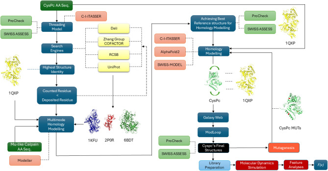
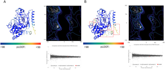
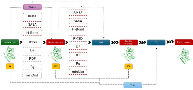
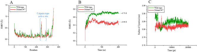
Verification
Claims the paper makes
The claims and statistics of this paper haven’t been checked yet.
Run the check — free, no account needed. It reads the open-access text and checks the sources it cites.
Self-check
Percentages, sums and reported differences re-computed from the paper’s own numbers.
The self-check needs the paper’s full text.
Citations
Cites
Highly accurate protein structure prediction with AlphaFold
Jumper, J., Evans, R., Pritzel, A., Green, T., Figurnov, M., Ronneberger, O., Tunyasuvunakool, K., Bates, R., Žídek, A., Potapenko, A., et al. (2021) · Nature · doi:10.1038/s41586-021-03819-2 · Cited by 43,764
Research works printed but not linked: 93
- Villaverde, A. Mar Carrió, M. Protein aggregation in recombinant bacteria: biological role of inclusion bodies. Biotechnol. Lett. 25, 1385–1395 (2003). · DOI
- Baneyx, F. & Mujacic, M. Recombinant protein folding and misfolding in Escherichia coli. Nat. Biotechnol. 22, 1399–1408 (2004). · DOI
- Pantophlet, R., Wilson, I. A. & Burton, D. R. Improved design of an antigen with enhanced specificity for the broadly HIV-neutralizing antibody b12. Protein Eng. Des. Selection. 17, 749–758 (2004). · DOI
- De Marco, A., Deuerling, E., Mogk, A., Tomoyasu, T. & Bukau, B. Chaperone-based procedure to increase yields of soluble recombinant proteins produced in E. coli. BMC Biotechnol 7, (2007). · DOI
- Gustafsson, C., Govindarajan, S. & Minshull, J. Codon bias and heterologous protein expression. Trends Biotechnol. 22, 346–353 (2004). · DOI · Paper page
- Chatterjee, D. K. & Esposito, D. Enhanced soluble protein expression using two new fusion tags. Protein Exp. Purif. 46, 122–129 (2006). · DOI
- Esposito, D. & Chatterjee, D. K. Enhancement of soluble protein expression through the use of fusion tags. Curr. Opin. Biotechnol. 17, 353–358 (2006). · DOI
- Sachdev, D. & Chirgwin, J. M. Properties of Soluble Fusions Between Mammalian Aspartic Proteinases and Bacterial Maltose-Binding Protein. J. Protein Chem. 18, 127–136 (1999). · DOI
- Agostini, F., Cirillo, D., Bolognesi, B. & Tartaglia, G. G. X-inactivation: quantitative predictions of protein interactions in the Xist network. Nucleic Acids Res. 41, e31–e31 (2013). · DOI
- Kulshreshtha, S., Chaudhary, V., Goswami, G. K. & Mathur, N. Computational approaches for predicting mutant protein stability. J. Comput. Aided Mol. Des. 30, 401–412 (2016). · DOI
- Damborsky, J. & Brezovsky, J. Computational tools for designing and engineering enzymes. Curr. Opin. Chem. Biol. 19, 8–16 (2014). · DOI
- Ebert, M. C. & Pelletier, J. N. Computational tools for enzyme improvement: why everyone can – and should – use them. Curr. Opin. Chem. Biol. 37, 89–96 (2017). · DOI
- Broom, A., Jacobi, Z., Trainor, K. & Meiering, E. M. Computational tools help improve protein stability but with a solubility tradeoff. J. Biol. Chem. 292, 14349–14361 (2017). · DOI
- Childers, M. C. & Daggett, V. Insights from molecular dynamics simulations for computational protein design. Mol. Syst. Des. Eng. 2, 9–33 (2017). · DOI
- Rouhani, M., Khodabakhsh, F., Norouzian, D., Cohan, R. A. & Valizadeh, V. Molecular dynamics simulation for rational protein engineering: Present and future prospectus. J. Mol. Graph. Model. 84, 43–53 (2018). · DOI
- Pikkemaat, M. G., Linssen, A. B. M., Berendsen, H. J. C. & Janssen, D. B. Molecular dynamics simulations as a tool for improving protein stability. Protein Eng. Des. Selection. 15, 185–192 (2002). · DOI
- Carballo-Amador, M. A., McKenzie, E. A., Dickson, A. J. & Warwicker, J. Surface patches on recombinant erythropoietin predict protein solubility: engineering proteins to minimise aggregation. BMC Biotechnol 19, (2019). · DOI
- Kumar, S., Kumar Bhardwaj, V., Singh, R. & Purohit, R. Explicit-solvent molecular dynamics simulations revealed conformational regain and aggregation inhibition of I113T SOD1 by Himalayan bioactive molecules. J. Mol. Liq. 339, 116798 (2021). · DOI
- Chennamsetty, N., Voynov, V., Kayser, V., Helk, B. & Trout, B. L. Prediction of Aggregation Prone Regions of Therapeutic Proteins. J. Phys. Chem. B. 114, 6614–6624 (2010). · DOI · Paper page
- Agrawal, N. J. et al. Aggregation in Protein-Based Biotherapeutics: Computational Studies and Tools to Identify Aggregation-Prone Regions. J. Pharm. Sci. 100, 5081–5095 (2011). · DOI
- Johnson, K. L., Faulkner, C., Jeffree, C. E. & Ingram, G. C. The Phytocalpain Defective Kernel 1 Is a Novel Arabidopsis Growth Regulator Whose Activity Is Regulated by Proteolytic Processing. Plant. Cell. 20, 2619–2630 (2008). · DOI
- Demko, V. et al. Genetic Analysis of DEFECTIVE KERNEL1 Loop Function in Three-Dimensional Body Patterning in Physcomitrella patens. Plant Physiol. 166, 903–919 (2014). · DOI
- Lid, S. E. et al. The defective kernel 1 (dek1) gene required for aleurone cell development in the endosperm of maize grains encodes a membrane protein of the calpain gene superfamily. Proc. Natl. Acad. Sci. U.S.A. 99, 5460–5465 (2002).
- Olsen, O. A., Perroud, P. F., Johansen, W. & Demko, V. DEK1; missing piece in puzzle of plant development. Trends Plant Sci. 20, 70–71 (2015). · DOI
- Johansen, W. et al. The DEK1 calpain Linker functions in three-dimensional body patterning in Physcomitrella patens. Plant Physiol. pp.00925. (2016) (2016). 10.1104/pp.16.00925 · DOI
- Demko, V. et al. Regulation of developmental gatekeeping and cell fate transition by the calpain protease DEK1 in Physcomitrium patens. Commun. Biol. 7, 261 (2024). · DOI
- Ako, A. E. et al. An intragenic mutagenesis strategy in Physcomitrella patens to preserve intron splicing. Sci. Rep. 7, 5111 (2017). · DOI
- Perroud, P. et al. Defective Kernel 1 (DEK 1) is required for three-dimensional growth in P hyscomitrella patens. New Phytol. 203, 794–804 (2014). · DOI
- Navarro, S. & Ventura, S. Computational re-design of protein structures to improve solubility. Expert Opin. Drug Discov. 14, 1077–1088 (2019). · DOI
- Trainor, K., Broom, A. & Meiering, E. M. Exploring the relationships between protein sequence, structure and solubility. Curr. Opin. Struct. Biol. 42, 136–146 (2017). · DOI
- Gupta, J., Nunes, C., Vyas, S. & Jonnalagadda, S. Prediction of Solubility Parameters and Miscibility of Pharmaceutical Compounds by Molecular Dynamics Simulations. J. Phys. Chem. B. 115, 2014–2023 (2011). · DOI · Paper page
- Ganugapati, J. & Akash, S. Multi-template homology based structure prediction and molecular docking studies of protein ‘L’ of Zaire ebolavirus (EBOV). Inf. Med. Unlocked. 9, 68–75 (2017). · DOI
- Lu, H., Cheng, Z., Hu, Y. & Tang, L. V. What Can De Novo Protein Design Bring to the Treatment of Hematological Disorders? Biology 12, 166 (2023).
- Roy, A., Yang, J. & Zhang, Y. COFACTOR: an accurate comparative algorithm for structure-based protein function annotation. Nucleic Acids Res. 40, W471–W477 (2012). · DOI
- Holm, L., Laiho, A., Törönen, P. & Salgado, M. DALI shines a light on remote homologs: One hundred discoveries. Protein Sci. 32, e4519 (2023). · DOI
- Chan, P., Curtis, R. A. & Warwicker, J. Soluble expression of proteins correlates with a lack of positively-charged surface. Sci. Rep. 3, 3333 (2013). · DOI
- Studer, G. et al. QMEANDisCo—distance constraints applied on model quality estimation. Bioinformatics 36, 1765–1771 (2020). · DOI
- Van Den Bedem, H. & Fraser, J. S. Integrative, dynamic structural biology at atomic resolution—it’s about time. Nat. Methods. 12, 307–318 (2015). · DOI
- Heo, L. & Feig, M. High-accuracy protein structures by combining machine‐learning with physics‐based refinement. Proteins 88, 637–642 (2020). · DOI
- Lobanov, M. Y., Bogatyreva, N. S. & Galzitskaya, O. V. Radius of gyration as an indicator of protein structure compactness. Mol. Biol. 42, 623–628 (2008). · DOI
- Abouzied, A. S. et al. Structural and free energy landscape analysis for the discovery of antiviral compounds targeting the cap-binding domain of influenza polymerase PB2. Sci. Rep. 14, 25441 (2024). · DOI
- Pace, C. N. et al. Contribution of hydrogen bonds to protein stability. Protein Sci. 23, 652–661 (2014). · DOI
- Jiang, L. & Lai, L. CH···O Hydrogen Bonds at Protein-Protein Interfaces. J. Biol. Chem. 277, 37732–37740 (2002). · DOI
- Tsumoto, K. et al. Role of Arginine in Protein Refolding, Solubilization, and Purification. Biotechnol. Prog. 20, 1301–1308 (2004). · DOI · Paper page
- Strub, C. et al. Mutation of exposed hydrophobic amino acids to arginine to increase protein stability. BMC Biochem. 5, 9 (2004). · DOI
- Warwicker, J., Charonis, S. & Curtis, R. A. Lysine and Arginine Content of Proteins: Computational Analysis Suggests a New Tool for Solubility Design. Mol. Pharm. 11, 294–303 (2014). · DOI
- Kramer, R. M., Shende, V. R., Motl, N., Pace, C. N. & Scholtz, J. M. Toward a Molecular Understanding of Protein Solubility: Increased Negative Surface Charge Correlates with Increased Solubility. Biophys. J. 102, 1907–1915 (2012). · DOI · Paper page
- Mills, B. J. & Laurence Chadwick, J. S. Effects of localized interactions and surface properties on stability of protein-based therapeutics. J. Pharm. Pharmacol. 70, 609–624 (2018). · DOI
- Trevino, S. R., Scholtz, J. M. & Pace, C. N. Measuring and Increasing Protein Solubility. J. Pharm. Sci. 97, 4155–4166 (2008). · DOI
- Kuhn, A. B. et al. Improved Solution-State Properties of Monoclonal Antibodies by Targeted Mutations. J. Phys. Chem. B. 121, 10818–10827 (2017). · DOI
- Ghahremanian, S., Rashidi, M. M., Raeisi, K. & Toghraie, D. Molecular dynamics simulation approach for discovering potential inhibitors against SARS-CoV-2: A structural review. J. Mol. Liq. 354, 118901 (2022). · DOI
- Xiao, S. et al. Rational modification of protein stability by targeting surface sites leads to complicated results. Proc. Natl. Acad. Sci. U.S.A. 110, 11337–11342 (2013).
- Kabsch, W. & Sander, C. Dictionary of protein secondary structure: Pattern recognition of hydrogen-bonded and geometrical features. Biopolymers 22, 2577–2637 (1983). · DOI · Paper page
- Mokmak, W., Chunsrivirot, S., Assawamakin, A., Choowongkomon, K. & Tongsima, S. Molecular dynamics simulations reveal structural instability of human trypsin inhibitor upon D50E and Y54H mutations. J. Mol. Model. 19, 521–528 (2013). · DOI
- Zhou, H. X. & Pang, X. Electrostatic Interactions in Protein Structure, Folding, Binding, and Condensation. Chem. Rev. 118, 1691–1741 (2018). · DOI
- Gregory, K. P. et al. Understanding specific ion effects and the Hofmeister series. Phys. Chem. Chem. Phys. 24, 12682–12718 (2022). · DOI
- Hyde, A. M. et al. General Principles and Strategies for Salting-Out Informed by the Hofmeister Series. Org. Process. Res. Dev. 21, 1355–1370 (2017). · DOI
- Tadeo, X., López-Méndez, B., Castaño, D., Trigueros, T. & Millet, O. Protein Stabilization and the Hofmeister Effect: The Role of Hydrophobic Solvation. Biophys. J. 97, 2595–2603 (2009). · DOI
- Tadeo, X., Pons, M. & Millet, O. Influence of the Hofmeister Anions on Protein Stability As Studied by Thermal Denaturation and Chemical Shift Perturbation. Biochemistry 46, 917–923 (2007). · DOI
- Sammond, D. W. et al. Structure-based Protocol for Identifying Mutations that Enhance Protein–Protein Binding Affinities. J. Mol. Biol. 371, 1392–1404 (2007). · DOI
- Xu, D., Tsai, C. J. & Nussinov, R. Hydrogen bonds and salt bridges across protein-protein interfaces. Protein Eng. Des. Selection. 10, 999–1012 (1997). · DOI
- Goldenzweig, A. & Fleishman, S. J. Principles of Protein Stability and Their Application in Computational Design. Annu. Rev. Biochem. 87, 105–129 (2018). · DOI
- Rosano, G. L. & Ceccarelli, E. A. Recombinant protein expression in Escherichia coli: advances and challenges. Front Microbiol 5, (2014). · DOI · Paper page
- Zheng, W. et al. Folding non-homologous proteins by coupling deep-learning contact maps with I-TASSER assembly simulations. Cell. Rep. Methods. 1, 100014 (2021). · DOI
- Zhang, C., Freddolino, L. & Zhang, Y. COFACTOR: improved protein function prediction by combining structure, sequence and protein–protein interaction information. Nucleic Acids Res. 45, W291–W299 (2017). · DOI · Paper page
- Šali, A. & Blundell, T. L. Comparative Protein Modelling by Satisfaction of Spatial Restraints. J. Mol. Biol. 234, 779–815 (1993). · DOI · Paper page
- Fiser, A., Šali, A. & Modeller Generation and Refinement of Homology-Based Protein Structure Models. in Methods in Enzymology vol. 374 461–491 (Elsevier, (2003).
- Mirdita, M. et al. ColabFold: making protein folding accessible to all. Nat. Methods. 19, 679–682 (2022). · DOI · Paper page
- Waterhouse, A. M. et al. The structure assessment web server: for proteins, complexes and more. Nucleic Acids Res. 52, W318–W323 (2024). · DOI
- Zheng, W. et al. Deep-learning-based single-domain and multidomain protein structure prediction with D-I-TASSER. Nat. Biotechnol. 10.1038/s41587-025-02654-4 (2025). · DOI
- Ko, J., Park, H., Heo, L. & Seok, C. GalaxyWEB server for protein structure prediction and refinement. Nucleic Acids Res. 40, W294–W297 (2012). · DOI
- Fiser, A. & Sali, A. ModLoop: automated modeling of loops in protein structures. Bioinformatics 19, 2500–2501 (2003). · DOI
- Lüthy, R., Bowie, J. U. & Eisenberg, D. Assessment of protein models with three-dimensional profiles. Nature 356, 83–85 (1992). · DOI
- Zhang, C., Shine, M., Pyle, A. M. & Zhang, Y. US-align: universal structure alignments of proteins, nucleic acids, and macromolecular complexes. Nat. Methods. 19, 1109–1115 (2022). · DOI · Paper page
- Xu, J. & Zhang, Y. How significant is a protein structure similarity with TM-score = 0.5? Bioinformatics 26, 889–895 (2010). · DOI · Paper page
- Wiederstein, M. & Sippl, M. J. ProSA-web: interactive web service for the recognition of errors in three-dimensional structures of proteins. Nucleic Acids Res. 35, W407–W410 (2007). · DOI · Paper page
- Uziela, K., Menéndez Hurtado, D., Shu, N., Wallner, B. & Elofsson, A. ProQ3D: improved model quality assessments using deep learning. Bioinformatics 33, 1578–1580 (2017). · DOI
- Ramachandran, G. N., Ramakrishnan, C. & Sasisekharan, V. Stereochemistry of polypeptide chain configurations. J. Mol. Biol. 7, 95–99 (1963). · DOI
- Lovell, S. C. et al. Structure validation by Cα geometry: ϕ,ψ and Cβ deviation. Proteins 50, 437–450 (2003). · DOI · Paper page
- Williams, C. J. et al. MolProbity: More and better reference data for improved all-atom structure validation. Protein Sci. 27, 293–315 (2018). · DOI · Paper page
- Brooks, B. R. et al. The biomolecular simulation program. J. Comput. Chem. 30, 1545–1614 (2009). · DOI
- MacKerell, A. D. et al. All-Atom Empirical Potential for Molecular Modeling and Dynamics Studies of Proteins. J. Phys. Chem. B. 102, 3586–3616 (1998). · DOI
- Lee, J. et al. CHARMM-GUI Input Generator for NAMD, GROMACS, AMBER, OpenMM, and CHARMM/OpenMM Simulations Using the CHARMM36 Additive Force Field. J. Chem. Theory Comput. 12, 405–413 (2016). · DOI
- Galm, L., Amrhein, S. & Hubbuch, J. Predictive approach for protein aggregation: Correlation of protein surface characteristics and conformational flexibility to protein aggregation propensity. Biotech Bioengineering. 114, 1170–1183 (2017). · DOI
- Soares, C. M., Teixeira, V. H. & Baptista, A. M. Protein Structure and Dynamics in Nonaqueous Solvents: Insights from Molecular Dynamics Simulation Studies. Biophys. J. 84, 1628–1641 (2003). · DOI · Paper page
- Friedman, R., Nachliel, E. & Gutman, M. Molecular Dynamics of a Protein Surface: Ion-Residues Interactions. Biophys. J. 89, 768–781 (2005). · DOI
- Zhang, Y. & Cremer, P. Interactions between macromolecules and ions: the Hofmeister series. Curr. Opin. Chem. Biol. 10, 658–663 (2006). · DOI
- Jurrus, E. et al. Improvements to the APBS biomolecular solvation software suite. Protein Sci. 27, 112–128 (2018). · DOI
- Sormanni, P., Aprile, F. A. & Vendruscolo, M. The CamSol Method of Rational Design of Protein Mutants with Enhanced Solubility. J. Mol. Biol. 427, 478–490 (2015). · DOI · Paper page
- Hebditch, M., Carballo-Amador, M. A., Charonis, S., Curtis, R. & Warwicker, J. Protein–Sol: a web tool for predicting protein solubility from sequence. Bioinformatics 33, 3098–3100 (2017). · DOI
- Van Der Spoel, D., Van Maaren, P. J., Larsson, P. & Tîmneanu, N. Thermodynamics of Hydrogen Bonding in Hydrophilic and Hydrophobic Media. J. Phys. Chem. B. 110, 4393–4398 (2006). · DOI
- van der Bondi, A. Waals Volumes and Radii. J. Phys. Chem. 68, 441–451 (1964). · DOI
- Schulman, J., Wolski, F., Dhariwal, P., Radford, A. & Klimov, O. Proximal Policy Optimization Algorithms. Preprint at (2017). https://doi.org/10.48550/ARXIV.1707.06347 · DOI · Paper page
Metadata
- Authors
- Dabiri, M., Levarski, Z., Struharnanska, E., Demko, V., Benes, V., Turna, J., & Stuchlík, S.
- Affiliations
- Comenius University Bratislava (SK)
- Comenius University in Bratislava
- Slovak Academy of Sciences (SK)
- European Molecular Biology Laboratory (DE)
- Corresponding author
- Dabiri, Mohammad
- Levarski, Zdenko
- Venue
- Scientific Reports
- Published
- 2025-10-03
- Volume
- 16
- Issue
- 1
- ISSN
- 2045-2322 (ISSN-L)
- PMC
- PMC12948991
- OpenAlex
- W7128355779
- Cited by
- 0
- References
- 1
- FWCI
- 0.00
- Type
- journal article
- Language
- en
- Open access
- gold
- doi.org
- Licence
- Locations
- Scientific Reports (publishedVersion, cc-by-nc-nd)
- Research Square (acceptedVersion, cc-by)
- PubMed (publishedVersion)
- PubMed Central (publishedVersion, cc-by-nc-nd)
- DOAJ (DOAJ: Directory of Open Access Journals) (submittedVersion)
- Funders
- Univerzita Komenského v Bratislave
- Agentúra na Podporu Výskumu a Vývoja (APVV-22-0161, APVV-21-0227, APVV-22-, APVV-21-, APVV-21-0215)
- HORIZON EUROPE Framework Programme
- Topics
- Life Sciences › Biochemistry, Genetics and Molecular Biology › Molecular Biology › Protein Structure and Dynamics
- Life Sciences › Biochemistry, Genetics and Molecular Biology › Molecular Biology › Protein purification and stability
- Life Sciences › Biochemistry, Genetics and Molecular Biology › Structural Biology › Enzyme Structure and Function
- Keywords
- protein solubility, targeted mutagenesis, protein aggregation, Physcomitrella patens, protein topology prediction, molecular dynamics simulation, recombinant protein expression, protein structure prediction, computational protein design
- MeSH terms
- Protein Domains, Calpain, Models, Molecular, Mutation, Plant Proteins, Solubility, Mutagenesis, Bryopsida, Molecular Dynamics Simulation
About these checks
Registry, retraction, Cites and statistics results are computed, not judged, with one exception: a printed reference that no index lists is read once by a model before it counts as a citation error. Claims are extracted by a model and kept only when their quote appears verbatim in the text; each cited source is then checked, with a model’s help, for whether it says what the claim relies on. Those verdicts can be wrong — read the claim’s quote and the cited source before relying on one.
Statistics and arithmetic are recomputed, not judged.
Confidence bands are shown only for fields calibrated on our annotated test set. None is calibrated yet, so no band is shown.
What counts as a claim ›How we measure confidence ›How we check this ›
Are you an author, or do you see a problem with this page? Request a review
