Cited by 111

The DOI matches its Crossref record; 2 of the 2 references it cites were found in a registry or an index.

Targeted delivery of doxorubicin to HER2 positive tumor models

Gomari, Hosna, Forouzandeh Moghadam, Mehdi, Soleimani, Masoud, Ghavami, Mahlegha, Khodashenas, Shabanali·2019·International Journal of Nanomedicine

Overview

The claims and statistics of this paper haven’t been checked yet.

Run the check — free, no account needed. It reads the open-access text and checks the sources it cites.

A few free checks a day · papers already verified open instantly · results are public

Integrity signals

Registry

Crossref · 10.2147/ijn.s210731

Cites

2 of 2 references verified0 citation errors · 0 not checkable

Statistics

Figures

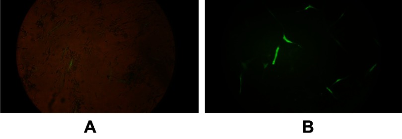
Figure 1 MSCs transduction with viral GFP. (A) Fluorescent and phase contrast images were merged. (B) Fluorescent image of the same frame. Low efficiency of transduction can be observed. Abbreviations: MSCs, mesenchymal stem cells; GFP, green fluorescent protein.
Figure 1 MSCs transduction with viral GFP. (A) Fluorescent and phase contrast images were merged. (B) Fluorescent image of the same frame. Low efficiency of transduction can be observed. Abbreviations: MSCs, mesenchymal stem cells; GFP, green fluorescent protein.CC BY-NC · PubMed Central
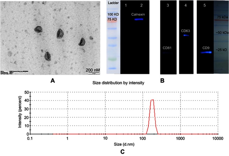
Figure 2 Characterization of exosomes. (A) TEM images of exosomes. Typical structure of exosomes was shown in this image (scale bar 200 nm). (B) Characterization of exosomes by ECL Western blotting. Exosome preparations were negative for endoplasmic reticulum marker (calnexin) in lane 1; lane 2 is the cell lysate which is positive for calnexin. The positive results for exosomal markers are displayed in lane 3 (CD81), lane 4 (CD63), and lane 5 (CD9). (C) Size distribution of exosomes was measured by a zeta-sizer. The peak diameter was about 100 nm. Abbreviations: ECL, enhanced chemiluminescence; TEM, transmission electron microscopy.
Figure 2 Characterization of exosomes. (A) TEM images of exosomes. Typical structure of exosomes was shown in this image (scale bar 200 nm). (B) Characterization of exosomes by ECL Western blotting. Exosome preparations were negative for endoplasmic reticulum marker (calnexin) in lane 1; lane 2 is the cell lysate which is positive for calnexin. The positive results for exosomal markers are displayed in lane 3 (CD81), lane 4 (CD63), and lane 5 (CD9). (C) Size distribution of exosomes was measured by a zeta-sizer. The peak diameter was about 100 nm. Abbreviations: ECL, enhanced chemiluminescence; TEM, transmission electron microscopy.CC BY-NC · PubMed Central
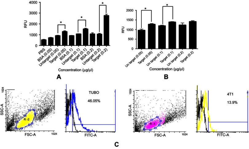
Figure 3 Fluorimetry and flow cytometry analysis of binding ability of targeted and untargeted exosomes. Binding of PKH-67 labeled targeted and untargeted exosomes to (A) coated HER2 protein and (B) HER2-positive SKBR3 cells with different concentrations. Fluorescence of each well was assessed by fluorimetry after removing unbound exosomes. BSA and PBS were used as controls. (B) Binding of PKH-67 labeled targeted and untargeted exosomes to HER2-positive SKBR3 cells. There were significant differences both in HER2 protein and HER2-positive cell immobilization; yet, no significant difference was observed at 0.2 µg/µL in binding to SKBR3 cells. (C) Binding and entrance of targeted labeled exosomes were quantified by flow cytometry. The results show a significant increase in binding of targeted exosomes to TUBO cells (46.05%) as HER2-positive murine cells compared to 4T1 cells (13.9%). Targeted exosomes showed significantly higher binding to HER2-positive cells compared to HER2-negative cells. Each error bar represents the mean±SD of three replicates. *P<0.05. Abbreviations: RFU, relative fluorescence units; BSA, bovine serum albumin.
Figure 3 Fluorimetry and flow cytometry analysis of binding ability of targeted and untargeted exosomes. Binding of PKH-67 labeled targeted and untargeted exosomes to (A) coated HER2 protein and (B) HER2-positive SKBR3 cells with different concentrations. Fluorescence of each well was assessed by fluorimetry after removing unbound exosomes. BSA and PBS were used as controls. (B) Binding of PKH-67 labeled targeted and untargeted exosomes to HER2-positive SKBR3 cells. There were significant differences both in HER2 protein and HER2-positive cell immobilization; yet, no significant difference was observed at 0.2 µg/µL in binding to SKBR3 cells. (C) Binding and entrance of targeted labeled exosomes were quantified by flow cytometry. The results show a significant increase in binding of targeted exosomes to TUBO cells (46.05%) as HER2-positive murine cells compared to 4T1 cells (13.9%). Targeted exosomes showed significantly higher binding to HER2-positive cells compared to HER2-negative cells. Each error bar represents the mean±SD of three replicates. *P<0.05. Abbreviations: RFU, relative fluorescence units; BSA, bovine serum albumin.CC BY-NC · PubMed Central
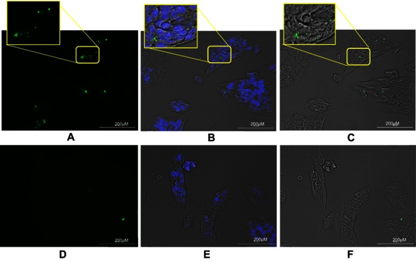
Figure 4 Binding of PKH-67 labeled exosomes to TUBO cells. Exosomes isolated from MSCs were labeled with PKH-67 and incubated with TUBO cells for 24 hours. (A) Fluorescent image of binding of PKH-67 targeted exosomes which showed significantly higher binding compared to PKH-67 untargeted exosomes. (B) Merged fluorescent image of DAPI, PKH-67, and phase contrast. (C) Merged bright field and PKH-67 fluorescent images of TUBO cells. (D) Fluorescent image of untargeted exosomes. (E) Merged DAPI, PKH-67, and bright field image of untargeted exosomes. (F) Merged bright field and PKH-67 fluorescent image of untargeted exosomes binding to TUBO cells. Labeled exosomes were colored green and nuclei colored by DAPI were observed in blue. Abbreviation: MSCs, mesenchymal stem cells.
Figure 4 Binding of PKH-67 labeled exosomes to TUBO cells. Exosomes isolated from MSCs were labeled with PKH-67 and incubated with TUBO cells for 24 hours. (A) Fluorescent image of binding of PKH-67 targeted exosomes which showed significantly higher binding compared to PKH-67 untargeted exosomes. (B) Merged fluorescent image of DAPI, PKH-67, and phase contrast. (C) Merged bright field and PKH-67 fluorescent images of TUBO cells. (D) Fluorescent image of untargeted exosomes. (E) Merged DAPI, PKH-67, and bright field image of untargeted exosomes. (F) Merged bright field and PKH-67 fluorescent image of untargeted exosomes binding to TUBO cells. Labeled exosomes were colored green and nuclei colored by DAPI were observed in blue. Abbreviation: MSCs, mesenchymal stem cells.CC BY-NC · PubMed Central
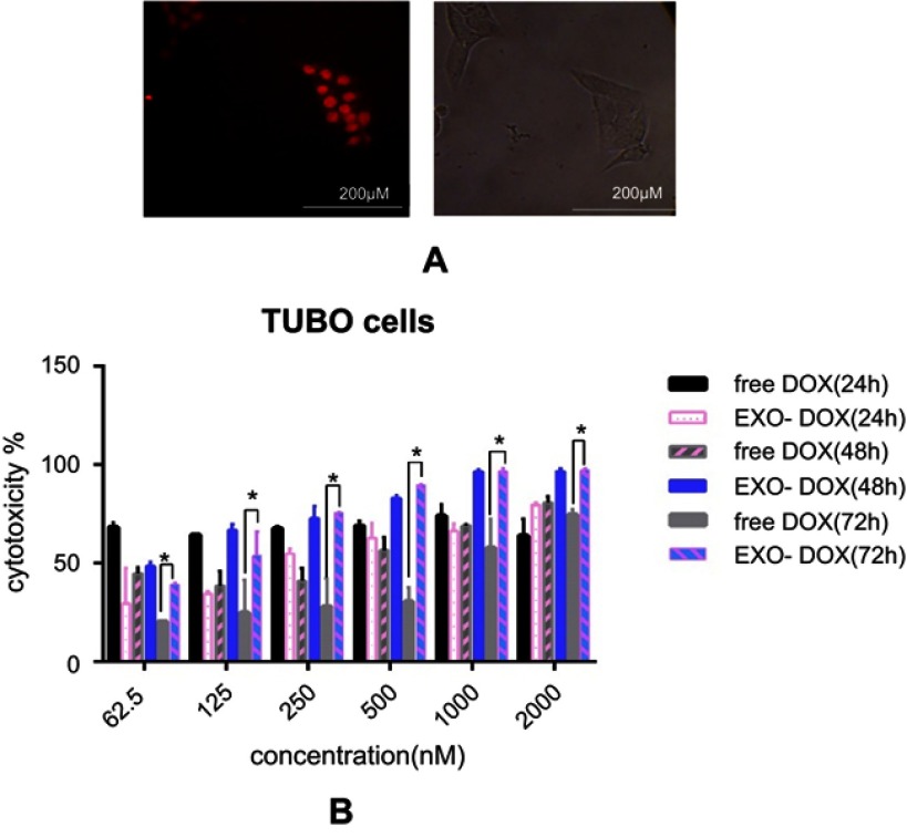
Figure 5 DOX delivery to TUBO cells and in vitro cellular cytotoxicity. (A) TUBO cells were incubated with 200 nM of exo-DOX for 4 hours. (B) TUBO cells were incubated with free DOX, targeted and untargeted exo-DOX for 24 hours, 48 hours, and 72 hours. Cell cytotoxicity was assessed using MTT assay. Exo-Dox showed similar cytotoxicity with free Dox after24 hours and 48 hours, with more cytotoxicity at 72 hours. The Student’s t-test was used to assess the significance of difference in cytotoxicity of the tested groups. Each error bar represents the mean±SD of three replicates. *P<0.05. Abbreviations: DOX, doxorubicin; exo-DOX, doxorubicin-loaded exosome.
Figure 5 DOX delivery to TUBO cells and in vitro cellular cytotoxicity. (A) TUBO cells were incubated with 200 nM of exo-DOX for 4 hours. (B) TUBO cells were incubated with free DOX, targeted and untargeted exo-DOX for 24 hours, 48 hours, and 72 hours. Cell cytotoxicity was assessed using MTT assay. Exo-Dox showed similar cytotoxicity with free Dox after24 hours and 48 hours, with more cytotoxicity at 72 hours. The Student’s t-test was used to assess the significance of difference in cytotoxicity of the tested groups. Each error bar represents the mean±SD of three replicates. *P<0.05. Abbreviations: DOX, doxorubicin; exo-DOX, doxorubicin-loaded exosome.CC BY-NC · PubMed Central
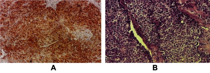
Figure 6 Immunohistochemistry of the TUBO tumor. (A) Brown colors indicate the HER2 receptor on the cell surface, (B) H&E staining of TUBO tumor.
Figure 6 Immunohistochemistry of the TUBO tumor. (A) Brown colors indicate the HER2 receptor on the cell surface, (B) H&E staining of TUBO tumor.CC BY-NC · PubMed Central
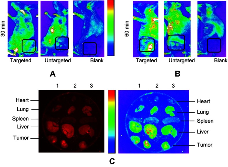
Figure 7 Biodistribution of exo-DOX in mice bearing TUBO tumor. In vivo targeting efficiency of targeted exo-DOX were assessed in a murine TUBO tumor model. These models were administered with a single 70-µg injection of targeted or untargeted exo-DOX. (A) In vivo fluorescent signals were recorded at 30 minutes and (B) 60 minutes post-injection. Fluorescence was detected at the tumor sites (black boxes), with gradual increasing in signal intensity at 60 minutes. Enhanced permeability and retention (EPR) effect causes accumulation of untargeted exo-DOX in tumor site, while it was lower in comparison to targeted exo-DOX. PBS was injected as blank and no fluorescence was detected in these mice. (C) Ex vivo fluorescent imaging of major organs of the tumor model after 60 minutes. Lanes 1, 2, and 3 were sequentially related to untargeted, targeted, and PBS intravenous injections. Accumulation of targeted and untargeted exo-DOX was mainly in the liver and lung, while, there was no accumulation in the heart after 60 minutes. Fluorescent intensity of untargeted exo-DOX was lower than targeted exo-DOX. Abbreviations: DOX, doxorubicin; exo-DOX, doxorubicin-loaded exosome DOX.
Figure 7 Biodistribution of exo-DOX in mice bearing TUBO tumor. In vivo targeting efficiency of targeted exo-DOX were assessed in a murine TUBO tumor model. These models were administered with a single 70-µg injection of targeted or untargeted exo-DOX. (A) In vivo fluorescent signals were recorded at 30 minutes and (B) 60 minutes post-injection. Fluorescence was detected at the tumor sites (black boxes), with gradual increasing in signal intensity at 60 minutes. Enhanced permeability and retention (EPR) effect causes accumulation of untargeted exo-DOX in tumor site, while it was lower in comparison to targeted exo-DOX. PBS was injected as blank and no fluorescence was detected in these mice. (C) Ex vivo fluorescent imaging of major organs of the tumor model after 60 minutes. Lanes 1, 2, and 3 were sequentially related to untargeted, targeted, and PBS intravenous injections. Accumulation of targeted and untargeted exo-DOX was mainly in the liver and lung, while, there was no accumulation in the heart after 60 minutes. Fluorescent intensity of untargeted exo-DOX was lower than targeted exo-DOX. Abbreviations: DOX, doxorubicin; exo-DOX, doxorubicin-loaded exosome DOX.CC BY-NC · PubMed Central
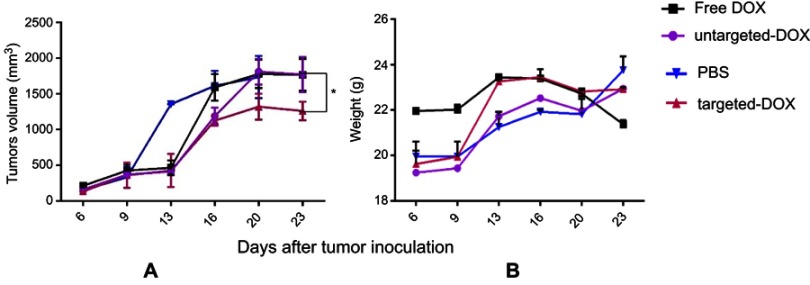
Figure 8 Therapeutic effects of exo-DOX in b6 nude mice bearing TUBO tumor. Mice bearing TUBO tumors (>100 mm3) were administrated intravenously with 1.5 mg/kg of different reagents (PBS, free DOX, targeted and untargeted exo-DOX), ttwice a week for a total of six injections. (A) Tumor growth was measured by digital caliper after each injection, and the antitumor activity of each group was assessed via tumor volume measurements. Targeted exo-DOX treatment dramatically reduced the rate of tumor growth compared to the control group (*P<0.05). At the experimental dose, free DOX and untargeted exo-DOX showed no significant effects on tumor growth (B) No significant difference was detected in body mass among the mice in these groups. Abbreviations: DOX, doxorubicin; exo-DOX, doxorubicin-loaded exosome DOX.
Figure 8 Therapeutic effects of exo-DOX in b6 nude mice bearing TUBO tumor. Mice bearing TUBO tumors (>100 mm3) were administrated intravenously with 1.5 mg/kg of different reagents (PBS, free DOX, targeted and untargeted exo-DOX), ttwice a week for a total of six injections. (A) Tumor growth was measured by digital caliper after each injection, and the antitumor activity of each group was assessed via tumor volume measurements. Targeted exo-DOX treatment dramatically reduced the rate of tumor growth compared to the control group (*P<0.05). At the experimental dose, free DOX and untargeted exo-DOX showed no significant effects on tumor growth (B) No significant difference was detected in body mass among the mice in these groups. Abbreviations: DOX, doxorubicin; exo-DOX, doxorubicin-loaded exosome DOX.CC BY-NC · PubMed Central

Verification

Claims the paper makes

The claims and statistics of this paper haven’t been checked yet.

Run the check — free, no account needed. It reads the open-access text and checks the sources it cites.

A few free checks a day · papers already verified open instantly · results are public

Claims other papers make about this one

Each is checked against this paper’s own text: does it say what the citing paper says it does?

Self-check

Percentages, sums and reported differences re-computed from the paper’s own numbers.

The self-check needs the paper’s full text.

Citations

Cites

Show all 2 references

Research works printed but not linked: 28

  1. [1]Hood JL. Post isolation modification of exosomes for nanomedicine applications. Nanomedicine. 2016; 11(13): 1745– 1756. doi: 10.2217/nnm-2016-0102 27348448 PMC4941124 · DOI
  2. [2]Zhao W, Song Zhuang X-RQ. Comparative study of the in vitro and in vivo characteristics of cationic and neutral liposomes. Int J Nanomedicine. 2011; 6: 3087. doi: 10.2147/IJN.S25646 22163162 PMC3235029 · DOI
  3. [3]Zhao J, Xie P, Ye C, et al. Outside-in synthesis of mesoporous silica/molybdenum disulfide nanoparticles for antitumor application. Chem Eng J. 2018; 351: 157– 168. doi: 10.1016/j.cej.2018.06.101 · DOI
  4. [4]Yang H, Zhao J, Wu C, Ye C, Zou D, Wang S. Facile synthesis of colloidal stable MoS2 nanoparticles for combined tumor therapy. Chem Eng J. 2018; 351: 548– 558. doi: 10.1016/j.cej.2018.06.100 · DOI
  5. [5]Guo B, Zhao J, Wu C, et al. One-pot synthesis of polypyrrole nanoparticles with tunable photothermal conversion and drug loading capacity. Colloids Surf B Biointerfaces. 2019; 177: 346– 355. doi: 10.1016/j.colsurfb.2019.02.016 30772669 · DOI
  6. [6]Yang Y, Chen Y, Zhang F, Zhao Q, Zhong H. Increased anti-tumour activity by exosomes derived from doxorubicin-treated tumour cells via heat stress. Int J Hyperthermia. 2015; 31(5): 498– 506. doi: 10.3109/02656736.2015.1036384 25955015 · DOI
  7. [8]Yuan Z, Kolluri KK, Gowers KH, Janes SM. TRAIL delivery by MSC-derived extracellular vesicles is an effective anticancer therapy. J Extracell Vesicles. 2017; 6(1): 1265291. doi: 10.1080/20013078.2017.1265291 28326166 PMC5328331 · DOI
  8. [9]Klopp AH, Spaeth EL, Dembinski JL, et al. Tumor irradiation increases the recruitment of circulating mesenchymal stem cells into the tumor microenvironment. Cancer Res. 2007; 67(24): 11687– 11695. doi: 10.1158/0008-5472.CAN-07-1406 18089798 PMC4329784 · DOI
  9. [10]Khodashenas Limoni S, Salimi F, Forouzandeh Moghaddam M. Designing pLEX-LAMP-DARPin lentiviral vector for exression of HER2 targeted DARPin on exosome surface. J Mazandaran Univ Med Sci. 2017; 27(151): 12– 23.
  10. [11]Limoni SK, Moghadam MF, Moazzeni SM, Gomari H, Salimi F. Engineered exosomes for targeted transfer of siRNA to HER2 positive breast cancer cells. #ABAB-D-18-00049R1, ed Appl Biochem Biotechnol. 2019;187(1):352–364. doi: 10.1007/s12010-018-2813-4 29951961 · DOI
  11. [12]Gomari H, Moghadam MF, Soleimani M. Targeted cancer therapy using engineered exosome as a natural drug delivery vehicle. Onco Targets Ther. 2018; 11: 5753– 5762. doi: 10.2147/OTT 30254468 PMC6140699 · DOI
  12. [13]Jalali SA, Sankian M, Tavakkol-Afshari J, Jaafari MR. Induction of tumor-specific immunity by multi-epitope rat HER2/neu-derived peptides encapsulated in LPD nanoparticles. Nanomedicine. 2012; 8(5): 692– 701. doi: 10.1016/j.nano.2011.09.010 22024191 · DOI
  13. [14]Razazan A, Behravan J, Arab A, et al. Conjugated nanoliposome with the HER2/neu-derived peptide GP2 as an effective vaccine against breast cancer in mice xenograft model. PLoS One. 2017; 12(10): e0185099. doi: 10.1371/journal.pone.0185099 29045460 PMC5646774 · DOI
  14. [15]Escrevente C, Keller S, Altevogt P, Costa J. Interaction and uptake of exosomes by ovarian cancer cells. BMC Cancer. 2011; 11(1): 108. doi: 10.1186/1471-2407-11-108 21439085 PMC3072949 · DOI
  15. [16]Tian Y, Li S, Song J, et al. A doxorubicin delivery platform using engineered natural membrane vesicle exosomes for targeted tumor therapy. Biomaterials. 2014; 35(7): 2383– 2390. doi: 10.1016/j.biomaterials.2013.11.083 24345736 · DOI
  16. [17]Tan CY, Lai RC, Wong W, Dan YY, Lim S-K, Ho HK. Mesenchymal stem cell-derived exosomes promote hepatic regeneration in drug-induced liver injury models. Stem Cell Res Ther. 2014; 5(3): 76. doi: 10.1186/scrt465 24915963 PMC4229780 · DOI · Paper page
  17. [18]Kim MS, Haney MJ, Zhao Y, et al. Development of exosome-encapsulated paclitaxel to overcome MDR in cancer cells. Nanomedicine. 2016; 12(3): 655– 664. doi: 10.1016/j.nano.2015.10.012 26586551 PMC4809755 · DOI · Paper page
  18. [19]Li W, Mu D, Tian F, et al. Exosomes derived from Rab27a‑overexpressing tumor cells elicit efficient induction of antitumor immunity. Mol Med Rep. 2013; 8(6): 1876– 1882. doi: 10.3892/mmr.2013.1738 24146068 · DOI
  19. [20]Bellavia D, Raimondo S, Calabrese G, et al. Interleukin 3-receptor targeted exosomes inhibit in vitro and in vivo chronic myelogenous leukemia cell growth. Theranostics. 2017; 7(5): 1333. doi: 10.7150/thno.17092 28435469 PMC5399597 · DOI · Paper page
  20. [21]Rivoltini L, Chiodoni C, Squarcina P, et al. TNF-related apoptosis-inducing ligand (TRAIL)–armed exosomes deliver proapoptotic signals to tumor site. Clin Cancer Res. 2016; 22(14): 3499– 3512. doi: 10.1158/1078-0432.CCR-15-2170 26944067 · DOI · Paper page
  21. [22]Antimisiaris S, Mourtas S, Marazioti A. Exosomes and exosome-inspired vesicles for targeted drug delivery. Pharmaceutics. 2018; 10(4): 218. doi: 10.3390/pharmaceutics10040218 PMC6321407 30404188 · DOI
  22. [23]Hadla M, Palazzolo S, Corona G, et al. Exosomes increase the therapeutic index of doxorubicin in breast and ovarian cancer mouse models. Nanomedicine. 2016; 11(18): 2431– 2441. doi: 10.2217/nnm-2016-0154 27558906 · DOI
  23. [24]Rudnick-Glick S, Corem-Salkmon E, Grinberg I, Margel S. Targeted drug delivery of near IR fluorescent doxorubicin-conjugated poly (ethylene glycol) bisphosphonate nanoparticles for diagnosis and therapy of primary and metastatic bone cancer in a mouse model. J Nanobiotechnology. 2016; 14(1): 80. doi: 10.1186/s12951-016-0160-6 27919267 PMC5139040 · DOI
  24. [25]MacDiarmid JA, Langova V, Bailey D, et al. Targeted doxorubicin delivery to brain tumors via minicells: proof of principle using dogs with spontaneously occurring tumors as a model. PLoS One. 2016; 11(4): e0151832. doi: 10.1371/journal.pone.0151832 27050167 PMC4822833 · DOI
  25. [26]Toffoli G, Hadla M, Corona G, et al. Exosomal doxorubicin reduces the cardiac toxicity of doxorubicin. Nanomedicine. 2015; 10(19): 2963– 2971. doi: 10.2217/nnm.15.118 26420143 · DOI
  26. [27]Ohno SI, Takanashi M, Sudo K, et al. Systemically injected exosomes targeted to EGFR deliver antitumor microRNA to breast cancer cells. Mol Ther. 2013; 21(1): 185– 191. doi: 10.1038/mt.2012.180 23032975 PMC3538304 · DOI · Paper page
  27. [29]Rovero S, Amici A, Di Carlo E, et al. DNA vaccination against rat her-2/Neu p185 more effectively inhibits carcinogenesis than transplantable carcinomas in transgenic BALB/c mice. J Immunol. 2000; 165(9): 5133– 5142. doi: 10.4049/jimmunol.165.9.5133 11046045 · DOI
  28. [30]Pierpaoli E, Viola V, Barucca A, Orlando F, Galli F, Provinciali M. Effect of annatto-tocotrienols supplementation on the development of mammary tumors in HER-2/neu transgenic mice. Carcinogenesis. 2013; 34(6): 1352– 1360. doi: 10.1093/carcin/bgt064 23430951 · DOI

Cited by

Sorted by citations

Metadata

Authors
Gomari, H., Forouzandeh Moghadam, M., Soleimani, M., Ghavami, M., & Khodashenas, S.
Venue
International Journal of Nanomedicine
Published
2019-07
Volume
Volume 14
Pages
5679-5690
ISSN
1178-2013
Cited by
111
References
2
Language
en

About these checks

Registry, retraction, Cites and statistics results are computed, not judged, with one exception: a printed reference that no index lists is read once by a model before it counts as a citation error. Claims are extracted by a model and kept only when their quote appears verbatim in the text; each cited source is then checked, with a model’s help, for whether it says what the claim relies on. Those verdicts can be wrong — read the claim’s quote and the cited source before relying on one.

Statistics and arithmetic are recomputed, not judged.

Confidence bands are shown only for fields calibrated on our annotated test set. None is calibrated yet, so no band is shown.

What counts as a claim ›How we measure confidence ›How we check this ›

Are you an author, or do you see a problem with this page? Request a review