

The DOI matches its Crossref record; 7 of the 7 references it cites were found in a registry or an index.
RNA Sequencing Identifies Dysregulation of the Human Pancreatic Islet Transcriptome by the Saturated Fatty Acid Palmitate
Abstract
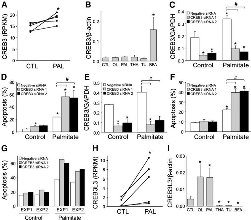
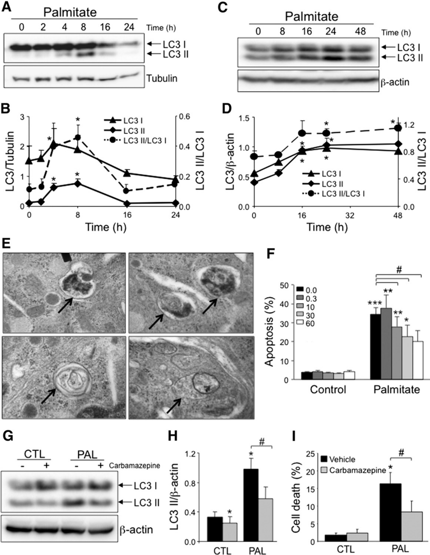
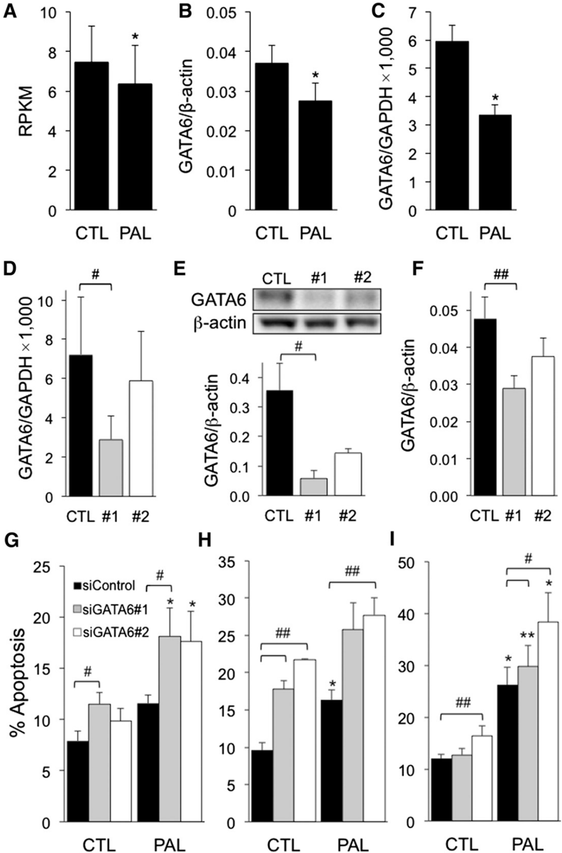
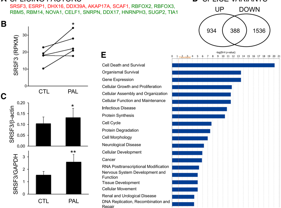
Pancreatic β-cell dysfunction and death are central in the pathogenesis of type 2 diabetes (T2D). Saturated fatty acids cause β-cell failure and contribute to diabetes development in genetically predisposed individuals. Here we used RNA sequencing to map transcripts expressed in five palmitate-treated human islet preparations, observing 1,325 modified genes. Palmitate induced fatty acid metabolism and endoplasmic reticulum (ER) stress. Functional studies identified novel mediators of adaptive ER stress signaling. Palmitate modified genes regulating ubiquitin and proteasome function, autophagy, and apoptosis. Inhibition of autophagic flux and lysosome function contributed to lipotoxicity. Palmitate inhibited transcription factors controlling β-cell phenotype, including PAX4 and GATA6. Fifty-nine T2D candidate genes were expressed in human islets, and 11 were modified by palmitate. Palmitate modified expression of 17 splicing factors and shifted alternative splicing of 3,525 transcripts. Ingenuity Pathway Analysis of modified transcripts and genes confirmed that top changed functions related to cell death. Database for Annotation, Visualization and Integrated Discovery (DAVID) analysis of transcription factor binding sites in palmitate-modified transcripts revealed a role for PAX4, GATA, and the ER stress response regulators XBP1 and ATF6. This human islet transcriptome study identified novel mechanisms of palmitate-induced β-cell dysfunction and death. The data point to cross talk between metabolic stress and candidate genes at the β-cell level.
Overview
The claims and statistics of this paper haven’t been checked yet.
Run the check — free, no account needed. It reads the open-access text and checks the sources it cites.
Integrity signals
Registry
Cites
Statistics
Figures

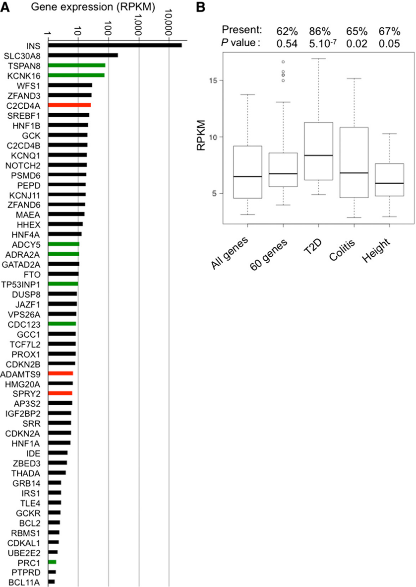
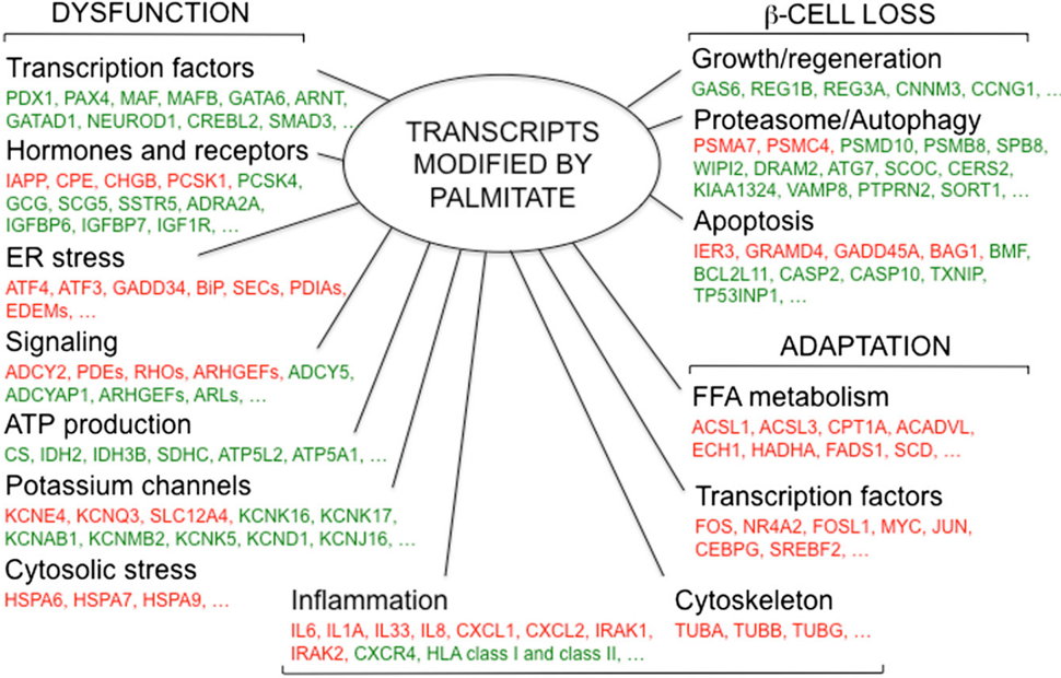
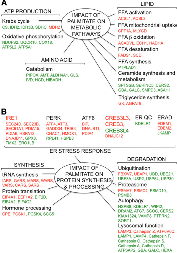
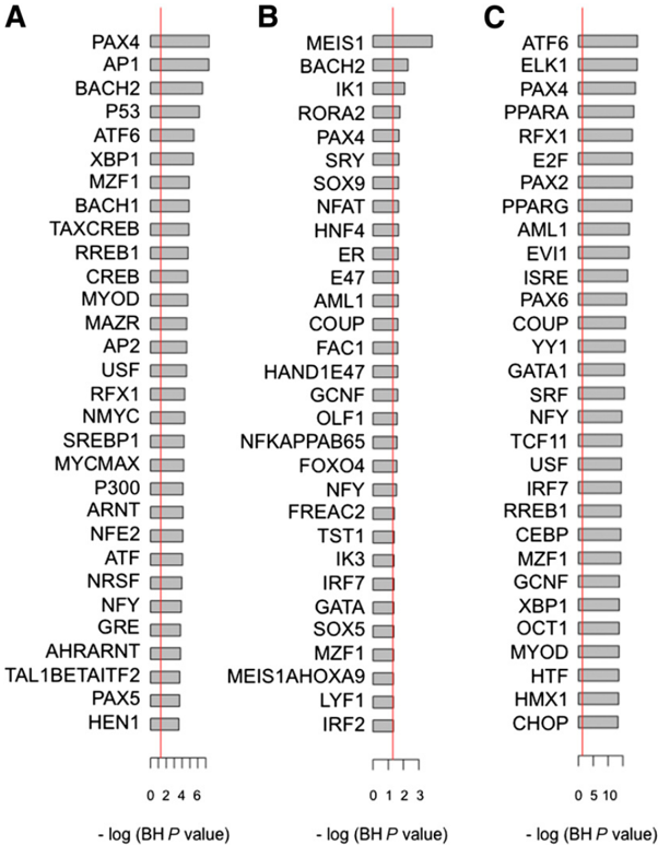
Verification
Claims the paper makes
The claims and statistics of this paper haven’t been checked yet.
Run the check — free, no account needed. It reads the open-access text and checks the sources it cites.
Claims other papers make about this one
Each is checked against this paper’s own text: does it say what the citing paper says it does?
The check wasn’t confident either way, so it gives no verdict — never disputed.
In the citing paper
“For example, SQSTM1 upregulation, and at least transient increases in the amount of SQSTM1, is seen in some situations where there is an increase in autophagic flux [571-573].”
From Guidelines for the use and interpretation of assays for monitoring autophagy (4th edition) 1 (2021) · cites this paper as [571]
Experimental — accuracy not yet measured
Self-check
Percentages, sums and reported differences re-computed from the paper’s own numbers.
The self-check needs the paper’s full text.
Citations
Cites
Mapping and quantifying mammalian transcriptomes by RNA-Seq
Mortazavi, A., Williams, B. A., McCue, K., Schaeffer, L., & Wold, B. (2008) · Nature Methods · doi:10.1038/nmeth.1226 · Cited by 13,696
Landscape of transcription in human cells
Djebali, S., Davis, C. A., Merkel, A., Dobin, A., Lassmann, T., Mortazavi, A., Tanzer, A., Lagarde, J., Lin, W., Schlesinger, F., et al. (2012) · Nature · doi:10.1038/nature11233 · Cited by 5,187
Self-citationTranscriptome genetics using second generation sequencing in a Caucasian population
Montgomery, S. B., Sammeth, M., Gutierrez-Arcelus, M., Lach, R. P., Ingle, C., Nisbett, J., Guigo, R., & Dermitzakis, E. T. (2010) · Nature · doi:10.1038/nature08903 · Cited by 797
Self-citationNCBI Reference Sequences: current status, policy and new initiatives
Pruitt, K. D., Tatusova, T., Klimke, W., & Maglott, D. R. (2009) · Nucleic Acids Research · doi:10.1093/nar/gkn721 · Cited by 736 · cited 1×
“Mapped reads were used to quantify transcripts from the RefSeq reference database (35), using the Flux Capacitor (http://flux.”
MethodRegulation of Insulin Secretion in Human Pancreatic Islets
Rorsman, P., & Braun, M. (2013) · Annual Review of Physiology · doi:10.1146/annurev-physiol-030212-183754 · Cited by 599
Volkmar, M., Dedeurwaerder, S., Cunha, D. A., Ndlovu, M. N., Defrance, M., Deplus, R., Calonne, E., Volkmar, U., Igoillo‐Esteve, M., Naamane, N., et al. (2012) · The EMBO Journal · doi:10.1038/emboj.2011.503 · Cited by 420 · cited 2×
“Epigenetic alterations, such as changes in DNA methylation, have been described in pancreatic islets from T2D patients (29) and may explain part of the missing heritability.”
BackgroundSelf-citationMitochondrial Networking Protects β-Cells From Nutrient-Induced Apoptosis
Molina, A. J. A., Wikstrom, J. D., Stiles, L., Las, G., Mohamed, H., Elorza, A., Walzer, G., Twig, G., Katz, S., Corkey, B. E., et al. (2009) · Diabetes · doi:10.2337/db07-1781 · Cited by 381
Research works printed but not linked: 60
- Cnop, 2010, The long lifespan and low turnover of human islet beta cells estimated by mathematical modelling of lipofuscin accumulation, Diabetologia, 53, 321, 10.1007/s00125-009-1562-x · DOI
- Cnop, 2012, Endoplasmic reticulum stress, obesity and diabetes, Trends Mol Med, 18, 59, 10.1016/j.molmed.2011.07.010 · DOI
- Cnop, 2008, Fatty acids and glucolipotoxicity in the pathogenesis of Type 2 diabetes, Biochem Soc Trans, 36, 348, 10.1042/BST0360348 · DOI
- Gargani, 2013, Adaptive changes of human islets to an obesogenic environment in the mouse, Diabetologia, 56, 350, 10.1007/s00125-012-2775-y · DOI
- Winzell, 2007, Temporal and dietary fat content-dependent islet adaptation to high-fat feeding-induced glucose intolerance in mice, Metabolism, 56, 122, 10.1016/j.metabol.2006.09.008 · DOI
- Kashyap, 2003, A sustained increase in plasma free fatty acids impairs insulin secretion in nondiabetic subjects genetically predisposed to develop type 2 diabetes, Diabetes, 52, 2461, 10.2337/diabetes.52.10.2461 · DOI · Paper page
- Sako, 1990, A 48-hour lipid infusion in the rat time-dependently inhibits glucose-induced insulin secretion and B cell oxidation through a process likely coupled to fatty acid oxidation, Endocrinology, 127, 1580, 10.1210/endo-127-4-1580 · DOI
- Cnop, 2001, Inverse relationship between cytotoxicity of free fatty acids in pancreatic islet cells and cellular triglyceride accumulation, Diabetes, 50, 1771, 10.2337/diabetes.50.8.1771 · DOI
- Kelpe, 2003, Palmitate inhibition of insulin gene expression is mediated at the transcriptional level via ceramide synthesis, J Biol Chem, 278, 30015, 10.1074/jbc.M302548200 · DOI
- Carlsson, 1999, Sodium palmitate induces partial mitochondrial uncoupling and reactive oxygen species in rat pancreatic islets in vitro, Endocrinology, 140, 3422, 10.1210/endo.140.8.6908 · DOI
- Hoppa, 2009, Chronic palmitate exposure inhibits insulin secretion by dissociation of Ca(2+) channels from secretory granules, Cell Metab, 10, 455, 10.1016/j.cmet.2009.09.011 · DOI · Paper page
- Shimabukuro, 1998, Lipoapoptosis in β-cells of obese prediabetic fa/fa rats. Role of serine palmitoyltransferase overexpression, J Biol Chem, 273, 32487, 10.1074/jbc.273.49.32487 · DOI
- Cunha, 2008, Initiation and execution of lipotoxic ER stress in pancreatic β-cells, J Cell Sci, 121, 2308, 10.1242/jcs.026062 · DOI
- Kharroubi, 2004, Free fatty acids and cytokines induce pancreatic β-cell apoptosis by different mechanisms: role of nuclear factor-κB and endoplasmic reticulum stress, Endocrinology, 145, 5087, 10.1210/en.2004-0478 · DOI
- Karaskov, 2006, Chronic palmitate but not oleate exposure induces endoplasmic reticulum stress, which may contribute to INS-1 pancreatic β-cell apoptosis, Endocrinology, 147, 3398, 10.1210/en.2005-1494 · DOI
- Laybutt, 2007, Endoplasmic reticulum stress contributes to beta cell apoptosis in type 2 diabetes, Diabetologia, 50, 752, 10.1007/s00125-006-0590-z · DOI · Paper page
- Eizirik, 2008, The role for endoplasmic reticulum stress in diabetes mellitus, Endocr Rev, 29, 42, 10.1210/er.2007-0015 · DOI · Paper page
- Cnop, 2007, Selective inhibition of eukaryotic translation initiation factor 2 α dephosphorylation potentiates fatty acid-induced endoplasmic reticulum stress and causes pancreatic β-cell dysfunction and apoptosis, J Biol Chem, 282, 3989, 10.1074/jbc.M607627200 · DOI
- Cunha, 2012, Death protein 5 and p53-upregulated modulator of apoptosis mediate the endoplasmic reticulum stress-mitochondrial dialog triggering lipotoxic rodent and human β-cell apoptosis, Diabetes, 61, 2763, 10.2337/db12-0123 · DOI · Paper page
- Busch, 2002, Expression profiling of palmitate- and oleate-regulated genes provides novel insights into the effects of chronic lipid exposure on pancreatic β-cell function, Diabetes, 51, 977, 10.2337/diabetes.51.4.977 · DOI
- Busch, 2005, Increased fatty acid desaturation and enhanced expression of stearoyl coenzyme A desaturase protects pancreatic β-cells from lipoapoptosis, Diabetes, 54, 2917, 10.2337/diabetes.54.10.2917 · DOI
- Xiao, 2001, The effect of chronic exposure to fatty acids on gene expression in clonal insulin-producing cells: studies using high density oligonucleotide microarray, Endocrinology, 142, 4777, 10.1210/endo.142.11.8483 · DOI
- Malmgren, 2013, Coordinate changes in histone modifications, mRNA levels, and metabolite profiles in clonal INS-1 832/13 β-cells accompany functional adaptations to lipotoxicity, J Biol Chem, 288, 11973, 10.1074/jbc.M112.422527 · DOI · Paper page
- Bikopoulos, 2008, Ex vivo transcriptional profiling of human pancreatic islets following chronic exposure to monounsaturated fatty acids, J Endocrinol, 196, 455, 10.1677/JOE-07-0174 · DOI
- Marioni, 2008, RNA-seq: an assessment of technical reproducibility and comparison with gene expression arrays, Genome Res, 18, 1509, 10.1101/gr.079558.108 · DOI · Paper page
- Eizirik, 2012, The human pancreatic islet transcriptome: expression of candidate genes for type 1 diabetes and the impact of pro-inflammatory cytokines, PLoS Genet, 8, e1002552, 10.1371/journal.pgen.1002552 · DOI · Paper page
- Nica, 2013, Cell-type, allelic, and genetic signatures in the human pancreatic β cell transcriptome, Genome Res, 23, 1554, 10.1101/gr.150706.112 · DOI · Paper page
- Morris, 2012, Large-scale association analysis provides insights into the genetic architecture and pathophysiology of type 2 diabetes, Nat Genet, 44, 981, 10.1038/ng.2383 · DOI · Paper page
- Marchetti, 2007, The endoplasmic reticulum in pancreatic beta cells of type 2 diabetes patients, Diabetologia, 50, 2486, 10.1007/s00125-007-0816-8 · DOI · Paper page
- Igoillo-Esteve, 2010, Palmitate induces a pro-inflammatory response in human pancreatic islets that mimics CCL2 expression by beta cells in type 2 diabetes, Diabetologia, 53, 1395, 10.1007/s00125-010-1707-y · DOI
- Ling, 1996, Prolonged exposure of human β cells to elevated glucose levels results in sustained cellular activation leading to a loss of glucose regulation, J Clin Invest, 98, 2805, 10.1172/JCI119108 · DOI
- Asfari, 1992, Establishment of 2-mercaptoethanol-dependent differentiated insulin-secreting cell lines, Endocrinology, 130, 167, 10.1210/endo.130.1.1370150 · DOI
- Cardozo, 2005, Cytokines downregulate the sarcoendoplasmic reticulum pump Ca2+ ATPase 2b and deplete endoplasmic reticulum Ca2+, leading to induction of endoplasmic reticulum stress in pancreatic β-cells, Diabetes, 54, 452, 10.2337/diabetes.54.2.452 · DOI
- Harrow, 2012, GENCODE: the reference human genome annotation for The ENCODE Project, Genome Res, 22, 1760, 10.1101/gr.135350.111 · DOI · Paper page
- Bernstein, 2012, An integrated encyclopedia of DNA elements in the human genome, Nature, 489, 57, 10.1038/nature11247 · DOI · Paper page
- Kharroubi, 2003, Expression of adiponectin receptors in pancreatic β cells, Biochem Biophys Res Commun, 312, 1118, 10.1016/j.bbrc.2003.11.042 · DOI
- Cardozo, 2003, IL-1β and IFN-γ induce the expression of diverse chemokines and IL-15 in human and rat pancreatic islet cells, and in islets from pre-diabetic NOD mice, Diabetologia, 46, 255, 10.1007/s00125-002-1017-0 · DOI
- Moore, 2012, Use of RNA interference to investigate cytokine signal transduction in pancreatic β cells, Methods Mol Biol, 820, 179, 10.1007/978-1-61779-439-1_11 · DOI
- Cnop, 2012, Central role and mechanisms of β-cell dysfunction and death in friedreich ataxia-associated diabetes, Ann Neurol, 72, 971, 10.1002/ana.23698 · DOI
- Taneera, 2012, A systems genetics approach identifies genes and pathways for type 2 diabetes in human islets, Cell Metab, 16, 122, 10.1016/j.cmet.2012.06.006 · DOI
- Anderson, 2011, Meta-analysis identifies 29 additional ulcerative colitis risk loci, increasing the number of confirmed associations to 47, Nat Genet, 43, 246, 10.1038/ng.764 · DOI
- Aulchenko, 2009, Predicting human height by Victorian and genomic methods, Eur J Hum Genet, 17, 1070, 10.1038/ejhg.2009.5 · DOI
- DenBoer, 2005, Luman is capable of binding and activating transcription from the unfolded protein response element, Biochem Biophys Res Commun, 331, 113, 10.1016/j.bbrc.2005.03.141 · DOI
- Zhang, 2006, Endoplasmic reticulum stress activates cleavage of CREBH to induce a systemic inflammatory response, Cell, 124, 587, 10.1016/j.cell.2005.11.040 · DOI · Paper page
- Ebato, 2008, Autophagy is important in islet homeostasis and compensatory increase of β cell mass in response to high-fat diet, Cell Metab, 8, 325, 10.1016/j.cmet.2008.08.009 · DOI · Paper page
- Masini, 2009, Autophagy in human type 2 diabetes pancreatic beta cells, Diabetologia, 52, 1083, 10.1007/s00125-009-1347-2 · DOI
- Las, 2011, Fatty acids suppress autophagic turnover in β-cells, J Biol Chem, 286, 42534, 10.1074/jbc.M111.242412 · DOI
- Hidvegi, 2010, An autophagy-enhancing drug promotes degradation of mutant α1-antitrypsin Z and reduces hepatic fibrosis, Science, 329, 229, 10.1126/science.1190354 · DOI
- Lee, 2011, Downregulation of mitochondrial lon protease impairs mitochondrial function and causes hepatic insulin resistance in human liver SK-HEP-1 cells, Diabetologia, 54, 1437, 10.1007/s00125-011-2074-z · DOI
- Gray, 2006, Activation of the extracellular calcium-sensing receptor initiates insulin secretion from human islets of Langerhans: involvement of protein kinases, J Endocrinol, 190, 703, 10.1677/joe.1.06891 · DOI
- Prentki, 2013, Metabolic signaling in fuel-induced insulin secretion, Cell Metab, 18, 162, 10.1016/j.cmet.2013.05.018 · DOI · Paper page
- Lango Allen, 2012, GATA6 haploinsufficiency causes pancreatic agenesis in humans, Nat Genet, 44, 20, 10.1038/ng.1035 · DOI
- Chen, 2010, Lack of TXNIP protects against mitochondria-mediated apoptosis but not against fatty acid-induced ER stress-mediated β-cell death, Diabetes, 59, 440, 10.2337/db09-0949 · DOI
- Oslowski, 2012, Thioredoxin-interacting protein mediates ER stress-induced β cell death through initiation of the inflammasome, Cell Metab, 16, 265, 10.1016/j.cmet.2012.07.005 · DOI
- Morán, 2012, Human β cell transcriptome analysis uncovers lncRNAs that are tissue-specific, dynamically regulated, and abnormally expressed in type 2 diabetes, Cell Metab, 16, 435, 10.1016/j.cmet.2012.08.010 · DOI
- Tang, 2009, mRNA-Seq whole-transcriptome analysis of a single cell, Nat Methods, 6, 377, 10.1038/nmeth.1315 · DOI · Paper page
- Sultan, 2008, A global view of gene activity and alternative splicing by deep sequencing of the human transcriptome, Science, 321, 956, 10.1126/science.1160342 · DOI · Paper page
- van Delft, 2012, RNA-Seq provides new insights in the transcriptome responses induced by the carcinogen benzo[a]pyrene, Toxicol Sci, 130, 427, 10.1093/toxsci/kfs250 · DOI
- Cunha, 2009, Glucagon-like peptide-1 agonists protect pancreatic β-cells from lipotoxic endoplasmic reticulum stress through upregulation of BiP and JunB, Diabetes, 58, 2851, 10.2337/db09-0685 · DOI
- Chen, 2013, Carbamazepine as a novel small molecule corrector of trafficking-impaired ATP-sensitive potassium channels identified in congenital hyperinsulinism, J Biol Chem, 288, 20942, 10.1074/jbc.M113.470948 · DOI
Cited by
Sorted by citations
Guidelines for the use and interpretation of assays for monitoring autophagy (3rd edition).
Klionsky, D. J., Abdelmohsen, K., Abe, A., Abedin, M. J., Abeliovich, H., Acevedo Arozena, A., Adachi, H., Adams, C. M., Adams, P. D., Adeli, K., et al. (2016) · Autophagy · doi:10.1080/15548627.2015.1100356 · Cited by 5,894
Self-citationGuidelines for the use and interpretation of assays for monitoring autophagy (4th edition) 1
Klionsky, D. J., Abdel-Aziz, A. K., Abdelfatah, S., Abdellatif, M., Abdoli, A., Abel, S., Abeliovich, H., Abildgaard, M. H., Abudu, Y. P., Acevedo-Arozena, A., et al. (2021) · Autophagy · doi:10.1080/15548627.2020.1797280 · Cited by 2,897
Molecular mechanisms of ROS production and oxidative stress in diabetes
Newsholme, P., Cruzat, V. F., Keane, K. N., Carlessi, R., & de Bittencourt, P. I. H. (2016) · Biochemical Journal · doi:10.1042/bcj20160503c · Cited by 878
Viral infections in type 1 diabetes mellitus — why the β cells?
Op de Beeck, A., & Eizirik, D. L. (2016) · Nature Reviews Endocrinology · doi:10.1038/nrendo.2016.30 · Cited by 291
Self-citationType 2 Diabetes: The Pathologic Basis of Reversible β-Cell Dysfunction
White, M. G., Shaw, J. A. M., & Taylor, R. (2016) · Diabetes Care · doi:10.2337/dc16-0619 · Cited by 174
Taylor, R., Barnes, A. C., Hollingsworth, K. G., Irvine, K. M., Solovyova, A. S., Clark, L., Kelly, T., Martin-Ruiz, C., Romeres, D., Koulman, A., et al. (2023) · Clinical Science · doi:10.1042/cs20230586 · Cited by 71
Metadata
- Authors
- Cnop, M., Abdulkarim, B., Bottu, G., Cunha, D. A., Igoillo-Esteve, M., Masini, M., Turatsinze, J.-V., Griebel, T., Villate, O., Santin, I., Bugliani, M., Ladriere, L., Marselli, L., McCarthy, M. I., Marchetti, P., Sammeth, M., & Eizirik, D. L.
- Affiliations
- Laboratory of Experimental Medicine, ULB Center for Diabetes Research, Université Libre de Bruxelles, Brussels, Belgium
- Department of Endocrinology and Metabolism, University of Pisa, Pisa, Italy
- Functional Bioinformatics, Centre Nacional d'Anàlisi Genòmica, Barcelona, Spain
- Oxford Centre for Diabetes, Endocrinology and Metabolism, Churchill Hospital, Oxford, U.K.
- Venue
- Diabetes
- Publisher
- American Diabetes Association
- Published
- 2014-05-15
- Volume
- 63
- Issue
- 6
- Pages
- 1978-1993
- ISSN
- 0012-1797
- 1939-327X
- Cited by
- 250
- References
- 7
- Language
- en
About these checks
Registry, retraction, Cites and statistics results are computed, not judged, with one exception: a printed reference that no index lists is read once by a model before it counts as a citation error. Claims are extracted by a model and kept only when their quote appears verbatim in the text; each cited source is then checked, with a model’s help, for whether it says what the claim relies on. Those verdicts can be wrong — read the claim’s quote and the cited source before relying on one.
Statistics and arithmetic are recomputed, not judged.
Confidence bands are shown only for fields calibrated on our annotated test set. None is calibrated yet, so no band is shown.
What counts as a claim ›How we measure confidence ›How we check this ›
Are you an author, or do you see a problem with this page? Request a review
