

The DOI matches its Crossref record; 3 of the 3 references it cites were found in a registry or an index.
Refining the domain architecture model of the replication origin firing factor Treslin/TICRR
Open accessPublished version of a preprint
Abstract
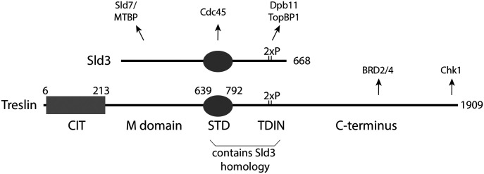
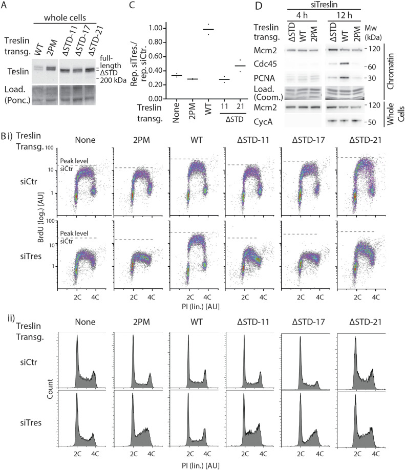
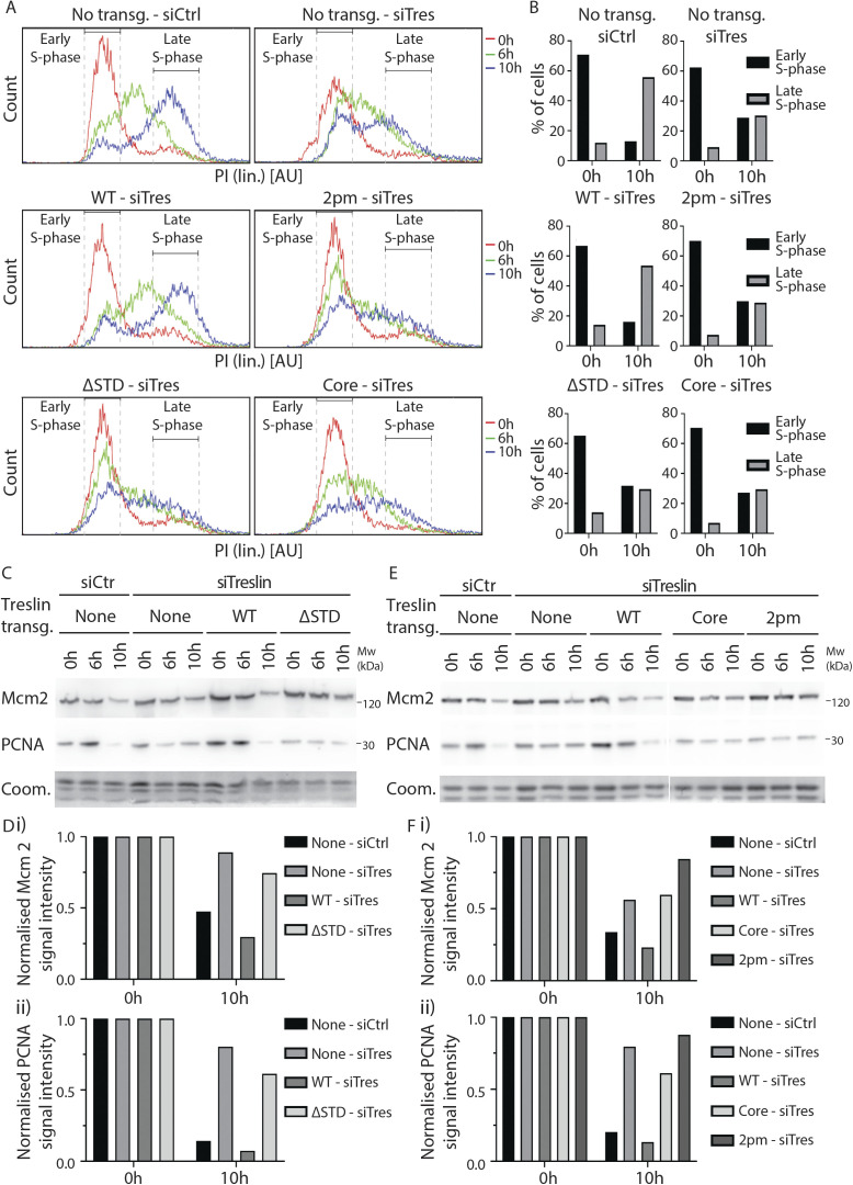
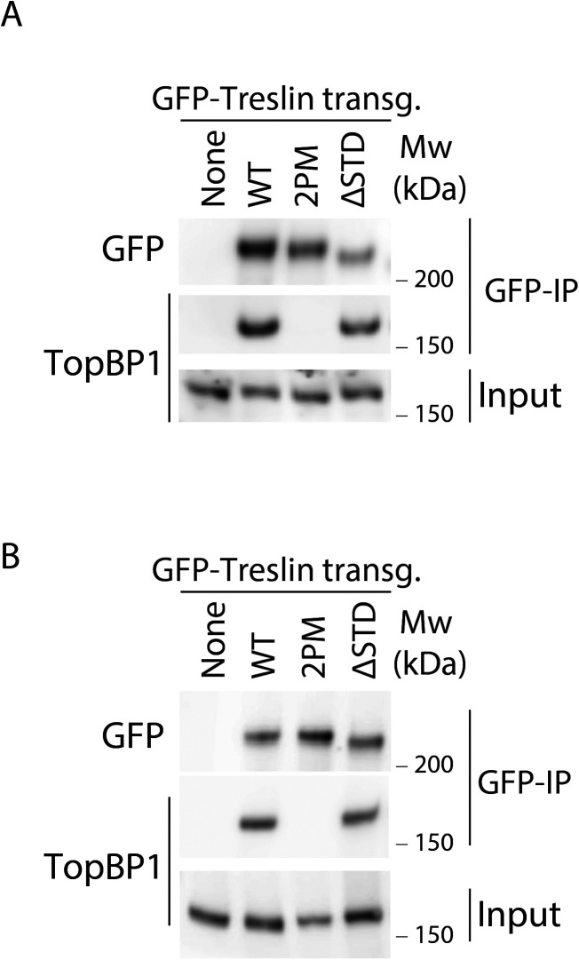
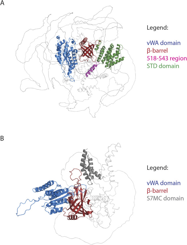
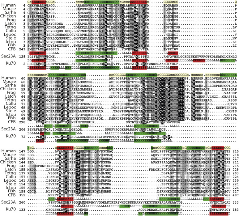
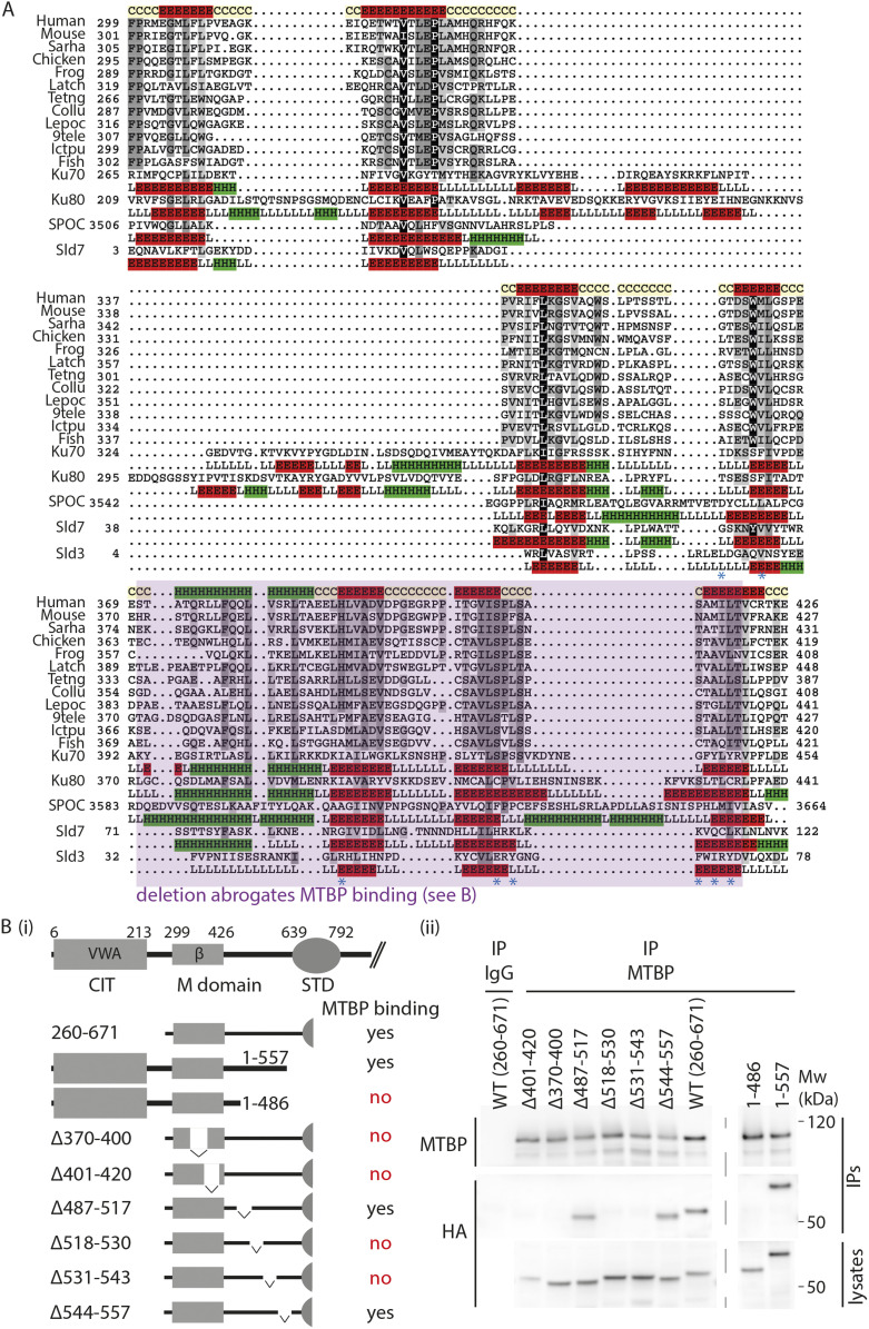
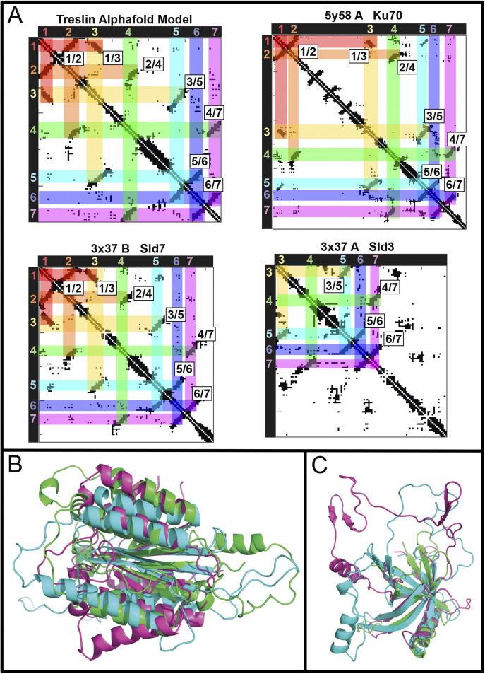
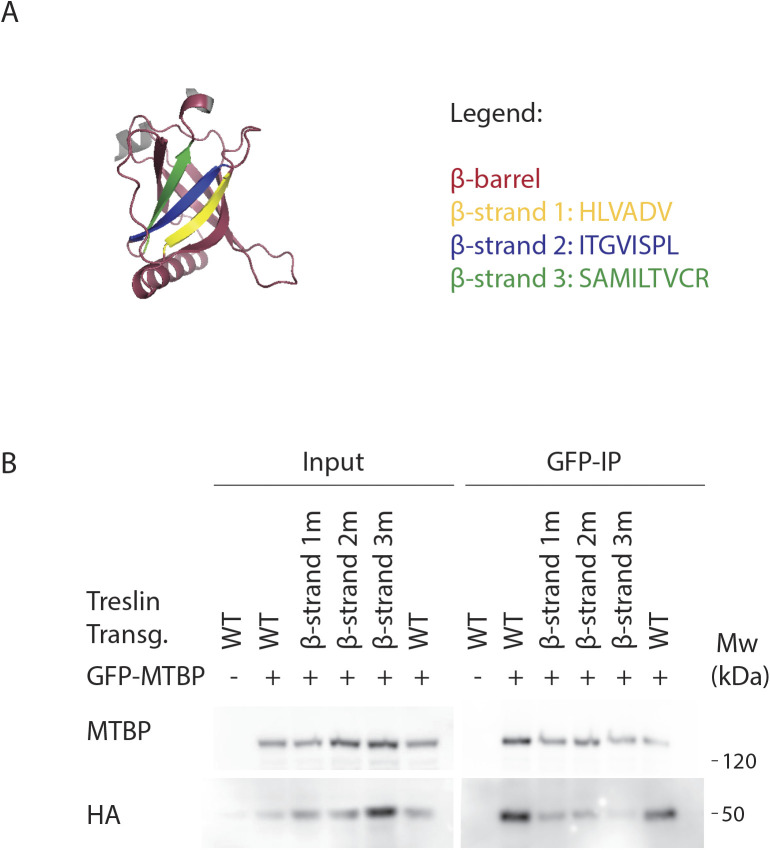
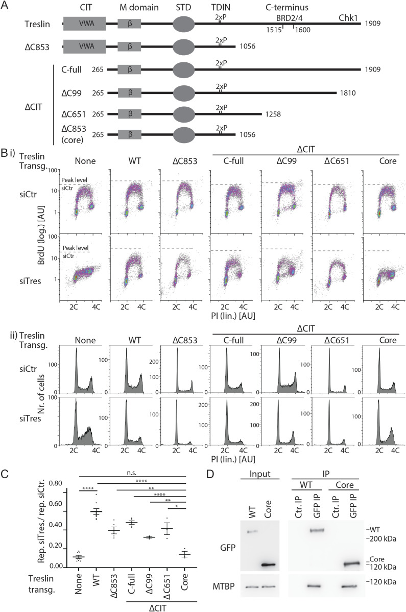
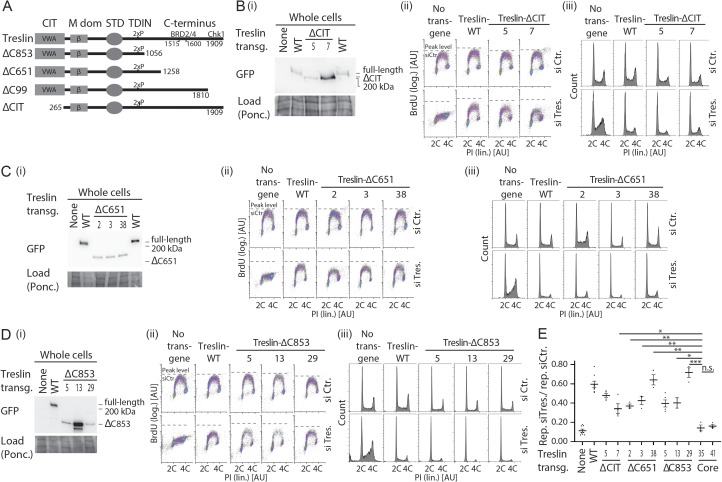
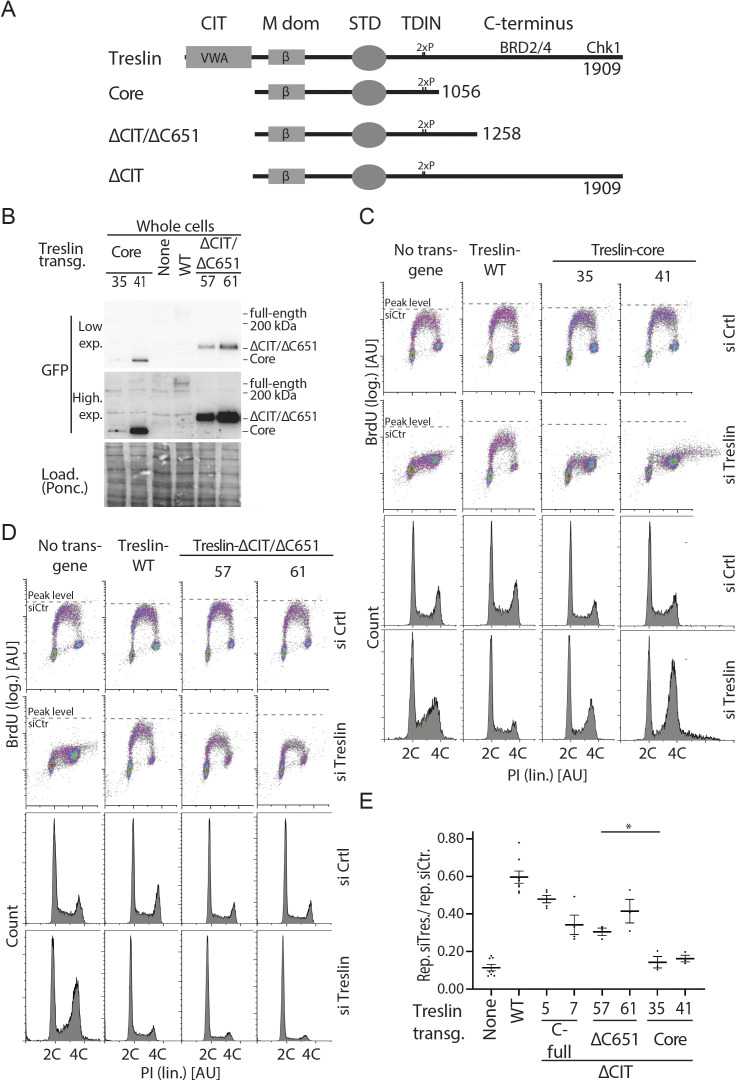
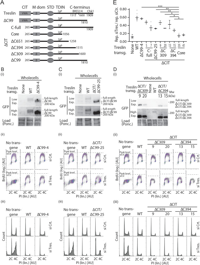
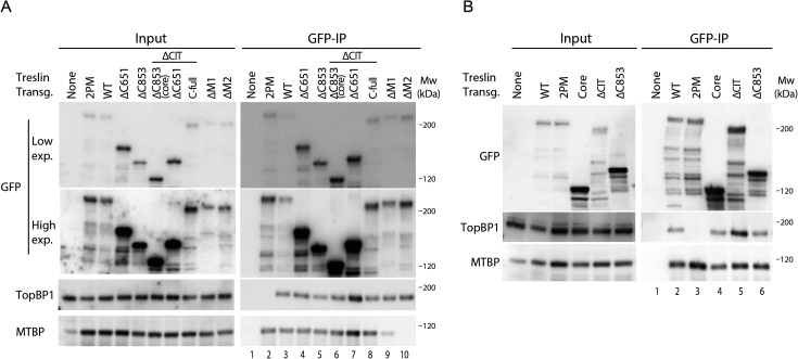
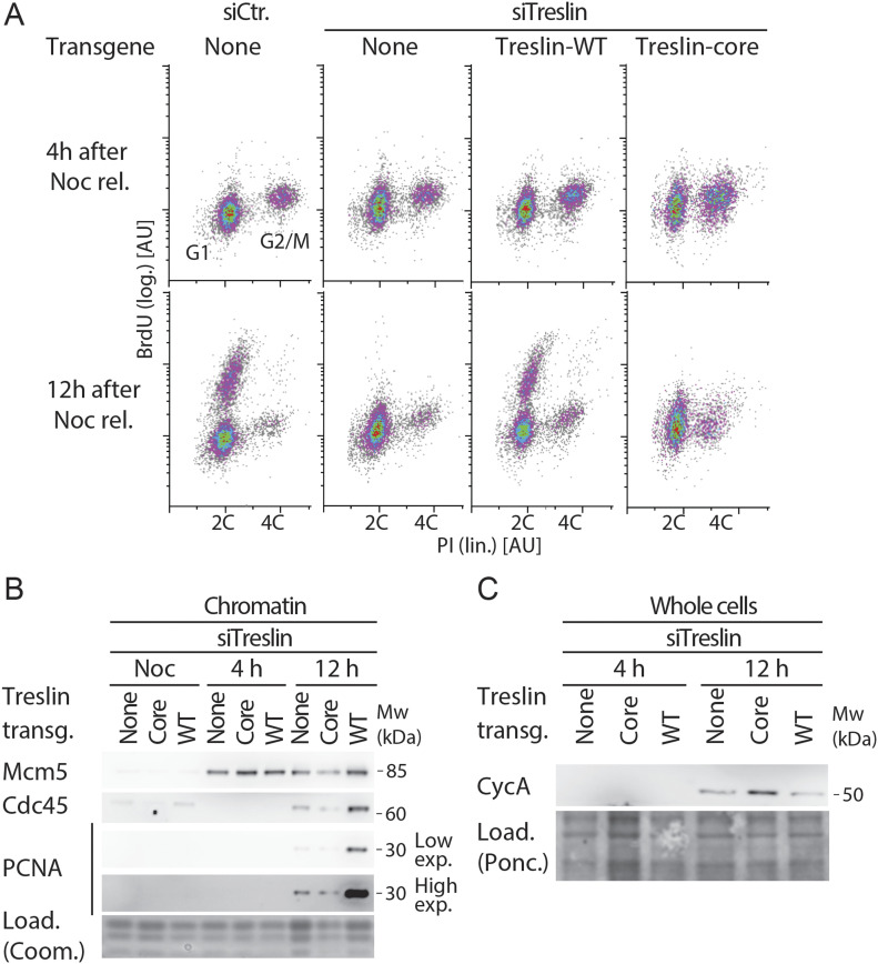
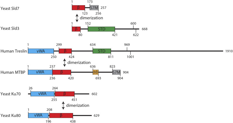
Faithful genome duplication requires appropriately controlled replication origin firing. The metazoan origin firing regulation hub Treslin/TICRR and its yeast orthologue Sld3 share the Sld3-Treslin domain and the adjacent TopBP1/Dpb11 interaction domain. We report a revised domain architecture model of Treslin/TICRR. Protein sequence analyses uncovered a conserved Ku70-homologous β-barrel fold in the Treslin/TICRR middle domain (M domain) and in Sld3. Thus, the Sld3-homologous Treslin/TICRR core comprises its three central domains, M domain, Sld3-Treslin domain, and TopBP1/Dpb11 interaction domain, flanked by non-conserved terminal domains, the CIT (conserved in Treslins) and the C terminus. The CIT includes a von Willebrand factor type A domain. Unexpectedly, MTBP, Treslin/TICRR, and Ku70/80 share the same N-terminal domain architecture, von Willebrand factor type A and Ku70-like β-barrels, suggesting a common ancestry. Binding experiments using mutants and the Sld3–Sld7 dimer structure suggest that the Treslin/Sld3 and MTBP/Sld7 β-barrels engage in homotypic interactions, reminiscent of Ku70-Ku80 dimerization. Cells expressing Treslin/TICRR domain mutants indicate that all Sld3-core domains and the non-conserved terminal domains fulfil important functions during origin firing in human cells. Thus, metazoa-specific and widely conserved molecular processes cooperate during metazoan origin firing.
Overview
The claims and statistics of this paper haven’t been checked yet.
Run the check — free, no account needed. It reads the open-access text and checks the sources it cites.
Integrity signals
Registry
Cites
Statistics
Figures
Verification
Claims the paper makes
The claims and statistics of this paper haven’t been checked yet.
Run the check — free, no account needed. It reads the open-access text and checks the sources it cites.
Self-check
Percentages, sums and reported differences re-computed from the paper’s own numbers.
The self-check needs the paper’s full text.
Citations
Cites
Highly accurate protein structure prediction with AlphaFold
Jumper, J., Evans, R., Pritzel, A., Green, T., Figurnov, M., Ronneberger, O., Tunyasuvunakool, K., Bates, R., Žídek, A., Potapenko, A., et al. (2021) · Nature · doi:10.1038/s41586-021-03819-2 · Cited by 43,764
Highly accurate protein structure prediction for the human proteome
Tunyasuvunakool, K., Adler, J., Wu, Z., Green, T., Zielinski, M., Žídek, A., Bridgland, A., Cowie, A., Meyer, C., Laydon, A., et al. (2021) · Nature · doi:10.1038/s41586-021-03828-1 · Cited by 3,025
Accurate De Novo Prediction of Protein Contact Map by Ultra-Deep Learning Model
Wang, S., Sun, S., Li, Z., Zhang, R., & Xu, J. (2017) · PLOS Computational Biology · doi:10.1371/journal.pcbi.1005324 · Cited by 931
Research works printed but not linked: 60
- 10.1038/s41467-017-02389-0 · DOI
- 10.1007/s004120050399 · DOI
- 10.3390/genes10030199 · DOI
- 10.1016/j.cub.2011.05.057 · DOI
- 10.1126/science.1237448 · DOI
- 10.1016/j.cell.2017.12.008 · DOI
- 10.15252/embj.201593552 · DOI
- 10.1101/gr.183699.114 · DOI
- 10.1038/nature25787 · DOI
- 10.1074/jbc.M110.190843 · DOI
- 10.1016/S0959-440X(96)80056-X · DOI
- 10.1073/pnas.0911500106 · DOI
- 10.1038/s41598-021-83287-w · DOI
- 10.1093/nar/gkr367 · DOI · Paper page
- 10.1016/j.molcel.2014.12.003 · DOI
- 10.1016/j.cell.2011.06.012 · DOI
- 10.1038/nsmb.2543 · DOI
- 10.1016/S0968-0004(00)89105-7 · DOI
- 10.1007/978-1-0716-0270-6_3 · DOI
- 10.1016/j.molcel.2009.12.030 · DOI
- 10.1016/j.str.2014.07.001 · DOI
- 10.1107/S1399004715010457 · DOI
- 10.1038/emboj.2009.184 · DOI
- 10.1006/jmbi.1999.3091 · DOI
- 10.1093/emboj/20.8.2097 · DOI
- 10.1371/journal.pbio.2006767 · DOI
- 10.1083/jcb.201102003 · DOI
- 10.1016/j.cell.2009.12.049 · DOI
- 10.1091/mbc.E17-07-0448 · DOI
- 10.1073/pnas.1418334111 · DOI
- 10.1038/nature09377 · DOI
- 10.1074/jbc.M102245200 · DOI
- 10.1038/emboj.2011.404 · DOI
- 10.15252/embr.201744206 · DOI
- 10.1128/MCB.00344-17 · DOI
- 10.1101/gad.1883410 · DOI
- 10.1006/jmbi.2000.4042 · DOI
- 10.1038/ncomms10208 · DOI
- 10.1016/S0065-3233(00)54007-8 · DOI
- 10.1016/j.cell.2009.10.015 · DOI
- 10.1016/j.celrep.2016.09.013 · DOI
- 10.1101/gr.099655.109 · DOI
- 10.1016/j.cub.2010.05.021 · DOI
- 10.1101/gad.306464.117 · DOI
- 10.1101/gad.1860310 · DOI
- 10.1093/nar/gki408 · DOI
- 10.1186/1471-2105-6-108 · DOI
- 10.1371/journal.pgen.1002136 · DOI
- 10.1016/j.cub.2011.11.038 · DOI
- 10.1038/nature05465 · DOI
- 10.1093/bioinformatics/btr484 · DOI
- 10.1038/35088000 · DOI
- 10.1091/mbc.E02-05-0259 · DOI
- 10.1093/nar/gkj161 · DOI
- 10.1038/nature14285 · DOI
- 10.1016/j.molcel.2016.11.017 · DOI
- 10.1038/nature09373 · DOI
- 10.1038/nature05432 · DOI
- 10.1007/s00412-014-0500-y · DOI
- 10.1126/science.280.5363.593 · DOI
Metadata
- Authors
- Ferreira, P., Sanchez-Pulido, L., Marko, A., Ponting, C. P., & Boos, D.
- Affiliations
- University of Duisburg-Essen (DE)
- Medical Research Council Human Genetics Unit, Institute of Genetics and Cancer, University of Edinburgh, Edinburgh, UK
- Institute of Genetics and Cancer (GB)
- Corresponding author
- Boos, Dominik
- Venue
- Life Science Alliance
- Publisher
- Life Science Alliance, LLC
- Published
- 2022-01-28
- Volume
- 5
- Issue
- 5
- Pages
- e202101088
- ISSN
- 2575-1077 (ISSN-L)
- PMC
- PMC8807876
- OpenAlex
- W4225260126
- Cited by
- 9
- References
- 3
- Citations by year
- 2026: 2 · 2025: 1 · 2024: 1 · 2022: 5
- FWCI
- 0.57
- Type
- journal article
- Language
- en
- Open access
- gold
- doi.org
- Licence
- Locations
- Life Science Alliance (publishedVersion, cc-by)
- bioRxiv (Cold Spring Harbor Laboratory) (acceptedVersion, cc-by-nc)
- PubMed Central (publishedVersion, cc-by)
- PubMed (publishedVersion)
- DOAJ (DOAJ: Directory of Open Access Journals) (submittedVersion, cc-by-sa)
- Edinburgh Research Explorer (University of Edinburgh) (submittedVersion, cc-by)
- Funders
- NRW Rückkehrerförderprogramm
- Topics
- Life Sciences › Biochemistry, Genetics and Molecular Biology › Molecular Biology › DNA Repair Mechanisms
- Life Sciences › Biochemistry, Genetics and Molecular Biology › Molecular Biology › Genomics and Chromatin Dynamics
- Life Sciences › Biochemistry, Genetics and Molecular Biology › Cell Biology › Microtubule and mitosis dynamics
- Keywords
- Sld3, replication origin firing, domain architecture, beta-barrel fold, von Willebrand factor type A domain, MTBP, whole-genome duplication
- MeSH terms
- Carrier Proteins, DNA Replication, DNA-Binding Proteins, Humans, Protein Conformation, Structure-Activity Relationship, Replication Origin, Cell Cycle Proteins, Cyclin-Dependent Kinases
About these checks
Registry, retraction, Cites and statistics results are computed, not judged, with one exception: a printed reference that no index lists is read once by a model before it counts as a citation error. Claims are extracted by a model and kept only when their quote appears verbatim in the text; each cited source is then checked, with a model’s help, for whether it says what the claim relies on. Those verdicts can be wrong — read the claim’s quote and the cited source before relying on one.
Statistics and arithmetic are recomputed, not judged.
Confidence bands are shown only for fields calibrated on our annotated test set. None is calibrated yet, so no band is shown.
What counts as a claim ›How we measure confidence ›How we check this ›
Are you an author, or do you see a problem with this page? Request a review
How to cite
Cite CoChat for the checks on this page, and the paper for its own findings.
- This page
- CoChat. (2026). Citation check: Refining the domain architecture model of the replication origin firing factor Treslin/TICRR. https://app.cochat.ai/papers/10.26508/lsa.202101088
- The paper
- Ferreira, P., Sanchez-Pulido, L., Marko, A., Ponting, C. P., & Boos, D. (2022). Refining the domain architecture model of the replication origin firing factor Treslin/TICRR. Life Science Alliance. https://doi.org/10.26508/lsa.202101088
