

The DOI matches its Crossref record; 2 of the 2 references it cites were found in a registry or an index.
Size and surface charge of gold nanoparticles determine absorption across intestinal barriers and accumulation in secondary target organs after oral administration
Overview
The claims and statistics of this paper haven’t been checked yet.
Run the check — free, no account needed. It reads the open-access text and checks the sources it cites.
Integrity signals
Registry
Cites
Statistics
Figures
Verification
Claims the paper makes
The claims and statistics of this paper haven’t been checked yet.
Run the check — free, no account needed. It reads the open-access text and checks the sources it cites.
Claims other papers make about this one
Each is checked against this paper’s own text: does it say what the citing paper says it does?
In the citing paper
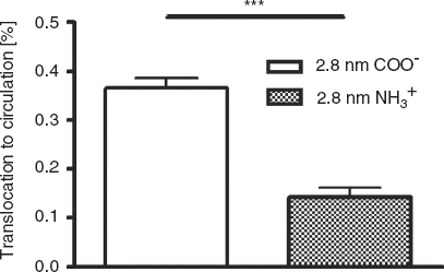
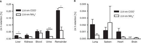
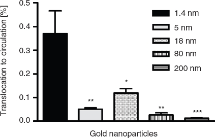
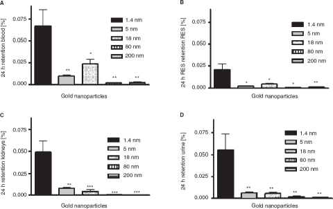
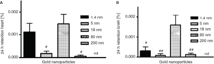
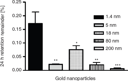
“Also, radiolabelled Au NPs in different sizes (1.4-200 nm) exposed by intra-oesophageal instillation into healthy adult female rats resulted in detectable NPs (ng/g organ) in the stomach, small intestine, liver, spleen, kidney, heart, lung, blood and brain after 24 h as measured by gamma-spectroscopy, with the highest accumulation in secondary organs being for the smallest particles, while the 18”
From Particle toxicology and health - where are we? (2019) · cites this paper as [132]
In the citing paper
“For example, in vivo studies using radiolabelled gold NPs in rats indicated that different exposure routes led to different biodistributions of the NPs, which is most likely a result of different biomolecule adsorption and thus different bioprocessing signals [131, 132].”
From Particle toxicology and health - where are we? (2019) · cites this paper as [132]
Self-check
Percentages, sums and reported differences re-computed from the paper’s own numbers.
The self-check needs the paper’s full text.
Citations
Cites
Nanoparticle Uptake by the Rat Gastrointestinal Mucosa: Quantitation and Particle Size Dependency
Jani, P., Halbert, G. W., Langridge, J., & Florence, A. T. (1990) · Journal of Pharmacy and Pharmacology · doi:10.1111/j.2042-7158.1990.tb07033.x · Cited by 920
Hariharan, S., Bhardwaj, V., Bala, I., Sitterberg, J., Bakowsky, U., & Ravi Kumar, M. N. V. (2006) · Pharmaceutical Research · doi:10.1007/s11095-005-8418-y · Cited by 257
Research works printed but not linked: 50
- 10.1016/j.addr.2009.03.009 · DOI · Paper page
- 10.1021/la0528450 · DOI
- 10.1021/ja050793v · DOI
- 10.1007/s11095-009-9965-4 · DOI
- 10.1016/j.biomaterials.2009.01.057 · DOI · Paper page
- 10.1016/j.toxlet.2009.04.017 · DOI
- 10.1002/smll.200400093 · DOI
- 10.1016/j.jconrel.2006.08.013 · DOI · Paper page
- 10.1016/j.ejps.2005.04.015 · DOI
- 10.1016/j.nano.2008.08.001 · DOI
- 10.1016/j.ijpharm.2006.10.043 · DOI
- 10.3109/10611869509015936 · DOI
- Florence AT. 2007. Pharmaceutical nanotechnology: More than size. Ten topics for research. Int J Pharm 339(1–2):1–2. · DOI
- 10.1016/S0168-3659(99)00237-0 · DOI
- 10.1016/S0074-7696(08)61346-7 · DOI
- 10.1289/ehp.8006 · DOI
- 10.1016/j.biomaterials.2010.01.065 · DOI · Paper page
- 10.3109/10611869409015904 · DOI
- 10.1002/jps.1143 · DOI
- Hirn S, Semmler-Behnke M, Schleh C, Wenk A, Lipka J, Schäffler M, Takenaka S, Möller W, Schmid G, Simon U, Kreyling WG. Particle size-dependent and surface charge-dependent biodistribution of gold nanoparticles after intravenous administration. Eur J Pharm Biopharm. 2010 Dec 31. [Epub ahead of print]
- 10.1023/A:1011981610840 · DOI
- Jani P, Halbert GW, Langridge J, Florence AT. 1989. The uptake and translocation of latex nanospheres and microspheres after oral administration to rats. J Pharm Pharmacol 41(12):809–812. · DOI
- 10.3109/10611869609046266 · DOI
- 10.1021/la100810z · DOI
- 10.1021/nn9011187 · DOI
- 10.1073/pnas.0805135105 · DOI · Paper page
- 10.1021/la0484725 · DOI
- Nassimi M Schleh C Lauenstein HD Hussein R Hoymann HG Koch W Pohlmann G Krug N Sewald K Rittinghausen S Braun A Muller-Goymann C. 2010. A toxicological evaluation of inhaled solid lipid nanoparticles used as a potential drug delivery system for the lung. Eur J Pharm Biopharm 75(2):107–116. · DOI
- 10.1080/08958370903005769 · DOI
- Oeff K, Konig A. 1955. Das Blutvolumen einiger Rattenorgane und ihre Restblutmenge nach entbluten bzw. durchspülung - Bestimmung mit P-32-markierten Erythrocyten. Naunyn-Schmiedebergs Archiv Fur Experimentelle Pathologie Und Pharmakologi 226(1):98–102.
- 10.1002/smll.200900466 · DOI
- 10.1002/smll.200700378 · DOI
- 10.1242/dev.110.4.1001 · DOI
- 10.1016/j.jaut.2009.11.006 · DOI
- 10.1016/j.vaccine.2004.11.007 · DOI
- 10.1016/j.jconrel.2009.10.032 · DOI
- 10.1016/j.biomaterials.2007.10.026 · DOI
- Sass W, Dreyer HP, Seifert J. 1990. Rapid insorption of small particles in the gut. Am J Gastroenterol 85(3):255–260.
- 10.1039/b713631p · DOI
- 10.1159/000171250 · DOI
- Seifert J, Haraszti B, Sass W. 1996. The influence of age and particle number on absorption of polystyrene particles from the rat gut. J Anat 189(Pt 3):483–486.
- 10.1002/smll.200800922 · DOI
- 10.1002/smll.200800995 · DOI
- 10.1016/j.ijpharm.2007.06.049 · DOI
- 10.1016/j.colsurfb.2008.02.013 · DOI
- 10.1039/b712170a · DOI
- 10.1246/cl.1996.1033 · DOI
- 10.1016/j.ijpharm.2004.12.028 · DOI
- 10.1021/la0497200 · DOI
- 10.1021/ja910675v · DOI
Cited by
Sorted by citations
Diverse Applications of Nanomedicine
Pelaz, B., Alexiou, C., Alvarez-Puebla, R. A., Alves, F., Andrews, A. M., Ashraf, S., Balogh, L. P., Ballerini, L., Bestetti, A., Brendel, C., et al. (2017) · ACS Nano · doi:10.1021/acsnano.6b06040 · Cited by 1,350
Particle toxicology and health - where are we?
Riediker, M., Zink, D., Kreyling, W., Oberdörster, G., Elder, A., Graham, U., Lynch, I., Duschl, A., Ichihara, G., Ichihara, S., et al. (2019) · Particle and Fibre Toxicology · doi:10.1186/s12989-019-0302-8 · Cited by 237
Self-citationIn Vitro Three‐Dimensional Liver Models for Nanomaterial DNA Damage Assessment
Llewellyn, S. V., Niemeijer, M., Nymark, P., Moné, M. J., van de Water, B., Conway, G. E., Jenkins, G. J. S., & Doak, S. H. (2021) · Small · doi:10.1002/smll.202006055 · Cited by 41
Metadata
- Authors
- Schleh, C., Semmler-Behnke, M., Lipka, J., Wenk, A., Hirn, S., Schäffler, M., Schmid, G., Simon, U., & Kreyling, W. G.
- Venue
- Nanotoxicology
- Publisher
- Informa UK Limited
- Published
- 2011-02-10
- Volume
- 6
- Issue
- 1
- Pages
- 36-46
- ISSN
- 1743-5390
- 1743-5404
- PMC
- PMC3267526
- Cited by
- 325
- References
- 2
- Language
- en
About these checks
Registry, retraction, Cites and statistics results are computed, not judged, with one exception: a printed reference that no index lists is read once by a model before it counts as a citation error. Claims are extracted by a model and kept only when their quote appears verbatim in the text; each cited source is then checked, with a model’s help, for whether it says what the claim relies on. Those verdicts can be wrong — read the claim’s quote and the cited source before relying on one.
Statistics and arithmetic are recomputed, not judged.
Confidence bands are shown only for fields calibrated on our annotated test set. None is calibrated yet, so no band is shown.
What counts as a claim ›How we measure confidence ›How we check this ›
Are you an author, or do you see a problem with this page? Request a review
How to cite
Cite CoChat for the checks on this page, and the paper for its own findings.
- This page
- CoChat. (2026). Citation check: Size and surface charge of gold nanoparticles determine absorption across intestinal barriers and accumulation in secondary target organs after oral administration. https://app.cochat.ai/papers/10.3109/17435390.2011.552811
- The paper
- Schleh, C., Semmler-Behnke, M., Lipka, J., Wenk, A., Hirn, S., Schäffler, M., Schmid, G., Simon, U., & Kreyling, W. G. (2012). Size and surface charge of gold nanoparticles determine absorption across intestinal barriers and accumulation in secondary target organs after oral administration. Nanotoxicology. https://doi.org/10.3109/17435390.2011.552811
