

Cellular abnormalities in depression: evidence from postmortem brain tissue
Open accessThe DOI matches its Crossref record; 91 of the 91 references it cites were found in a registry or an index.
Abstract
During the past two decades, in vivo neuroimaging studies have permitted significant insights into the general location of dysfunctional brain regions in depression. In parallel and often intersecting ways, neuroanatomical, pharmacological, and biochemical studies of postmortem brain tissue are permitting new insights into the pathophysiology of depression. In addition to long-recognized neurochemical abnormalities in depression, novel studies at the microscopic level support the contention that mood disorders are associated with abnormalities in cell morphology and distribution. In the past 6 years, cell-counting studies have identified changes in the density and size of both neurons and glia in a number of frontolimbic brain regions, including dorsolateral prefrontal, orbitofrontal, and anterior cingulate cortex, and the amygdala and hippocampus. Convergence of cellular changes at the microscopic level with neuroimaging changes detected in vivo provides a compelling integration of clinical and basic research for disentangling the pathophysiology of depression. The ultimate integration of these two research approaches will occur with premortem longitudinal clinical studies on well-characterized patients linked to postmortem studies of the same subjects.
Overview
The claims and statistics of this paper haven’t been checked yet.
Free: it reads the open-access text and checks the first 5 claims that cite a source we can check. Create a free account to check every claim.
Integrity signals
Registry
Cites
Statistics
Figures


Verification
Claims the paper makes
The claims and statistics of this paper haven’t been checked yet.
Free: it reads the open-access text and checks the first 5 claims that cite a source we can check. Create a free account to check every claim.
Claims other papers make about this one
Each is checked against this paper’s own text: does it say what the citing paper says it does?
In the citing paper
“Cell-counting studies incriminated neuronal and glial abnormalities for lower gray matter volume in MDD [37], while histopathological findings showed lower prefrontal synapse-related gene expression hinting at lower synaptic density in MDD [36].”
From Preliminary evidence for preserved synaptic density in late-life depression (2024) · cites this paper as [37]
Self-check
Percentages, sums and reported differences re-computed from the paper’s own numbers.
The self-check needs the paper’s full text.
Citations
Cites
Chronic antidepressant treatment increases neurogenesis in adult rat hippocampus
Malberg, J. E., Eisch, A. J., Nestler, E. J., & Duman, R. S. (2000) · The Journal of Neuroscience · doi:10.1523/jneurosci.20-24-09104.2000 · Cited by 3,111
Subgenual prefrontal cortex abnormalities in mood disorders
Drevets, W. C., Price, J. L., Simpson, J. R., Todd, R. D., Reich, T., Vannier, M., & Raichle, M. E. (1997) · Nature · doi:10.1038/386824a0 · Cited by 2,693 · cited 1×
“Glial reductions reported in this study may in fact reflect a true loss of glial cells since the neuroimaging studies in the same cortical region show a reduction in the volume of gray matter.(84) There are unquestionable limitations to the use of postmortem brain tissue in studying the mood disorders.”
ResultAlexander, G. E., Crutcher, M. D., & DeLong, M. R. (1990) · PubMed · Cited by 2,680
(1991) · Progress in Brain Research · doi:10.1016/s0079-6123(08)62678-3 · Cited by 1,961
Hippocampal atrophy in recurrent major depression
Sheline, Y. I., Wang, P. W., Gado, M. H., Csernansky, J. G., & Vannier, M. W. (1996) · Proceedings of the National Academy of Sciences · doi:10.1073/pnas.93.9.3908 · Cited by 1,932
Hippocampal Volume Reduction in Major Depression
Bremner, J. D., Narayan, M., Anderson, E. R., Staib, L. H., Miller, H. L., & Charney, D. S. (2000) · American Journal of Psychiatry · doi:10.1176/ajp.157.1.115 · Cited by 1,670
Glial reduction in the subgenual prefrontal cortex in mood disorders
Öngür, D., Drevets, W. C., & Price, J. L. (1998) · Proceedings of the National Academy of Sciences · doi:10.1073/pnas.95.22.13290 · Cited by 1,525 · cited 8×
“For example, a 24% to 41% reduction in the number of a general population of Nissl-stained glial cells is reported in the subgenual region of the anterior cingulate cortex (ventral part of Brodmann’s area 24) in a small subgroup of patients with familial MDD and familial BPD, as compared to control subjects.(1) Howeve…”
BackgroundResultInfluentialRajkowska, G., Miguel-Hidalgo, J. J., Wei, J., Dilley, G., Pittman, S. D., Meltzer, H. Y., Overholser, J. C., Roth, B. L., & Stockmeier, C. A. (1999) · Biological Psychiatry · doi:10.1016/s0006-3223(99)00041-4 · Cited by 1,477 · cited 3×
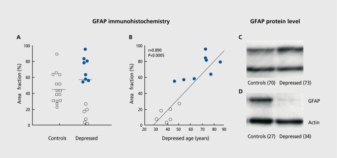
“An illustration of the pathology of glial cells found in the dorsolateral prefrontal cortex in MDD.(5,68) Reductions in the glial fibrillary acidic protein (GFAP) immunoreactive astroglia are found in a subgroup of young adults with major depression as compared to aged-matched control subjects and older subjects with…”
BackgroundSelf-citationSheline, Y. I., Sanghavi, M., Mintun, M. A., & Gado, M. H. (1999) · The Journal of Neuroscience · doi:10.1523/jneurosci.19-12-05034.1999 · Cited by 1,382
Neuroimaging studies of mood disorders
Drevets, W. C. (2000) · Biological Psychiatry · doi:10.1016/s0006-3223(00)01020-9 · Cited by 1,313
Research works printed but not linked: 4
- Sanacora G., 2000, Crit RevNeurobiol., 14, 23
- Stockmeier CA., 2003, Biol Psychiatry., 53, 198
- Chen B., 2001, BiolPsychia try., 50, 260
- 10.1002/1099-1166(200012)15:12<1138::aid-gps266>3.0.co;2-7 · doi:10.1002/1099-1166(200012)15:12<1138::aid-gps266>3.0.co;2-7
Cited by
Brites, D., & Fernandes, A. (2015) · Frontiers in Cellular Neuroscience · doi:10.3389/fncel.2015.00476 · Cited by 599
“In this article, we discuss the role of neuroinflammation in the emergence of depression, namely dynamic alterations in the status of microglia response to stimulation, and how their activation phenotypes may have an etiological role in neurodegeneneration, in particular in depressive-like behavior.”
BackgroundMolecular pathways of major depressive disorder converge on the synapse
Fries, G. R., Saldana, V. A., Finnstein, J., & Rein, T. (2023) · Molecular Psychiatry · doi:10.1038/s41380-022-01806-1 · Cited by 504
“In parallel, postmortem studies have reported changes in the density and size of neurons and glia in several brain regions of patients [10] along with reduced expression of pre- and postsynaptic genes [11, 12].”
BackgroundRadley, J., Morilak, D., Viau, V., & Campeau, S. (2015) · Neuroscience & Biobehavioral Reviews · doi:10.1016/j.neubiorev.2015.06.018 · Cited by 339
Maitra, M., Mitsuhashi, H., Rahimian, R., Chawla, A., Yang, J., Fiori, L. M., Davoli, M. A., Perlman, K., Aouabed, Z., Mash, D. C., et al. (2023) · Nature Communications · doi:10.1038/s41467-023-38530-5 · Cited by 159
Gonadal steroid regulation of mood: The lessons of premenstrual syndrome☆
Rubinow, D. R., & SCHMIDT, P. (2006) · Frontiers in Neuroendocrinology · doi:10.1016/j.yfrne.2006.02.003 · Cited by 194
Metadata
- Authors
- Stockmeier, C. A., & Rajkowska, G.
- Affiliations
- University of Mississippi Medical Center (US)
- The University of Mississippi Medical Center, Department of Psychiatry and Human Behavior, Jackson, Miss, USA
- Corresponding author
- Stockmeier, Craig A.
- ORCID
- Stockmeier, Craig A.
- Venue
- Dialogues in Clinical Neuroscience
- Publisher
- Informa UK Limited
- Published
- 2004-06-30
- Volume
- 6
- Issue
- 2
- Pages
- 185-197
- ISSN
- 1294-8322 (ISSN-L)
- 1958-5969
- PMC
- PMC3181793
- OpenAlex
- W139876275
- Cited by
- 67
- References
- 87
- Citations by year
- 2026: 4 · 2025: 8 · 2024: 3 · 2023: 11 · 2022: 9 · 2021: 5 · 2020: 1 · 2019: 1 · 2018: 2 · 2017: 4 · 2016: 3 · 2015: 4 · 2014: 3 · 2013: 2 · 2012: 1
- FWCI
- 0.42
- Type
- journal article
- Language
- en
- Open access
- diamond
- doi.org
- Licence
- Locations
- Dialogues in Clinical Neuroscience (publishedVersion, cc-by-nc-nd)
- Europe PMC (PubMed Central) (publishedVersion, cc-by-nc-nd)
- PubMed Central (publishedVersion, cc-by-nc-nd)
- PubMed (publishedVersion)
- Topics
- Health Sciences › Medicine › Pharmacology (medical) › Treatment of Major Depression
- Life Sciences › Neuroscience › Behavioral Neuroscience › Stress Responses and Cortisol
- Life Sciences › Neuroscience › Cellular and Molecular Neuroscience › Neurotransmitter Receptor Influence on Behavior
- Keywords
- depression, postmortem brain, cellular abnormalities, cell counting, neuronal density, glial density, dorsolateral prefrontal cortex, orbitofrontal cortex, anterior cingulate cortex, amygdala, hippocampus, neurochemical abnormalities, mood disorders, depression pathophysiology, neuroimaging
About these checks
Registry, retraction, Cites and statistics results are computed, not judged, with one exception: a printed reference that no index lists is read once by a model before it counts as a citation error. Claims are extracted by a model and kept only when their quote appears verbatim in the text; each cited source is then checked, with a model’s help, for whether it says what the claim relies on. Those verdicts can be wrong — read the claim’s quote and the cited source before relying on one.
Statistics and arithmetic are recomputed, not judged.
Confidence bands are shown only for fields calibrated on our annotated test set. None is calibrated yet, so no band is shown.
What counts as a claim ›How we measure confidence ›How we check this ›
Are you an author, or do you see a problem with this page? Request a review
How to cite
Cite CoChat for the checks on this page, and the paper for its own findings.
- This page
- CoChat. (2026). Citation check: Cellular abnormalities in depression: evidence from postmortem brain tissue. https://app.cochat.ai/papers/10.31887/dcns.2004.6.2/cstockmeier
- The paper
- Stockmeier, C. A., & Rajkowska, G. (2004). Cellular abnormalities in depression: evidence from postmortem brain tissue. Dialogues in Clinical Neuroscience. https://doi.org/10.31887/dcns.2004.6.2/cstockmeier
Data sources
Bibliographic data: Semantic Scholar, OpenAlex, Crossref.
Contains information from the Semantic Scholar Academic Graph Datasets, made available under the ODC Attribution License (ODC-BY 1.0).
Semantic Scholar’s data is described in: Kinney, R., et al. (2023). The Semantic Scholar Open Data Platform. arXiv:2301.10140.
