

Cellular abnormalities in depression: evidence from postmortem brain tissue
Open accessThe DOI matches its Crossref record; 91 of the 91 references it cites were found in a registry or an index.
Abstract
During the past two decades, in vivo neuroimaging studies have permitted significant insights into the general location of dysfunctional brain regions in depression. In parallel and often intersecting ways, neuroanatomical, pharmacological, and biochemical studies of postmortem brain tissue are permitting new insights into the pathophysiology of depression. In addition to long-recognized neurochemical abnormalities in depression, novel studies at the microscopic level support the contention that mood disorders are associated with abnormalities in cell morphology and distribution. In the past 6 years, cell-counting studies have identified changes in the density and size of both neurons and glia in a number of frontolimbic brain regions, including dorsolateral prefrontal, orbitofrontal, and anterior cingulate cortex, and the amygdala and hippocampus. Convergence of cellular changes at the microscopic level with neuroimaging changes detected in vivo provides a compelling integration of clinical and basic research for disentangling the pathophysiology of depression. The ultimate integration of these two research approaches will occur with premortem longitudinal clinical studies on well-characterized patients linked to postmortem studies of the same subjects.
Overview
The claims and statistics of this paper haven’t been checked yet.
Free: it reads the open-access text and checks the first 5 claims that cite a source we can check. Create a free account to check every claim.
Integrity signals
Registry
Cites
Statistics
Figures


Verification
Claims the paper makes
The claims and statistics of this paper haven’t been checked yet.
Free: it reads the open-access text and checks the first 5 claims that cite a source we can check. Create a free account to check every claim.
Claims other papers make about this one
Each is checked against this paper’s own text: does it say what the citing paper says it does?
In the citing paper
“Cell-counting studies incriminated neuronal and glial abnormalities for lower gray matter volume in MDD [37], while histopathological findings showed lower prefrontal synapse-related gene expression hinting at lower synaptic density in MDD [36].”
From Preliminary evidence for preserved synaptic density in late-life depression (2024) · cites this paper as [37]
Self-check
Percentages, sums and reported differences re-computed from the paper’s own numbers.
The self-check needs the paper’s full text.
Citations
Cites
Chronic antidepressant treatment increases neurogenesis in adult rat hippocampus
Malberg, J. E., Eisch, A. J., Nestler, E. J., & Duman, R. S. (2000) · The Journal of Neuroscience · doi:10.1523/jneurosci.20-24-09104.2000 · Cited by 3,111 · cited 1×
“Pharmacologically induced increases in neurogenesis in adult rodent brain have been reported in two independent studies.(77,78) Moreover, there is evidence that treatment with lithium induces an increase in the astrocytic protein GFAP in rodent hippocampus(79,80) and the neural lobe of the pituitary.”
BackgroundSubgenual prefrontal cortex abnormalities in mood disorders
Drevets, W. C., Price, J. L., Simpson, J. R., Todd, R. D., Reich, T., Vannier, M., & Raichle, M. E. (1997) · Nature · doi:10.1038/386824a0 · Cited by 2,693 · cited 1×
“Glial reductions reported in this study may in fact reflect a true loss of glial cells since the neuroimaging studies in the same cortical region show a reduction in the volume of gray matter.(84) There are unquestionable limitations to the use of postmortem brain tissue in studying the mood disorders.”
ResultAlexander, G. E., Crutcher, M. D., & DeLong, M. R. (1990) · PubMed · Cited by 2,680
(1991) · Progress in Brain Research · doi:10.1016/s0079-6123(08)62678-3 · Cited by 1,961
Hippocampal atrophy in recurrent major depression
Sheline, Y. I., Wang, P. W., Gado, M. H., Csernansky, J. G., & Vannier, M. W. (1996) · Proceedings of the National Academy of Sciences · doi:10.1073/pnas.93.9.3908 · Cited by 1,932
Hippocampal Volume Reduction in Major Depression
Bremner, J. D., Narayan, M., Anderson, E. R., Staib, L. H., Miller, H. L., & Charney, D. S. (2000) · American Journal of Psychiatry · doi:10.1176/ajp.157.1.115 · Cited by 1,670
Glial reduction in the subgenual prefrontal cortex in mood disorders
Öngür, D., Drevets, W. C., & Price, J. L. (1998) · Proceedings of the National Academy of Sciences · doi:10.1073/pnas.95.22.13290 · Cited by 1,525 · cited 8×
“For example, a 24% to 41% reduction in the number of a general population of Nissl-stained glial cells is reported in the subgenual region of the anterior cingulate cortex (ventral part of Brodmann’s area 24) in a small subgroup of patients with familial MDD and familial BPD, as compared to control subjects.(1) Howeve…”
BackgroundResultInfluentialRajkowska, G., Miguel-Hidalgo, J. J., Wei, J., Dilley, G., Pittman, S. D., Meltzer, H. Y., Overholser, J. C., Roth, B. L., & Stockmeier, C. A. (1999) · Biological Psychiatry · doi:10.1016/s0006-3223(99)00041-4 · Cited by 1,477 · cited 3×
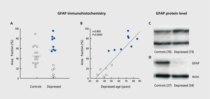
“An illustration of the pathology of glial cells found in the dorsolateral prefrontal cortex in MDD.(5,68) Reductions in the glial fibrillary acidic protein (GFAP) immunoreactive astroglia are found in a subgroup of young adults with major depression as compared to aged-matched control subjects and older subjects with…”
BackgroundSelf-citationSheline, Y. I., Sanghavi, M., Mintun, M. A., & Gado, M. H. (1999) · The Journal of Neuroscience · doi:10.1523/jneurosci.19-12-05034.1999 · Cited by 1,382
Neuroimaging studies of mood disorders
Drevets, W. C. (2000) · Biological Psychiatry · doi:10.1016/s0006-3223(00)01020-9 · Cited by 1,313 · cited 1×
“Abnormal regulation of glucose metabolism, regional cerebral blood flow, and high-energy phosphate metabolism are observed in the prefrontal and temporal cortex, basal ganglia, and amygdala in mood disorders.(88) Neuroimaging studies that examine neurochemical changes in the living brain provide further support for th…”
BackgroundIncreased hippocampal bdnf immunoreactivity in subjects treated with antidepressant medication
Chen, B., Dowlatshahi, D., MacQueen, G. M., Wang, J.-F., & Young, L. T. (2001) · Biological Psychiatry · doi:10.1016/s0006-3223(01)01083-6 · Cited by 1,164 · cited 1×
“In an immunohistochemical study of subjects with MDD and others with BPD or schizophrenia, the immunoreactivity of BDNF, as measured by optical density, is upregulated in the dentate gyrus and hilus only in subjects taking antidepressant medications (regardless of psychiatric diagnosis).(39) Chen et al(39) provide the…”
BackgroundMayberg, H. S., Brannan, S. K., Tekell, J. L., Silva, J. A., Mahurin, R. K., McGinnis, S., & Jerabek, P. A. (2000) · Biological Psychiatry · doi:10.1016/s0006-3223(00)01036-2 · Cited by 1,115
Czéh, B., Michaelis, T., Watanabe, T., Frahm, J., de Biurrun, G., van Kampen, M., Bartolomucci, A., & Fuchs, E. (2001) · Proceedings of the National Academy of Sciences · doi:10.1073/pnas.211427898 · Cited by 1,091 · cited 1×
“The NAA decrease in the animals exposed to repeated stressors was normalized by chronic treatment with the antidepressant tianeptine.(92) Increases in glutamate-glutamine-GABA metabolites in the adult anterior cingulate cortex of these animals were also observed 10 years after the stressors.”
BackgroundCourse of illness, hippocampal function, and hippocampal volume in major depression
MacQueen, G. M., Campbell, S., McEwen, B. S., Macdonald, K., Amano, S., Joffe, R. T., Nahmias, C., & Young, L. T. (2003) · Proceedings of the National Academy of Sciences · doi:10.1073/pnas.0337481100 · Cited by 980 · cited 12×
“On the other hand, recent immunohistochemical examination of glial fibrillary acidic protein (GFAP), a marker of reactive astroglia, in the dorsolateral prefrontal cortex implicates astrocytes in the overall glial pathology in MDD.68 Although no significant group dif- Postmortem abnormalities in depression - Stockmeie…”
BackgroundInfluentialOligodendrocyte dysfunction in schizophrenia and bipolar disorder
Tkachev, D., Mimmack, M. L., Ryan, M. M., Wayland, M., Freeman, T., Jones, P. B., Starkey, M., Webster, M. J., Yolken, R. H., & Bahn, S. (2003) · The Lancet · doi:10.1016/s0140-6736(03)14289-4 · Cited by 933 · cited 13×
“Moreover, key oligodendrocyte-related and myelin-related gene expression is reduced in the dorsolateral prefrontal cortex in BPD.(76) While these results are intriguing, further immunohistochemical and molecular studies are needed to definitively determine which specific glial cell types are compromised in BPD and whe…”
BackgroundInfluentialDwivedi, Y., Rizavi, H. S., Conley, R. R., Roberts, R. C., Tamminga, C. A., & Pandey, G. N. (2003) · Archives of General Psychiatry · doi:10.1001/archpsyc.60.8.804 · Cited by 875
Cotter, D., Mackay, D., Landau, S., Kerwin, R., & Everall, I. (2001) · Archives of General Psychiatry · doi:10.1001/archpsyc.58.6.545 · Cited by 766 · cited 3×
“In a manner more subtle than in MDD, reductions in neuronal soma size have been observed in BPD by some,(4,12) but not by all investigators.(1,2,8) In another study, a minor increase in the size of small nonpyramidal neurons was noted in the anterior cingulate cortex in BPD subjects.”
BackgroundThe anatomy of mood disorders—review of structural neuroimaging studies
Soares, J. C., & Mann, J. J. (1997) · Biological Psychiatry · doi:10.1016/s0006-3223(96)00006-6 · Cited by 760 · cited 1×
“Structural neuroimaging studies in mood disorders provide evidence of modest but intriguing volumetric changes that suggest cell loss and/or atrophy.(86) Some studies, but not all, report enlargement of the lateral and third ventricles in mood disorders(87) that may be indicative of atrophy of surrounding cortical and…”
BackgroundThe Functional Neuroanatomy of the Placebo Effect
Mayberg, H. S., Silva, J. A., Brannan, S. K., Tekell, J. L., Mahurin, R. K., McGinnis, S., & Jerabek, P. A. (2002) · American Journal of Psychiatry · doi:10.1176/appi.ajp.159.5.728 · Cited by 720
Kennedy, S. H., Evans, K. R., Krüger, S., Mayberg, H. S., Meyer, J. H., McCann, S., Arifuzzman, A. I., Houle, S., & Vaccarino, F. J. (2001) · American Journal of Psychiatry · doi:10.1176/appi.ajp.158.6.899 · Cited by 710
Raadsheer, F. C., Hoogendijk, W. J. G., Stam, F. C., Tilders, F. J. H., & Swaab, D. F. (2008) · Neuroendocrinology · doi:10.1159/000126778 · Cited by 667 · cited 1×
“Stereological investigation of specific types of hypothalamic neurons reveals an increase in the numbers of arginine-vasopressin (AVP)–immunoreactive neurons, oxytocin-expressing neurons, and corticotropin-releasing hormone (CRH) neurons in the paraventricular nucleus in subjects with BPD or MDD, compared to normal co…”
BackgroundNeural plasticity to stress and antidepressant treatment
Duman, R. S., Malberg, J., & Thome, J. (1999) · Biological Psychiatry · doi:10.1016/s0006-3223(99)00177-8 · Cited by 645
Sargent, P. A., Kjaer, K. H., Bench, C. J., Rabiner, E. A., Messa, C., Meyer, J., Gunn, R. N., Grasby, P. M., & Cowen, P. J. (2000) · Archives of General Psychiatry · doi:10.1001/archpsyc.57.2.174 · Cited by 642 · cited 1×
“In a neuroimaging study, the authors find that radioligand binding to serotonin-1A receptors is decreased in medication-free subjects with MDD in several cortical regions, including medial temporal cortex, the temporal pole, orbitofrontal cortex, anterior cingulate cortex, insula and dorsolateral prefrontal cortex.(58…”
BackgroundCotter, D. (2002) · Cerebral Cortex · doi:10.1093/cercor/12.4.386 · Cited by 634 · cited 1×
“Neuronal abnormalities in mood disorders are not immediately evident, inasmuch as there is no significant reduction in the density of Nissl-stained neurons measured across all cortical laminae.(1,3,4) However, when neurons are assessed within individual cortical layers or in subgroups determined by size or immunohisto…”
BackgroundMann, J. J., Huang, Y.-Y., Underwood, M. D., Kassir, S. A., Oppenheim, S., Kelly, T. M., Dwork, A. J., & Arango, V. (2000) · Archives of General Psychiatry · doi:10.1001/archpsyc.57.8.729 · Cited by 613 · cited 1×
“Expression of another component of serotonin neurotransmission, the serotonin transporter, is also decreased in the dorsolateral prefrontal and ventral/orbitofrontal cortex in postmortem brains from depressed suicide victims.(59,60) Detailed laminar analysis of the density of serotonin transporter–immunoreactive axons…”
BackgroundEnhancement of Hippocampal Neurogenesis by Lithium
Chen, G., Rajkowska, G., Du, F., Seraji‐Bozorgzad, N., & Manji, H. K. (2000) · Journal of Neurochemistry · doi:10.1046/j.1471-4159.2000.0751729.x · Cited by 599 · cited 1×
“Pharmacologically induced increases in neurogenesis in adult rodent brain have been reported in two independent studies.(77,78) Moreover, there is evidence that treatment with lithium induces an increase in the astrocytic protein GFAP in rodent hippocampus(79,80) and the neural lobe of the pituitary.”
BackgroundSelf-citationRajkowska, G., Halaris, A., & Selemon, L. D. (2001) · Biological Psychiatry · doi:10.1016/s0006-3223(01)01080-0 · Cited by 570 · cited 14×
“In addition, changes in the shape of glial nuclei to a less rounded conformation are detected in the dorsolateral prefrontal cortex in BPD.(4) Reductions in glial density, paralleled by an increase in the size of glial nuclei, suggest that some compensatory mechanisms may take place in mood disorders.”
BackgroundSelf-citationAuer, D. P., Pütz, B., Kraft, E., Lipinski, B., Schill, J., & Holsboer, F. (2000) · Biological Psychiatry · doi:10.1016/s0006-3223(99)00159-6 · Cited by 564 · cited 1×
“These findings in postmortem brain tissue coincide with an in vivo proton magnetic resonance spectroscopy study in the anterior cingulate cortex revealing a reduction in glutamate levels in depression.(65) There is increasing preclinical and clinical evidence that antidepressant drugs directly or indirectly reduce the…”
ResultRajkowska, G., & Goldman-Rakic, P. S. (1995) · Cerebral Cortex · doi:10.1093/cercor/5.4.323 · Cited by 562
Self-citationGlutamate and GABA systems as targets for novel antidepressant and mood-stabilizing treatments
Krystal, J. H., Sanacora, G., Blumberg, H., Anand, A., Charney, D. S., Marek, G., Epperson, C. N., Goddard, A., & Mason, G. F. (2002) · Molecular Psychiatry · doi:10.1038/sj.mp.4001021 · Cited by 553 · cited 2×
“There is increasing preclinical and clinical evidence that antidepressant drugs directly or indirectly reduce the function of N-methyl-D-aspartate (NMDA) glutamate receptors.(66) Depression-related decreases in glutamate levels or the density of glutamatergic pyramidal neurons may alter in cortex and elsewhere the glu…”
BackgroundJohnston-Wilson, N. L., Sims, C. D., Hofmann, J.-P., Anderson, L., Shore, A. D., Torrey, E. F., Yolken, R. H., & the Stanley Neuropathology Consortium (2000) · Molecular Psychiatry · doi:10.1038/sj.mp.4000696 · Cited by 531 · cited 1×
“Alterations in GFAP in both BPD and MDD are also suggested by a proteomic study in which different forms of GFAP proteins displayed disease-specific abnormalities.(73) Oligodendrocytes may also be involved in the cellular pathology of depression.”
BackgroundHippocampal Changes in Patients With a First Episode of Major Depression
Frodl, T., Meisenzahl, E. M., Zetzsche, T., Born, C., Groll, C., Jäger, M., Leinsinger, G., Bottlender, R., Hahn, K., & Möller, H.-J. (2002) · American Journal of Psychiatry · doi:10.1176/appi.ajp.159.7.1112 · Cited by 526 · cited 1×
“However, hippocampal volume is generally considered to be significantly decreased only in older depressed subjects with multiple episodes of depression.(25,26) Few studies have structurally examined the postmortem human hippocampus in depression.”
BackgroundLow glial numbers in the amygdala in major depressive disorder
Bowley, M. P., Drevets, W. C., Öngür, D., & Price, J. L. (2002) · Biological Psychiatry · doi:10.1016/s0006-3223(02)01404-x · Cited by 503 · cited 14×
“This last observation coincides with reports on reductions in the density of layer II nonpyramidal neurons that are identified with an antibody against the calciumbinding protein, calbindin, in the anterior cingulate cortex7 and dorsolateral prefrontal cortex9 in BPD. Calbindin immunoreactive neurons are known to colo…”
BackgroundResultInfluentialElectron microscopy of oligodendroglia in severe mental illness
Uranova, N., Orlovskaya, D., Vikhreva, O., Zimina, I., Kolomeets, N., Vostrikov, V., & Rachmanova, V. (2001) · Brain Research Bulletin · doi:10.1016/s0361-9230(01)00528-7 · Cited by 495 · cited 1×
“In both the dorsolateral prefrontal and anterior frontal cortex in subjects with BPD or MDD, there are ultrastructural changes in oligodendrocytes and there is a reduction in the density and immunoreactivity of these cells.(74,75) Moreover, key oligodendrocyte-related and myelin-related gene expression is reduced in t…”
BackgroundA reduction of nonpyramidal cells in sector CA2 of schizophrenics and manic depressives
Benes, F. M., Kwok, E. W., Vincent, S. L., & Todtenkopf, M. S. (1998) · Biological Psychiatry · doi:10.1016/s0006-3223(98)00138-3 · Cited by 470
Arango, V., Underwood, M. D., Gubbi, A. V., & Mann, J. J. (1995) · Brain Research · doi:10.1016/0006-8993(95)00523-s · Cited by 459
Reduced Levels of Norepinephrine Transporters in the Locus Coeruleus in Major Depression
Klimek, V., Stockmeier, C., Overholser, J., Meltzer, H. Y., Kalka, S., Dilley, G., & Ordway, G. A. (1997) · The Journal of Neuroscience · doi:10.1523/jneurosci.17-21-08451.1997 · Cited by 445 · cited 1×
“Another study in a larger number of subjects found no differences in the number of pigmented neurons in the locus ceruleus between subjects with MDD (most were suicides) and control subjects.(52) Although the number of neurons in the locus ceruleus does not appear altered in MDD, CRH immunoreactivity is increased in t…”
BackgroundSelf-citationFatemi, S. H., Earle, J. A., & McMenomy, T. (2000) · Molecular Psychiatry · doi:10.1038/sj.mp.4000783 · Cited by 445 · cited 13×
“This last observation coincides with reports on reductions in the density of layer II nonpyramidal neurons that are identified with an antibody against the calciumbinding protein, calbindin, in the anterior cingulate cortex7 and dorsolateral prefrontal cortex9 in BPD. Calbindin immunoreactive neurons are known to colo…”
BackgroundInfluentialPurba, J. S. (1996) · Archives of General Psychiatry · doi:10.1001/archpsyc.1996.01830020055007 · Cited by 433 · cited 1×
“Stereological investigation of specific types of hypothalamic neurons reveals an increase in the numbers of arginine-vasopressin (AVP)–immunoreactive neurons, oxytocin-expressing neurons, and corticotropin-releasing hormone (CRH) neurons in the paraventricular nucleus in subjects with BPD or MDD, compared to normal co…”
BackgroundStockmeier, C. A., Shapiro, L. A., Dilley, G. E., Kolli, T. N., Friedman, L., & Rajkowska, G. (1998) · The Journal of Neuroscience · doi:10.1523/jneurosci.18-18-07394.1998 · Cited by 429
Self-citation(1995) · American Journal of Psychiatry · doi:10.1176/ajp.152.9.1372 · Cited by 411 · cited 1×
“Moreover, increases in CRH mRNA, and in the number of CRH neurons colocalizing AVP are also found in depressed patients.(46,47) These findings of increases in specific immunoreactive neurons are consistent with the evidence of activation of the hypothalamicpituitary-adrenal (HPA) axis in some subsets of depressed pati…”
ResultMoore, G. J., Bebchuk, J. M., Hasanat, K., Chen, G., Seraji-Bozorgzad, N., Wilds, I. B., Faulk, M. W., Koch, S., Glitz, D. A., Jolkovsky, L., et al. (2000) · Biological Psychiatry · doi:10.1016/s0006-3223(00)00252-3 · Cited by 401 · cited 13×
“This last observation coincides with reports on reductions in the density of layer II nonpyramidal neurons that are identified with an antibody against the calciumbinding protein, calbindin, in the anterior cingulate cortex7 and dorsolateral prefrontal cortex9 in BPD. Calbindin immunoreactive neurons are known to colo…”
BackgroundInfluentialShah, P. J., Ebmeier, K. P., Glabus, M. F., & Goodwin, G. M. (1998) · British Journal of Psychiatry · doi:10.1192/bjp.172.6.527 · Cited by 395
Quantitative MRI of the hippocampus and amygdala in severe depression
MERVAALA, E., FÖHR, J., KÖNÖNEN, M., VALKONEN-KORHONEN, M., VAINIO, P., PARTANEN, K., PARTANEN, J., TIIHONEN, J., VIINAMÄKI, H., KARJALAINEN, A.-K., et al. (2000) · Psychological Medicine · doi:10.1017/s0033291799001567 · Cited by 382
Hippocampal volume in geriatric depression
Steffens, D. C., Byrum, C. E., McQuoid, D. R., Greenberg, D. L., Payne, M. E., Blitchington, T. F., MacFall, J. R., & Krishnan, K. R. R. (2000) · Biological Psychiatry · doi:10.1016/s0006-3223(00)00829-5 · Cited by 379
Hippocampal volume in primary unipolar major depression: a magnetic resonance imaging study
Vakili, K., Pillay, S. S., Lafer, B., Fava, M., Renshaw, P. F., Bonello-Cintron, C. M., & Yurgelun-Todd, D. A. (2000) · Biological Psychiatry · doi:10.1016/s0006-3223(99)00296-6 · Cited by 363
Miguel-Hidalgo, J. J., Baucom, C., Dilley, G., Overholser, J. C., Meltzer, H. Y., Stockmeier, C. A., & Rajkowska, G. (2000) · Biological Psychiatry · doi:10.1016/s0006-3223(00)00999-9 · Cited by 336 · cited 2×
“An illustration of the pathology of glial cells found in the dorsolateral prefrontal cortex in MDD.(5,68) Reductions in the glial fibrillary acidic protein (GFAP) immunoreactive astroglia are found in a subgroup of young adults with major depression as compared to aged-matched control subjects and older subjects with…”
BackgroundSelf-citationBenes, F. M., Vincent, S. L., & Todtenkopf, M. (2001) · Biological Psychiatry · doi:10.1016/s0006-3223(01)01084-8 · Cited by 327 · cited 1×
“In another study, a minor increase in the size of small nonpyramidal neurons was noted in the anterior cingulate cortex in BPD subjects.(6) Factors leading to a reduction in the size of neuronal soma are not known.”
BackgroundDecreased dorsolateral prefrontal N-acetyl aspartate in bipolar disorder
Winsberg, M. E., Sachs, N., Tate, D. L., Adalsteinsson, E., Spielman, D., & Ketter, T. A. (2000) · Biological Psychiatry · doi:10.1016/s0006-3223(99)00183-3 · Cited by 294
Lucassen, P. J., Müller, M. B., Holsboer, F., Bauer, J., Holtrop, A., Wouda, J., Hoogendijk, W. J. G., De Kloet, E. R., & Swaab, D. F. (2001) · The American Journal of Pathology · doi:10.1016/s0002-9440(10)63988-0 · Cited by 286 · cited 3×
“In most of the depressives, there was evidence for a slight increase in fragmented DNA associated with apoptosis and necrotic neuron death detected in the dentate gyrus, CA1 and CA4.”
BackgroundMethodEastwood, S. L., & Harrison, P. J. (2001) · Brain Research Bulletin · doi:10.1016/s0361-9230(01)00530-5 · Cited by 263
Nowak, G., Ordway, G. A., & Paul, I. A. (1995) · Brain Research · doi:10.1016/0006-8993(95)00057-w · Cited by 262
Elkis, H. (1995) · Archives of General Psychiatry · doi:10.1001/archpsyc.1995.03950210029008 · Cited by 259
Müller, M. B., Lucassen, P. J., Yassouridis, A., Hoogendijk, W. J. G., Holsboer, F., & Swaab, D. F. (2001) · European Journal of Neuroscience · doi:10.1046/j.0953-816x.2001.01784.x · Cited by 249 · cited 2×
“Decreases in astrocytic immunoreactivity for cellular GFAP and the neuron-specific phosphoprotein B50 (or GAP-45) were detected in CA1 and CA2 in depression.(31) The authors suggest that apoptosis may only be a minor contributor to volume changes in the hippocampus in depression, while patterns of reactive astrogliosi…”
BackgroundMethodChana, G., Landau, S., Beasley, C., Everall, I. P., & Cotter, D. (2003) · Biological Psychiatry · doi:10.1016/s0006-3223(03)00114-8 · Cited by 246 · cited 12×
“On the other hand, recent immunohistochemical examination of glial fibrillary acidic protein (GFAP), a marker of reactive astroglia, in the dorsolateral prefrontal cortex implicates astrocytes in the overall glial pathology in MDD.68 Although no significant group dif- Postmortem abnormalities in depression - Stockmeie…”
BackgroundInfluentialSynaptic and plasticity-associated proteins in anterior frontal cortex in severe mental illness
Honer, W. G., Falkai, P., Chen, C., Arango, V., Mann, J. J., & Dwork, A. J. (1999) · Neuroscience · doi:10.1016/s0306-4522(98)00679-4 · Cited by 244
Neuropathological studies of synaptic connectivity in the hippocampal formation in schizophrenia
Harrison, P. J., & Eastwood, S. L. (2001) · Hippocampus · doi:10.1002/hipo.1067 · Cited by 223 · cited 13×
“This last observation coincides with reports on reductions in the density of layer II nonpyramidal neurons that are identified with an antibody against the calciumbinding protein, calbindin, in the anterior cingulate cortex7 and dorsolateral prefrontal cortex9 in BPD. Calbindin immunoreactive neurons are known to colo…”
ResultInfluentialBernstein, H.-G., Stanarius, A., Baumann, B., Henning, H., Krell, D., Danos, P., Falkai, P., & Bogerts, B. (1998) · Neuroscience · doi:10.1016/s0306-4522(97)00461-2 · Cited by 223 · cited 13×
“This last observation coincides with reports on reductions in the density of layer II nonpyramidal neurons that are identified with an antibody against the calciumbinding protein, calbindin, in the anterior cingulate cortex7 and dorsolateral prefrontal cortex9 in BPD. Calbindin immunoreactive neurons are known to colo…”
BackgroundInfluentialCotter, D., Landau, S., Beasley, C., Stevenson, R., Chana, G., MacMillan, L., & Everall, I. (2002) · Biological Psychiatry · doi:10.1016/s0006-3223(01)01243-4 · Cited by 211
Anterior cingulate cortex pathology in schizophrenia and bipolar disorder
Bouras, C., Kövari, E., Hof, P. R., Riederer, B. M., & Giannakopoulos, P. (2001) · Acta Neuropathologica · doi:10.1007/s004010100392 · Cited by 209 · cited 3×
“In a manner more subtle than in MDD, reductions in neuronal soma size have been observed in BPD by some,(4,12) but not by all investigators.(1,2,8) In another study, a minor increase in the size of small nonpyramidal neurons was noted in the anterior cingulate cortex in BPD subjects.”
BackgroundResultHigh-Dimensional Mapping of the Hippocampus in Depression
Posener, J. A., Wang, L., Price, J. L., Gado, M. H., Province, M. A., Miller, M. I., Babb, C. M., & Csernansky, J. G. (2003) · American Journal of Psychiatry · doi:10.1176/appi.ajp.160.1.83 · Cited by 200
Clinical and phenomenological comparisons of late-onset and early-onset depression
(1995) · American Journal of Psychiatry · doi:10.1176/ajp.152.5.785 · Cited by 178
Dopaminergic abnormalities in amygdaloid nuclei in major depression: a postmortem study
Klimek, V., Schenck, J. E., Han, H., Stockmeier, C. A., & Ordway, G. A. (2002) · Biological Psychiatry · doi:10.1016/s0006-3223(02)01383-5 · Cited by 177 · cited 1×
“Postmortem neurochemical studies in MDD report alterations in noradrenergic α2-adrenergic receptors and the norepinephrine transporter, as well as levels of tyrosine hydroxylase in the locus ceruleus,(52,61,62) serotonin-1A receptors in the midbrain dorsal raphe nucleus,(63) and dopaminergic receptors and transporters…”
BackgroundSelf-citationFunctional neuroanatomy and neuropathology of the human hypothalamus
Swaab, D. F., Hofman, M. A., Lucassen, P. J., Purba, J. S., Raadsheer, F. C., & van de Nes, J. A. P. (1993) · Anatomy and Embryology · doi:10.1007/bf00185889 · Cited by 173 · cited 1×
“Moreover, increases in CRH mRNA, and in the number of CRH neurons colocalizing AVP are also found in depressed patients.(46,47) These findings of increases in specific immunoreactive neurons are consistent with the evidence of activation of the hypothalamicpituitary-adrenal (HPA) axis in some subsets of depressed pati…”
ResultHansson, E., Muyderman, H., Leonova, J., Allansson, L., Sinclair, J., Blomstrand, F., Thorlin, T., Nilsson, M., & Rönnbäck, L. (2000) · Neurochemistry International · doi:10.1016/s0197-0186(00)00033-4 · Cited by 172
Morphometry of the dorsal raphe nucleus serotonergic neurons in suicide victims
Underwood, M. D., Khaibulina, A. A., Ellis, S. P., Moran, A., Rice, P. M., Mann, J. J., & Arango, V. (1999) · Biological Psychiatry · doi:10.1016/s0006-3223(99)00043-8 · Cited by 165
Hippocampal morphometry in depressed patients and control subjects: relations to anxiety symptoms
Rusch, B. D., Abercrombie, H. C., Oakes, T. R., Schaefer, S. M., & Davidson, R. J. (2001) · Biological Psychiatry · doi:10.1016/s0006-3223(01)01248-3 · Cited by 164
Fewer pigmented locus coeruleus neurons in suicide victims: Preliminary results
Arango, V., Underwood, M. D., & Mann, J. J. (1996) · Biological Psychiatry · doi:10.1016/0006-3223(95)00107-7 · Cited by 163
Austin, M. C., Janosky, J. E., & Murphy, H. A. (2003) · Molecular Psychiatry · doi:10.1038/sj.mp.4001250 · Cited by 162 · cited 1×
“Although the number of neurons in the locus ceruleus does not appear altered in MDD, CRH immunoreactivity is increased in the locus ceruleus and pontine dorsal and median raphe nuclei.(53,54) No changes in neuronal densities were detected in amygdala in subjects with either MDD or BPD, as compared to normal controls.”
BackgroundHolsboer, F., Spengler, D., & Heuser, I. (1992) · Progress in Brain Research · doi:10.1016/s0079-6123(08)64586-0 · Cited by 159 · cited 1×
“These findings of increases in specific immunoreactive neurons are consistent with the evidence of activation of the hypothalamicpituitary-adrenal (HPA) axis in some subsets of depressed patients.(48) On the other hand, decreased number and density of nitric oxide synthase–containing neurons in the paraventricular hyp…”
ResultAgonist Binding to α2‐Adrenoceptors Is Elevated in the Locus Coeruleus from Victims of Suicide
Ordway, G. A., Widdowson, P. S., Smith, K. S., & Halaris, A. (1994) · Journal of Neurochemistry · doi:10.1046/j.1471-4159.1994.63020617.x · Cited by 156
Elevated levels of tyrosine hydroxylase in the locus coeruleus in major depression
Zhu, M.-Y., Klimek, V., Dilley, G. E., Haycock, J. W., Stockmeier, C., Overholser, J. C., Meltzer, H. Y., & Ordway, G. A. (1999) · Biological Psychiatry · doi:10.1016/s0006-3223(99)00135-3 · Cited by 137
Self-citationElevated Concentrations of CRF in the Locus Coeruleus of Depressed Subjects
Bissette, G., Klimek, V., Pan, J., Stockmeier, C., & Ordway, G. (2003) · Neuropsychopharmacology · doi:10.1038/sj.npp.1300191 · Cited by 136 · cited 1×
“Although the number of neurons in the locus ceruleus does not appear altered in MDD, CRH immunoreactivity is increased in the locus ceruleus and pontine dorsal and median raphe nuclei.(53,54) No changes in neuronal densities were detected in amygdala in subjects with either MDD or BPD, as compared to normal controls.”
BackgroundSelf-citationImpairment of GABAergic Transmission in Depression: New Insights from Neuroimaging Studies
Sanacora, G., Mason, G. F., & Krystal, J. H. (2000) · Critical Reviews™ in Neurobiology · doi:10.1615/critrevneurobiol.v14.i1.20 · Cited by 132 · cited 4×
“The depression-related decrease in calbindin immunoreactive neurons, which colocalize GABA, may be closely related to in vivo clinical evidence suggesting that MDD is associated with decreased levels of GABA in cerebral cortex.(11) Another manifestation of neuronal pathology in cerebral cortex in mood disorders is the…”
BackgroundInfluentialBertolino, A., Knable, M. B., Saunders, R. C., Callicott, J. H., Kolachana, B. S., Mattay, V. S., Bachevalier, J., Frank, J. A., Egan, M. F., & Weinberger, D. R. (1999) · Biological Psychiatry · doi:10.1016/s0006-3223(98)00380-1 · Cited by 113
Beckmann, H., & Jakob, H. (1991) · Journal of Neural Transmission · doi:10.1007/bf01249120 · Cited by 112 · cited 1×
“Two other studies conducted on the postmortem hippocampal formation in a small sample of subjects with BPD reveal a decrease in the density and size of nonpyramidal neurons in the CA2 region and some disorganization in neuronal clusters in layers II and III of the entorhinal cortex.(34,35) Neuronal and glial cell pack…”
ResultAustin, M. C., Whitehead, R. E., Edgar, C. L., Janosky, J. E., & Lewis, D. A. (2002) · Neuroscience · doi:10.1016/s0306-4522(02)00289-0 · Cited by 109 · cited 2×
“Expression of another component of serotonin neurotransmission, the serotonin transporter, is also decreased in the dorsolateral prefrontal and ventral/orbitofrontal cortex in postmortem brains from depressed suicide victims.(59,60) Detailed laminar analysis of the density of serotonin transporter–immunoreactive axons…”
BackgroundBernstein, H.-G., Krell, D., Baumann, B., Dános, P., Falkai, P., Diekmann, S., Henning, H. J., & Bogerts, B. (1998) · Schizophrenia Research · doi:10.1016/s0920-9964(98)00071-1 · Cited by 86
Morphological Brain Changes in Depression: Can Antidepressants Reverse Them?
Miguel-Hidalgo, J. J., & Rajkowska, G. (2002) · CNS Drugs · doi:10.2165/00023210-200216060-00001 · Cited by 64
Self-citationYohannes, A. M., Baldwin, R. C., & Connolly, M. J. (2000) · International Journal of Geriatric Psychiatry · doi:10.1002/1099-1166(200012)15:12 · Cited by 58
Relationship of Age, Age at Onset, and Sex to Depression in Older Adults
Lavretsky, H., Lesser, I. M., Wohl, M. A., & Miller, B. L. (1998) · American Journal of Geriatric Psychiatry · doi:10.1097/00019442-199800630-00007 · Cited by 58
Relationship of Age, Age at Onset, and Sex to Depression in Older Adults
Lavretsky, H., Lesser, I. M., Wohl, M., & Miller, B. L. (1998) · American Journal of Geriatric Psychiatry · doi:10.1097/00019442-199808000-00007 · Cited by 56
Rocha, E., & Rodnight, R. (1994) · Journal of Neurochemistry · doi:10.1046/j.1471-4159.1994.63041582.x · Cited by 38
Orlovskaya, D. D., Vm, V., Rachmanova, V. I., & Uranova, N. A. (2000) · Schizophrenia Research · doi:10.1016/s0920-9964(00)90551-6 · Cited by 35 · cited 1×
“In both the dorsolateral prefrontal and anterior frontal cortex in subjects with BPD or MDD, there are ultrastructural changes in oligodendrocytes and there is a reduction in the density and immunoreactivity of these cells.(74,75) Moreover, key oligodendrocyte-related and myelin-related gene expression is reduced in t…”
BackgroundLithium treatment causes gliosis and modifies the morphology of hippocampal astrocytes in rats
Rocha, E., Achaval, M., Santos, P., & Rodnight, R. (1998) · NeuroReport · doi:10.1097/00001756-199812010-00037 · Cited by 35
The importance of a human 3D database and atlas for studies of prefrontal and thalamic functions
Uylings, H. B. M., Sanz‐Arigita, E., de Vos, K., Smeets, W. J. A. J., Pool, C. W., Amunts, K., Rajkowska, G., & Zilles, K. (2000) · Progress in brain research · doi:10.1016/s0079-6123(00)26024-x · Cited by 27
Self-citationGlial Pathology in Major Psychiatric Disorders
Cotter, D., Pariante, C. M., & Rajkowska, G. (2002) · Neurobiological foundation of aberrant behaviors · doi:10.1007/978-1-4757-3631-1_5 · Cited by 22 · cited 15×
“This last observation coincides with reports on reductions in the density of layer II nonpyramidal neurons that are identified with an antibody against the calciumbinding protein, calbindin, in the anterior cingulate cortex7 and dorsolateral prefrontal cortex9 in BPD. Calbindin immunoreactive neurons are known to colo…”
BackgroundSelf-citationProliferation of glial cells in vivo induced in the neural lobe of the rat pituitary by lithium
Levine, S., Saltzman, A., & Klein, A. W. (2000) · Cell Proliferation · doi:10.1046/j.1365-2184.2000.00170.x · Cited by 14 · cited 1×
“Moreover, there is evidence that treatment with lithium induces an increase in the astrocytic protein GFAP in rodent hippocampus(79,80) and the neural lobe of the pituitary.(81) However, whether these increases represent a protective or compensatory effect of these medications, and the mechanisms underlying the regula…”
BackgroundReynolds, G. P., Zhang, Z. J., Patten, I., & Beasley, C. L. (2000) · Schizophrenia Research · doi:10.1016/s0920-9964(00)90944-7 · Cited by 11
Stockmeier, C. A., & Jurjus, G. (2002) · Neurobiological foundation of aberrant behaviors · doi:10.1007/978-1-4757-3631-1_20 · Cited by 2 · cited 2×
“Studies in postmortem brain tissue identify alterations in serotonin and norepinephrine receptors and transporters in the dorsolateral prefrontal cortex and ventrolateral/orbitofrontal cortex in brains from suicide victims with or without clinical depression.(56) These cortical regions also exhibit abnormal cell densi…”
BackgroundSelf-citationRosoklija, G., Toomayan, G., Ellis, S. P., Keilp, J., Mann, J. J., Latov, N., Hays, A. P., & Dwork, A. J. (2000) · Archives of General Psychiatry · doi:10.1001/archpsyc.57.4.349 · Cited by 1
Research works printed but not linked: 1
- Stockmeier CA. Mahajan GJ. Konick L. et al. Neural and glial density is increased and neural soma size is decreased in hippocampus in major depressive disorder. Biol Psychiatry. 2003 53(suppl 8) 198
Cited by
Brites, D., & Fernandes, A. (2015) · Frontiers in Cellular Neuroscience · doi:10.3389/fncel.2015.00476 · Cited by 599
“In this article, we discuss the role of neuroinflammation in the emergence of depression, namely dynamic alterations in the status of microglia response to stimulation, and how their activation phenotypes may have an etiological role in neurodegeneneration, in particular in depressive-like behavior.”
BackgroundMolecular pathways of major depressive disorder converge on the synapse
Fries, G. R., Saldana, V. A., Finnstein, J., & Rein, T. (2023) · Molecular Psychiatry · doi:10.1038/s41380-022-01806-1 · Cited by 504
“In parallel, postmortem studies have reported changes in the density and size of neurons and glia in several brain regions of patients [10] along with reduced expression of pre- and postsynaptic genes [11, 12].”
BackgroundRadley, J., Morilak, D., Viau, V., & Campeau, S. (2015) · Neuroscience & Biobehavioral Reviews · doi:10.1016/j.neubiorev.2015.06.018 · Cited by 339
“Post mortem histological analyses of brains from depressed individuals generally corroborate volumetric data in observing reductions in neuron and glial cell numbers in the same cortical subfields (Cotter et al., 2002; Cotter et al., 2001; Rajkowska et al., 1999; Stockmeier and Rajkowska, 2004).”
Maitra, M., Mitsuhashi, H., Rahimian, R., Chawla, A., Yang, J., Fiori, L. M., Davoli, M. A., Perlman, K., Aouabed, Z., Mash, D. C., et al. (2023) · Nature Communications · doi:10.1038/s41467-023-38530-5 · Cited by 159
“Classic cytological experiments from the turn of the century identi fi ed abnormalities in morphology and distribution of cell types, but alsoput into question cell number, size, and neuropil density, particularly for neurons and astrocytes 30 – 34 .”
Gonadal steroid regulation of mood: The lessons of premenstrual syndrome☆
Rubinow, D. R., & SCHMIDT, P. (2006) · Frontiers in Neuroendocrinology · doi:10.1016/j.yfrne.2006.02.003 · Cited by 194
“Similarly, many areas of the brain that have been implicated in depression, including prefrontal cortex (PFC), amygdala, hippocampus, striatum, and thalamus [17,21,22,52,53,66,74], contain estrogen receptors and are modulated by estradiol [1,10,15,41].”
Background
Metadata
- Authors
- Stockmeier, C. A., & Rajkowska, G.
- Affiliations
- University of Mississippi Medical Center (US)
- The University of Mississippi Medical Center, Department of Psychiatry and Human Behavior, Jackson, Miss, USA
- Corresponding author
- Stockmeier, Craig A.
- ORCID
- Stockmeier, Craig A.
- Venue
- Dialogues in Clinical Neuroscience
- Publisher
- Informa UK Limited
- Published
- 2004-06-30
- Volume
- 6
- Issue
- 2
- Pages
- 185-197
- ISSN
- 1294-8322 (ISSN-L)
- 1958-5969
- PMC
- PMC3181793
- OpenAlex
- W139876275
- Cited by
- 67
- References
- 87
- Citations by year
- 2026: 4 · 2025: 8 · 2024: 3 · 2023: 11 · 2022: 9 · 2021: 5 · 2020: 1 · 2019: 1 · 2018: 2 · 2017: 4 · 2016: 3 · 2015: 4 · 2014: 3 · 2013: 2 · 2012: 1
- FWCI
- 0.42
- Type
- journal article
- Language
- en
- Open access
- diamond
- doi.org
- Licence
- Locations
- Dialogues in Clinical Neuroscience (publishedVersion, cc-by-nc-nd)
- Europe PMC (PubMed Central) (publishedVersion, cc-by-nc-nd)
- PubMed Central (publishedVersion, cc-by-nc-nd)
- PubMed (publishedVersion)
- Topics
- Health Sciences › Medicine › Pharmacology (medical) › Treatment of Major Depression
- Life Sciences › Neuroscience › Behavioral Neuroscience › Stress Responses and Cortisol
- Life Sciences › Neuroscience › Cellular and Molecular Neuroscience › Neurotransmitter Receptor Influence on Behavior
- Keywords
- depression, postmortem brain, cellular abnormalities, cell counting, neuronal density, glial density, dorsolateral prefrontal cortex, orbitofrontal cortex, anterior cingulate cortex, amygdala, hippocampus, neurochemical abnormalities, mood disorders, depression pathophysiology, neuroimaging
About these checks
Registry, retraction, Cites and statistics results are computed, not judged, with one exception: a printed reference that no index lists is read once by a model before it counts as a citation error. Claims are extracted by a model and kept only when their quote appears verbatim in the text; each cited source is then checked, with a model’s help, for whether it says what the claim relies on. Those verdicts can be wrong — read the claim’s quote and the cited source before relying on one.
Statistics and arithmetic are recomputed, not judged.
Confidence bands are shown only for fields calibrated on our annotated test set. None is calibrated yet, so no band is shown.
What counts as a claim ›How we measure confidence ›How we check this ›
Are you an author, or do you see a problem with this page? Request a review
Data sources
Bibliographic data: Semantic Scholar, OpenAlex, Crossref.
Contains information from the Semantic Scholar Academic Graph Datasets, made available under the ODC Attribution License (ODC-BY 1.0).
Semantic Scholar’s data is described in: Kinney, R., et al. (2023). The Semantic Scholar Open Data Platform. arXiv:2301.10140.
