

The DOI matches its Crossref record; 117 of the 117 references it cites were found in a registry or an index.
Corticostriatal circuitry
Open accessAbstract
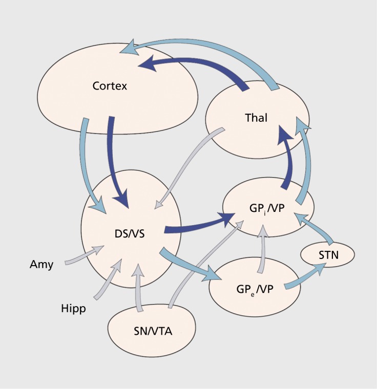
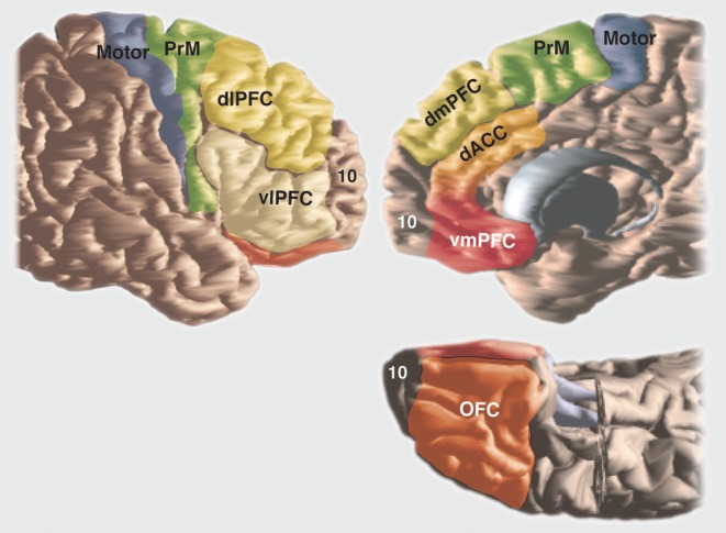
Corticostriatal connections play a central role in developing appropriate goal-directed behaviors, including the motivation and cognition to develop appropriate actions to obtain a specific outcome. The cortex projects to the striatum topographically. Thus, different regions of the striatum have been associated with these different functions: the ventral striatum with reward; the caudate nucleus with cognition; and the putamen with motor control. However, corticostriatal connections are more complex, and interactions between functional territories are extensive. These interactions occur in specific regions in which convergence of terminal fields from different functional cortical regions are found. This article provides an overview of the connections of the cortex to the striatum and their role in integrating information across reward, cognitive, and motor functions. Emphasis is placed on the interface between functional domains within the striatum.
Overview
The claims and statistics of this paper haven’t been checked yet.
Free: it reads the open-access text and checks the first 5 claims that cite a source we can check. Create a free account to check every claim.
Integrity signals
Registry
Cites
Statistics
Figures


Verification
Claims the paper makes
The claims and statistics of this paper haven’t been checked yet.
Free: it reads the open-access text and checks the first 5 claims that cite a source we can check. Create a free account to check every claim.
Claims other papers make about this one
Each is checked against this paper’s own text: does it say what the citing paper says it does?
The cited source says what the claim attributes to it.
In the citing paper
“For example, the substantia nigra makes extensive connections with the striatum, cortex, thalamus and neighboring brainstem regions, allowing dopaminergic outputs to exert far-reaching effects on the flow of CSTC information (Haber, 2016).”
From Cortico-striatal-thalamic loop circuits of the salience network: A central pathway in psychiatric disease and treatment (2016) · cites this paper as Haber, 2016
The cited source says what the claim attributes to it.
In the citing paper
“Importantly, the mesolimbic and nigrostriatal dopaminergic pathway have been implicated in the encoding of “saliency prediction errors”, underscoring their broader contributions to the attribution of salience to environmental stimuli (Kapur, 2003; Haber, 2016).”
From Cortico-striatal-thalamic loop circuits of the salience network: A central pathway in psychiatric disease and treatment (2016) · cites this paper as Haber, 2016
The cited source says what the claim attributes to it.
In the citing paper
“Corticostriatal circuitry is essential to processing reward, cognition, and movement (Haber, 2016)”
From Developmental origins of sex differences in the neural circuitry of play (2020) · cites this paper as Haber, 2016
In the citing paper
“Specifically, the dACC projects primarily to the dorsal caudate nucleus and ventral striatum, overlapping partially but not completely with frontostriatal projections from the dlPFC (Haber, 2016).”
From Cortico-striatal-thalamic loop circuits of the salience network: A central pathway in psychiatric disease and treatment (2016) · cites this paper as Haber, 2016
Self-check
Percentages, sums and reported differences re-computed from the paper’s own numbers.
The self-check needs the paper’s full text.
Citations
Cites
The Brain's Default Network: Anatomy, Function, and Relevance to Disease
Buckner, R. L., Andrews‐Hanna, J. R., & Schacter, D. L. (2008) · Annals of the New York Academy of Sciences · doi:10.1196/annals.1440.011 · Cited by 9,761
Koob, G. F., & Volkow, N. D. (2010) · Neuropsychopharmacology · doi:10.1038/npp.2009.110 · Cited by 5,340
Predictive reward signal of dopamine neurons
Schultz, W. (1998) · Journal of Neurophysiology · doi:10.1152/jn.1998.80.1.1 · Cited by 4,564
Functional architecture of basal ganglia circuits: neural substrates of parallel processing
Alexander, G. E., & Crutcher, M. D. (1990) · Trends in Neurosciences · doi:10.1016/0166-2236(90)90107-l · Cited by 4,131
Functional anatomy of the basal ganglia. I. The cortico-basal ganglia-thalamo-cortical loop
Parent, A., & Hazrati, L.-N. (1995) · Brain Research Reviews · doi:10.1016/0165-0173(94)00007-c · Cited by 2,161
Wise, R. A., & Rompre, P.-P. (1989) · Annual Review of Psychology · doi:10.1146/annurev.ps.40.020189.001203 · Cited by 1,968
Haber, S. N., Fudge, J. L., & McFarland, N. R. (2000) · The Journal of Neuroscience · doi:10.1523/jneurosci.20-06-02369.2000 · Cited by 1,610
Self-citationNeurons in the orbitofrontal cortex encode economic value
Padoa-Schioppa, C., & Assad, J. A. (2006) · Nature · doi:10.1038/nature04676 · Cited by 1,598
Neurotransmitters and neuromodulators in the basal ganglia
Graybiel, A. M. (1990) · Trends in Neurosciences · doi:10.1016/0166-2236(90)90104-i · Cited by 1,576
Specificity in the projection patterns of accumbal core and shell in the rat
Heimer, L., Zahm, D. S., Churchill, L., Kalivas, P. W., & Wohltmann, C. (1991) · Neuroscience · doi:10.1016/0306-4522(91)90202-y · Cited by 1,219
Research works printed but not linked: 7
- Robbins TW., 1999, Fundamental Neuroscience. 2nd ed. San Diego, CA: Academic Press;, 1245
- Koob GF., 1999, Fundamental Neuroscience. New York, NY: Academic Press;, 1261
- Kalivas PW., 1993, Limbic Motor Circuits and Neuropsychiatry. Boca Raton, FL: CRC Press, Inc;, 237
- Heimer L., 1993, Limbic Motor Circuits and Neuropsychiatry. Boca Raton, FL: CRC Press, Inc;, 1
- Parent A., 1986, Comparative Neurobiology of the Basal Ganglia. New York, NY: John Wiley and Sons;
- McFarland NR., 1996, Soc Neurosci Abst., 22, 412
- Fibiger HC., 1986, Handbook of Physiology. Section I: the Nervous System. Vol 4. Bethesda,MD: American Physiological Society;, 647
Cited by
Schizophrenia, dopamine and the striatum: From biology to symptoms
McCutcheon, R. A., Abi-Dargham, A., & Howes, O. D. (2019) · Trends in Neurosciences · doi:10.1016/j.tins.2018.12.004 · Cited by 763
Faraone, S. V. (2018) · Neuroscience & Biobehavioral Reviews · doi:10.1016/j.neubiorev.2018.02.001 · Cited by 631
Peters, S. K., Dunlop, K., & Downar, J. (2016) · Frontiers in Systems Neuroscience · doi:10.3389/fnsys.2016.00104 · Cited by 611
“Extensive connections from these regions to the dorsal striatum may link reactional cognitive control to behavioral guidance, allowing for on-line behavior regulation in response to salient environmental stimuli (Botvinick et al., 2004; Haber, 2016).”
BackgroundInfluentialObsessive-compulsive disorder: Puzzles and prospects
Robbins, T. W., Vaghi, M. M., & Banca, P. (2019) · Neuron · doi:10.1016/j.neuron.2019.01.046 · Cited by 510
The graphical brain: Belief propagation and active inference
Friston, K. J., Parr, T., & de Vries, B. (2017) · Network Neuroscience · doi:10.1162/netn_a_00018 · Cited by 415
“This message passing could be mediated by corticostriatal projections, from layer 5 (and deep layer 3) pyramidal neurons, which are distributed in a patchy manner (Haber, 2016).”
Background
Metadata
- Authors
- Haber, S. N.
- Affiliations
- University of Rochester Medicine (US)
- Corresponding author
- Haber, Suzanne N.
- ORCID
- Haber, Suzanne N.
- Venue
- Dialogues in Clinical Neuroscience
- Publisher
- Informa UK Limited
- Published
- 2016-03-31
- Volume
- 18
- Issue
- 1
- Pages
- 7-21
- ISSN
- 1294-8322 (ISSN-L)
- 1958-5969
- PMC
- PMC4826773
- OpenAlex
- W4241221200
- Cited by
- 922
- References
- 116
- Citations by year
- 2026: 79 · 2025: 122 · 2024: 111 · 2023: 98 · 2022: 129 · 2021: 101 · 2020: 92 · 2019: 88 · 2018: 56 · 2017: 39 · 2016: 7
- FWCI
- 35.52
- Type
- journal article
- Language
- en
- Open access
- diamond
- doi.org
- Licence
- Locations
- Dialogues in Clinical Neuroscience (publishedVersion, cc-by-nc-nd)
- PubMed Central (publishedVersion, cc-by-nc-nd)
- PubMed (publishedVersion)
- Funders
- National Institute of Mental Health (R01MH045573, MH045573)
- Topics
- Life Sciences › Neuroscience › Cellular and Molecular Neuroscience › Neurotransmitter Receptor Influence on Behavior
- Life Sciences › Neuroscience › Cognitive Neuroscience › Neural and Behavioral Psychology Studies
- Health Sciences › Medicine › Clinical Neurology › Deep Brain Stimulation and Movement Disorders
- Keywords
- corticostriatal connections, striatum, ventral striatum, caudate nucleus, putamen, goal-directed behavior, reward, cognition, motor control, topographic projection, functional integration, terminal fields
- MeSH terms
- Animals, Cerebral Cortex, Cognition, Corpus Striatum, Humans, Nerve Net, Neural Pathways, Reward
About these checks
Registry, retraction, Cites and statistics results are computed, not judged, with one exception: a printed reference that no index lists is read once by a model before it counts as a citation error. Claims are extracted by a model and kept only when their quote appears verbatim in the text; each cited source is then checked, with a model’s help, for whether it says what the claim relies on. Those verdicts can be wrong — read the claim’s quote and the cited source before relying on one.
Statistics and arithmetic are recomputed, not judged.
Confidence bands are shown only for fields calibrated on our annotated test set. None is calibrated yet, so no band is shown.
What counts as a claim ›How we measure confidence ›How we check this ›
Are you an author, or do you see a problem with this page? Request a review
How to cite
Cite CoChat for the checks on this page, and the paper for its own findings.
- This page
- CoChat. (2026). Citation check: Corticostriatal circuitry. https://app.cochat.ai/papers/10.31887/dcns.2016.18.1/shaber
- The paper
- Haber, S. N. (2016). Corticostriatal circuitry. Dialogues in Clinical Neuroscience. https://doi.org/10.31887/dcns.2016.18.1/shaber
Data sources
Bibliographic data: Semantic Scholar, OpenAlex, Crossref.
Contains information from the Semantic Scholar Academic Graph Datasets, made available under the ODC Attribution License (ODC-BY 1.0).
Semantic Scholar’s data is described in: Kinney, R., et al. (2023). The Semantic Scholar Open Data Platform. arXiv:2301.10140.
