

The DOI matches its Crossref record.
Myeloproliferative and lymphoproliferative malignancies occurring in the same patient: a nationwide discovery cohort
Abstract
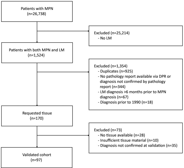
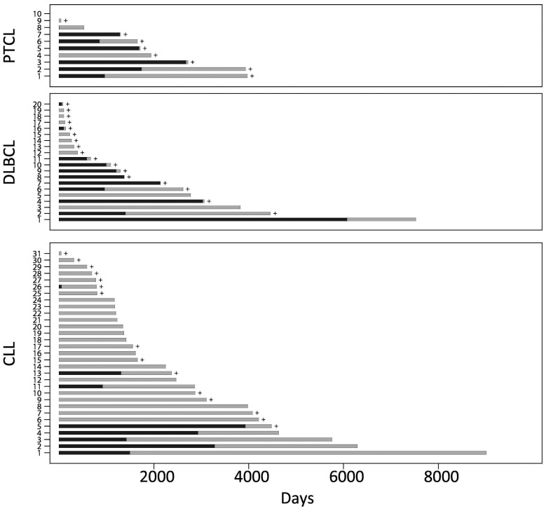
Myeloid and lymphoid malignancies are postulated to have distinct pathogenetic mechanisms. The recent observation that patients with a myeloproliferative neoplasm have an increased risk of developing lymphoproliferative malignancy has challenged this assumption. We collected a nationwide cohort of patients with both malignancies. Patients diagnosed in 1990-2015 were identified through the national Danish Pathology Registry. We identified 599 patients with myeloproliferative neoplasm and a concomitant or subsequent diagnosis of lymphoma. Histopathological review of the diagnostic samples from each patient led to a final cohort of 97 individuals with confirmed dual diagnoses of myeloproliferative neoplasm and lymphoma. The age range at diagnosis was 19-94 years (median: 71 years). To avoid the inclusion of cases of therapy-induced myeloproliferative neoplasm occurring in patients previously treated for lymphoma, only patients with myeloproliferative neoplasm diagnosed unequivocally before the development of lymphoma were included. The average time interval between the diagnoses of the two malignancies was 1.5 years. In the majority of patients (90%) both diagnoses were established within 5 years from each other. Among the lymphoma entities, the frequency of peripheral T-cell lymphomas was markedly increased. Interestingly, all but one of the T-cell lymphomas were of angioimmunoblastic type. These findings suggest that myeloproliferative neoplasm and lymphoproliferative malignancy developing in the same patient may have common pathogenetic events, possibly already at progenitor level. We believe that the molecular characterization of the newly developed biorepository will help to highlight the mechanisms driving the genesis and clonal evolution of these hematopoietic malignancies.
Overview
The claims and statistics of this paper haven’t been checked yet.
Run the check — free, no account needed. It reads the open-access text and checks the sources it cites.
Integrity signals
Registry
Cites
Statistics
Figures
Verification
Claims the paper makes
The claims and statistics of this paper haven’t been checked yet.
Run the check — free, no account needed. It reads the open-access text and checks the sources it cites.
Claims other papers make about this one
Each is checked against this paper’s own text: does it say what the citing paper says it does?
In the citing paper
“It has been noted that myeloid and lymphoid malignancies may co-occur in the same patients”
From Mutation analysis links angioimmunoblastic T-cell lymphoma to clonal hematopoiesis and smoking (2021) · cites this paper as Holst et al., 2020
Self-check
Percentages, sums and reported differences re-computed from the paper’s own numbers.
The self-check needs the paper’s full text.
Citations
Cites
No references could be resolved yet.
Research works printed but not linked: 34
- Dobay M, Lemonnier F, Missiaglia E. Integrative clinicopathological and molecular analyses of angioimmunoblastic T-cell lymphoma and other nodal lymphomas of follicular helper T-cell origin. Haematologica. 2017;201(4):e148-e151. · DOI
- Cairns RA, Iqbal J, Lemonnier F. IDH2 mutations are frequent in angioimmunoblastic T-cell lymphoma. Blood. 2012;119(8):1901-1903. · DOI
- Lemonnier F, Couronne L, Parrens M. Recurrent TET2 mutations in peripheral Tcell lymphomas correlate with TFH-like features and adverse clinical parameters. Blood. 2012;120(7):1466-1469. · DOI · Paper page
- Couronne L, Bastard C, Bernard OA. TET2 and DNMT3A mutations in human T-cell lymphoma. N Engl J Med. 2012;366(1):95-96. · DOI · Paper page
- Sakata-Yanagimoto M, Enami T, Yoshida K. Somatic RHOA mutation in angioimmunoblastic T cell lymphoma. Nat Genet. 2014;46(2):171-175. · DOI
- Kodali S, Chen C, Rathnasabapathy C, Wang JC. JAK2 mutation in a patient with CLL with coexistent myeloproliferative neoplasm (MPN). Leuk Res. 2009;33(12):236-239. · DOI
- Roncero AM, López-Nieva P, Cobos-Fernández MA. Contribution of JAK2 mutations to T-cell lymphoblastic lymphoma development. Leukemia. 2016;30(1):94-103. · DOI
- Swerdlow S, Campo E, Harris N. WHO Classification of Tumours of Haematopoietic and Lymphoid Tissues (revised 4th edition). Lyon. 2017.
- Frederiksen H, Farkas DK, Christiansen CF, Hasselbalch HC, Sørensen HT. Chronic myeloproliferative neoplasms and subsequent cancer risk: a Danish populationbased cohort study. Blood. 2011;118(25):6515-6520. · DOI
- Landtblom AR, Bower H, Andersson TM. Second malignancies in patients with myeloproliferative neoplasms: a population- based cohort study of 9379 patients. Leukemia. 2018;32(10):2203-2210. · DOI
- Marchetti M, Carobbio A, Capitoni E, Barbui T. Lymphoproliferative disorders in patients with chronic myeloproliferative neoplasms: a systematic review. Am J Hematol. 2018;93(5):698-703. · DOI
- Erichsen R, Lash TL, Hamilton-Dutoit SJ, Bjerregaard B, Vyberg M, Pedersen L. Existing data sources for clinical epidemiology: the Danish national pathology registry and data bank. Clin Epidemiol. 2010;2:51-56. · DOI
- Schmidt M, Pedersen L, Sørensen HT. The Danish civil registration system as a tool in epidemiology. Eur J Epidemiol. 2014;29(8):541-549. · DOI
- Arboe B, Josefsson P, Jørgensen J. Danish national lymphoma registry. Clin Epidemiol. 2016;8:577-581. · DOI
- Pedersen M, Hamilton-Dutoit S, Bendix K. DUSP22 and TP63 rearrangements predict outcome of ALK-negative anaplastic large cell lymphoma: a Danish cohort study. Blood. 2017;130(4):554-557. · DOI
- Rumi E, Passamonti F, Elena C. Increased risk of lymphoid neoplasms in patients with myeloproliferative neoplasms: a study of 1,915 patients. Haematologica. 2011;96(3):454-458. · DOI
- Vannucchi AM, Masala G, Antonioli E. Increased risk of lymphoid neoplasms in patients with Philadelphia chromosomenegative myeloproliferative neoplasms. Cancer Epidemiol Biomarkers Prev. 2009;18(7):2068-2073. · DOI
- Khanal N, Giri S, Upadhyay S, Shostrom VK, Pathak R, Bhatt VR. Risk of second primary malignancies and survival of adult patients with polycythemia vera: a United States population-based retrospective study. Leuk Lymphoma. 2016;57(1):129-133. · DOI
- Shrestha R, Giri S, Pathak R, Bhatt VR. Risk of second primary malignancies in a population- based study of adult patients with essential thrombocythemia. World J Clin Oncol. 2016;7(4):324-330. · DOI
- Vose J, Armitage J, Weisenburger D. International peripheral T-cell and natural killer/T-cell lymphoma study: pathology findings and clinical outcomes. J Clin Oncol. 2008;26(25):4124-4130. · DOI
- Laurent C, Baron M, Amara N. Impact of expert pathologic review of lymphoma diagnosis: study of patients from the French Lymphopath Network. J Clin Oncol. 2017;35(18):2008-2017. · DOI
- Federico M, Rudiger T, Bellei M. Clinicopathologic characteristics of angioimmunoblastic T-cell lymphoma: analysis of the International Peripheral Tcell Lymphoma Project. J Clin Oncol. 2013;31(2):240-246. · DOI
- de Leval L, Rickman DS, Thielen C. The gene expression profile of nodal peripheral T-cell lymphoma demonstrates a molecular link between angioimmunoblastic T-cell lymphoma (AITL) and follicular helper T (TFH ) cells. Blood. 2007;109(11):4952-4963. · DOI
- Odejide O, Weigert O, Lane AA. A targeted mutational landscape of angioimmunoblastic T-cell lymphoma. Blood. 2014;123(9):1293-1296. · DOI · Paper page
- Palomero T, Couronné L, Khiabanian H. Recurrent mutations in epigenetic regulators, RHOA and FYN kinase in peripheral T cell lymphomas. Nat Genet. 2014;46(2):166-170. · DOI
- Vallois D, Dobay MPD, Morin RD. Activating mutations in genes related to TCR signaling in angioimmunoblastic and other follicular helper T-cell-derived lymphomas. Blood. 2016;128(11):1490-1502. · DOI
- Rosenquist R, Rosenwald A, Du M-Q. Clinical impact of recurrently mutated genes on lymphoma diagnostics: state-ofthe- art and beyond. Haematologica. 2016;101(9):1002-1009. · DOI
- Nguyen TB, Sakata-Yanagimoto M, Asabe Y. Identification of cell-type-specific mutations in nodal T-cell lymphomas. Blood Cancer J. 2017;7(1):1-10. · DOI · Paper page
- Zang S, Li J, Yang H. Mutations in 5- methylcytosine oxidase TET2 and RhoA cooperatively disrupt T cell homeostasis. J Clin Invest. 2017;127(8):2998-3012. · DOI
- Ng SY, Brown L, Stevenson K. RhoA G17V is sufficient to induce autoimmunity and promotes T-cell lymphomagenesis in mice. Blood. 2018;132(9):935-947. · DOI
- Cortes JR, Ambesi-Impiombato A, Couronné L. RHOA G17V induces T follicular helper cell specification and promotes lymphomagenesis. Cancer Cell. 2018;33(2):259-273. · DOI · Paper page
- Lemonnier F, Dupuis J, Sujobert P. Treatment with 5-azacytidine induces a sustained response in patients with angioimmunoblastic T-cell lymphoma. Blood. 2018;132(21):2305-2309. · DOI
- Frederiksen H, Farkas DK, Christiansen CF. Survival of patients with chronic myeloproliferative neoplasms and new primary cancers: a population-based cohort study. Lancet Haematol. 2015;2(7):e289-e296. · DOI
- Porpaczy E, Tripolt S, Hoelbl-Kovacic A. Aggressive B-cell lymphomas in patients with myelofibrosis receiving JAK1/2 inhibitor therapy. Blood. 2018;132(7):694-706. · DOI · Paper page
Cited by
Mutation analysis links angioimmunoblastic T-cell lymphoma to clonal hematopoiesis and smoking
Cheng, S., Zhang, W., Inghirami, G., & Tam, W. (2021) · eLife · doi:10.7554/elife.66395 · Cited by 37
Self-citation
Metadata
- Authors
- Holst, J. M., Plesner, T. L., Pedersen, M. B., Frederiksen, H., Møller, M. B., Clausen, M. R., Hansen, M. C., Hamilton-Dutoit, S. J., Nørgaard, P., Johansen, P., Eberlein, T. R., Mortensen, B. K., Mathiasen, G., Øvlisen, A., Wang, R., Wang, C., Zhang, W., Ommen, H. B., Stentoft, J., Ludvigsen, M., Tam, W., Chan, W. C., Inghirami, G., & d'Amore, F.
- Venue
- Haematologica
- Published
- 2019-11-28
- Volume
- 105
- Issue
- 10
- Pages
- 2432-2439
- ISSN
- 1592-8721
- 0390-6078
- PMC
- PMC7556673
- Cited by
- 25
- References
- 0
About these checks
Registry, retraction, Cites and statistics results are computed, not judged, with one exception: a printed reference that no index lists is read once by a model before it counts as a citation error. Claims are extracted by a model and kept only when their quote appears verbatim in the text; each cited source is then checked, with a model’s help, for whether it says what the claim relies on. Those verdicts can be wrong — read the claim’s quote and the cited source before relying on one.
Statistics and arithmetic are recomputed, not judged.
Confidence bands are shown only for fields calibrated on our annotated test set. None is calibrated yet, so no band is shown.
What counts as a claim ›How we measure confidence ›How we check this ›
Are you an author, or do you see a problem with this page? Request a review
