

The DOI matches its Crossref record; 3 of the 3 references it cites were found in a registry or an index.
Tau phosphorylation and PAD exposure in regulation of axonal growth
Abstract
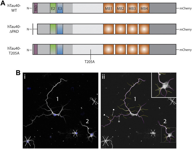
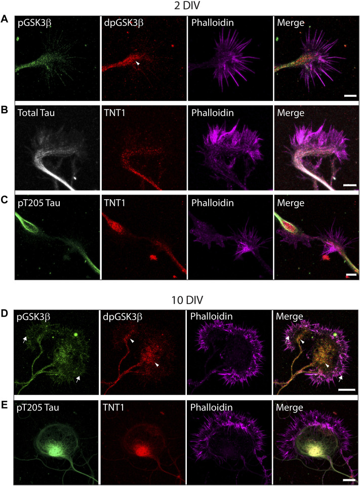
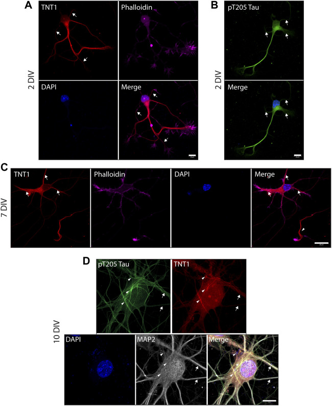
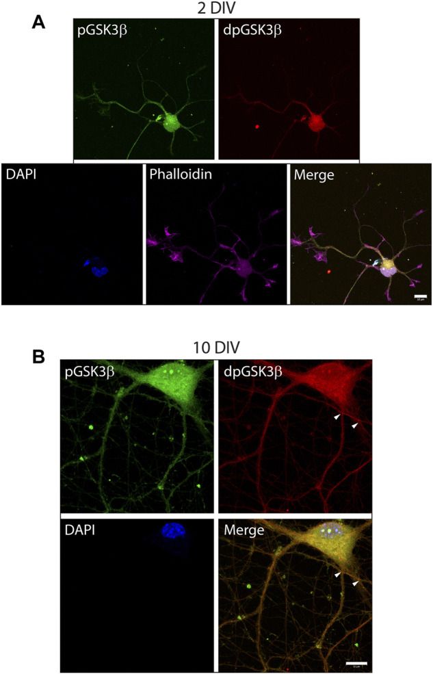
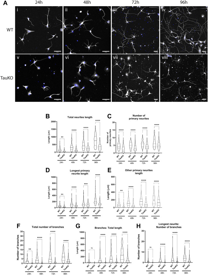
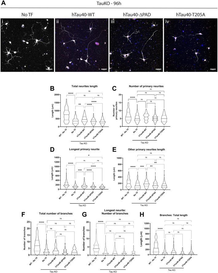
Introduction: Tau is a microtubule associated phosphoprotein found principally in neurons. Prevailing dogma continues to define microtubule stabilization as the major function of tau in vivo, despite several lines of evidence suggesting this is not the case. Most importantly, tau null mice have deficits in axonal outgrowth and neuronal migration while still possessing an extensive microtubule network. Instead, mounting evidence suggests that tau may have a major function in the regulation of fast axonal transport (FAT) through activation of neuronal signaling pathways. Previous studies identified a phosphatase activating domain (PAD) at the tau N-terminal that is normally sequestered, but is constitutively exposed in tauopathies. When exposed, the PAD activates a signaling cascade involving PP1 and GSK3β which affects cellular functions including release of cargo from kinesin. Furthermore, we discovered that PAD exposure can be regulated by a single phosphorylation at T205. Exposure of the PAD is an early event in multiple tauopathies and a major contributing factor to neurodegeneration associated with tau hyperphosphorylation. However, effects of tau PAD exposure on anterograde FAT raised the interesting possibility that this pathway may be a mechanism for physiological regulation of cargo delivery through site-specific phosphorylation of tau and transient activation of PP1 and GSK3β. Significantly, there is already evidence of local control of PP1 and GSK3β at sites which require cargo delivery. Methods: To investigate this hypothesis, first we evaluated cellular localization of tau PAD exposure, pT205 tau phosphorylation, and active GSK3β in primary hippocampal neurons during development. Second, we analyzed the axonal outgrowth of tau knockout neurons following transfection with full length hTau40-WT, hTau40-ΔPAD, or hTau40-T205A. Results and Discussion: The results presented here suggest that transient activation of a PP1-GSK3β signaling pathway through locally regulated PAD exposure is a mechanism for cargo delivery, and thereby important for neurite outgrowth of developing neurons.
Overview
The claims and statistics of this paper haven’t been checked yet.
Run the check — free, no account needed. It reads the open-access text and checks the sources it cites.
Integrity signals
Registry
Cites
Statistics
Figures
Verification
Claims the paper makes
The claims and statistics of this paper haven’t been checked yet.
Run the check — free, no account needed. It reads the open-access text and checks the sources it cites.
Claims other papers make about this one
Each is checked against this paper’s own text: does it say what the citing paper says it does?
In the citing paper
“hTau overexpression rescued neurite outgrowth defects in tau knockout neurons, and phosphorylation at tau T205 was necessary to rescue this defect43.”
From Multi-omic phenotyping of MAPT V337M neurons reveals early changes in axonogenesis and tau phosphorylation (2026) · cites this paper as [43]
In the citing paper
“tau knockout in primary hippocampal neurons and human iPSC-derived neurons reduces neurite outgrowth43–45.”
From Multi-omic phenotyping of MAPT V337M neurons reveals early changes in axonogenesis and tau phosphorylation (2026) · cites this paper as [43]
Self-check
Percentages, sums and reported differences re-computed from the paper’s own numbers.
The self-check needs the paper’s full text.
Citations
Cites
Kinesin superfamily motor proteins and intracellular transport
Hirokawa, N., Noda, Y., Tanaka, Y., & Niwa, S. (2009) · Nature Reviews Molecular Cell Biology · doi:10.1038/nrm2774 · Cited by 1,859 · cited 1×
“KIF5 motors have been suggested to have functions in both axons and dendrites (Setou et al., 2002; Kanai et al., 2004; Hirokawa et al., 2009), although other studies have found that KIF5 is excluded from dendrites (Jacobson et al.”
ResultTanaka, Y., Kanai, Y., Okada, Y., Nonaka, S., Takeda, S., Harada, A., & Hirokawa, N. (1998) · Cell · doi:10.1016/s0092-8674(00)81459-2 · Cited by 654
Tau Is Enriched on Dynamic Microtubules in the Distal Region of Growing Axons
Black, M. M., Slaughter, T., Moshiach, S., Obrocka, M., & Fischer, I. (1996) · The Journal of Neuroscience · doi:10.1523/jneurosci.16-11-03601.1996 · Cited by 270
Research works printed but not linked: 62
- Ahmari, 2000, Assembly of presynaptic active zones from cytoplasmic transport packets, Nat. Neurosci., 3, 445, 10.1038/74814 · DOI
- Ahmari, 2002, Knowing a nascent synapse when you see it, Neuron, 34, 333, 10.1016/s0896-6273(02)00685-2 · DOI
- Bartlett, 1984, An electron microscopic study of the development of axons and dendrites by hippocampal neurons in culture. II. Synaptic relationships, J. Neurosci. Off. J. Soc. Neurosci., 4, 1954, 10.1523/JNEUROSCI.04-08-01954.1984 · DOI · Paper page
- Bibow, 2011, Structural impact of proline-directed pseudophosphorylation at AT8, AT100, and PHF1 epitopes on 441-residue tau, J. Am. Chem. Soc., 133, 15842, 10.1021/ja205836j · DOI
- Bolte, 2006, A guided tour into subcellular colocalization analysis in light microscopy, J. Microsc., 224, 213, 10.1111/j.1365-2818.2006.01706.x · DOI
- Bury, 2011, Coordinated trafficking of synaptic vesicle and active zone proteins prior to synapse formation, Neural Dev., 6, 24, 10.1186/1749-8104-6-24 · DOI
- Caceres, 1990, Inhibition of neurite polarity by tau antisense oligonucleotides in primary cerebellar neurons, Nature, 343, 461, 10.1038/343461a0 · DOI · Paper page
- Caceres, 1991, The effect of tau antisense oligonucleotides on neurite formation of cultured cerebellar macroneurons, J. Neurosci. Off. J. Soc. Neurosci., 11, 1515, 10.1523/JNEUROSCI.11-06-01515.1991 · DOI · Paper page
- Combs, 2016, Pathological conformations involving the amino terminus of tau occur early in Alzheimer’s disease and are differentially detected by monoclonal antibodies, Neurobiol. Dis., 94, 18, 10.1016/j.nbd.2016.05.016 · DOI
- Combs, 2017, Exposure of the amino terminus of tau is a pathological event in multiple tauopathies, Am. J. Pathol., 187, 1222, 10.1016/j.ajpath.2017.01.019 · DOI
- Cox, 2016, Analysis of isoform-specific tau aggregates suggests a common toxic mechanism involving similar pathological conformations and axonal transport inhibition, Neurobiol. Aging, 47, 113, 10.1016/j.neurobiolaging.2016.07.015 · DOI
- Craig, 1995, Preferential addition of newly synthesized membrane protein at axonal growth cones, Nature, 375, 592, 10.1038/375592a0 · DOI
- Dawson, 2001, Inhibition of neuronal maturation in primary hippocampal neurons from tau deficient mice, J. Cell Sci., 114, 1179, 10.1242/jcs.114.6.1179 · DOI · Paper page
- de Waegh, 1992, Local modulation of neurofilament phosphorylation, axonal caliber, and slow axonal transport by myelinating Schwann cells, Cell, 68, 451, 10.1016/0092-8674(92)90183-D · DOI
- DeBoer, 2008, Conventional kinesin holoenzymes are composed of heavy and light chain homodimers, Biochemistry, 47, 4535, 10.1021/bi702445j · DOI
- Devaux, 2012, Specific serine-proline phosphorylation and glycogen synthase kinase 3β-directed subcellular targeting of stathmin 3/Sclip in neurons, J. Biol. Chem., 287, 22341, 10.1074/jbc.M112.344044 · DOI
- DiTella, 1996, MAP-1B/TAU functional redundancy during laminin-enhanced axonal growth, J. Cell Sci., 109, 467, 10.1242/jcs.109.2.467 · DOI
- Dotti, 1988, The establishment of polarity by hippocampal neurons in culture, J. Neurosci. Off. J. Soc. Neurosci., 8, 1454, 10.1523/JNEUROSCI.08-04-01454.1988 · DOI · Paper page
- Dubourg, 2011, Clinical and molecular characterization of 17q21.31 microdeletion syndrome in 14 French patients with mental retardation, Eur. J. Med. Genet., 54, 144, 10.1016/j.ejmg.2010.11.003 · DOI
- Fiock, 2020, Increased tau expression correlates with neuronal maturation in the developing human cerebral cortex, eNeuro, 7, ENEURO.0058, 10.1523/ENEURO.0058-20.2020 · DOI
- Fletcher, 1989, The establishment of polarity by hippocampal neurons: The relationship between the stage of a cell’s development in situ and its subsequent development in culture, Dev. Biol., 136, 446, 10.1016/0012-1606(89)90269-8 · DOI
- Futerman, 1996, The economics of neurite outgrowth — The addition of new membrane to growing axons, Trends Neurosci., 19, 144, 10.1016/S0166-2236(96)80025-7 · DOI
- Grabinski, 2016, Novel non-phosphorylated serine 9/21 gsk3β/α antibodies: Expanding the tools for studying GSK3 regulation, Front. Mol. Neurosci., 9, 123, 10.3389/fnmol.2016.00123 · DOI
- Harada, 1994, Altered microtubule organization in small-calibre axons of mice lacking tau protein, Nature, 369, 488, 10.1038/369488a0 · DOI · Paper page
- Hsieh, 1994, Regional modulation of neurofilament organization by myelination in normal axons, J. Neurosci. Off. J. Soc. Neurosci., 14, 6392, 10.1523/JNEUROSCI.14-11-06392.1994 · DOI
- Ikegami, 2000, Muscle weakness, hyperactivity, and impairment in fear conditioning in tau-deficient mice, Neurosci. Lett., 279, 129, 10.1016/S0304-3940(99)00964-7 · DOI
- Iworima, 2016, Kif5 regulates mitochondrial movement, morphology, function and neuronal survival, Mol. Cell. Neurosci., 72, 22, 10.1016/j.mcn.2015.12.014 · DOI
- Jacobson, 2006, A change in the selective translocation of the kinesin-1 motor domain marks the initial specification of the axon, Neuron, 49, 797, 10.1016/j.neuron.2006.02.005 · DOI
- Jeganathan, 2008, Proline-directed pseudo-phosphorylation at AT8 and PHF1 epitopes induces a compaction of the paperclip folding of tau and generates a pathological (MC-1) conformation, J. Biol. Chem., 283, 32066, 10.1074/jbc.M805300200 · DOI · Paper page
- Kanaan, 2016, Characterization of early pathological tau conformations and phosphorylation in chronic traumatic encephalopathy, J. Neuropathol. Exp. Neurol., 75, 19, 10.1093/jnen/nlv001 · DOI
- Kanaan, 2011, Pathogenic forms of tau inhibit kinesin-dependent axonal transport through a mechanism involving activation of axonal phosphotransferases, J. Neurosci. Off. J. Soc. Neurosci., 31, 9858, 10.1523/JNEUROSCI.0560-11.2011 · DOI
- Kanaan, 2012, Phosphorylation in the amino terminus of tau prevents inhibition of anterograde axonal transport, Neurobiol. Aging, 33, e15, 10.1016/j.neurobiolaging.2011.06.006 · DOI · Paper page
- Kanai, 2004, Kinesin transports RNA: Isolation and characterization of an RNA-transporting granule, Neuron, 43, 513, 10.1016/j.neuron.2004.07.022 · DOI
- Karasmanis, 2018, Polarity of neuronal membrane traffic requires sorting of kinesin motor cargo during entry into dendrites by a microtubule-associated septin, Dev. Cell, 46, 204, 10.1016/j.devcel.2018.06.013 · DOI
- Karle, 2012, Axonal transport deficit in a KIF5A(-/-) mouse model, Neurogenetics, 13, 169, 10.1007/s10048-012-0324-y · DOI
- LaPointe, 2009, The amino terminus of tau inhibits kinesin-dependent axonal transport: Implications for filament toxicity, J. Neurosci. Res., 87, 440, 10.1002/jnr.21850 · DOI
- Li, 2004, A syntaxin 1, galphao, and N-type calcium channel complex at a presynaptic nerve terminal: Analysis by quantitative immunocolocalization, J. Neurosci., 24, 4070, 10.1523/jneurosci.0346-04.2004 · DOI
- Liu, 2013, Profiling murine tau with 0N, 1N and 2N isoform-specific antibodies in brain and peripheral organs reveals distinct subcellular localization, with the 1N isoform being enriched in the nucleus, PLOS ONE, 8, e84849, 10.1371/journal.pone.0084849 · DOI
- Liu, 2005, Contributions of protein phosphatases PP1, PP2A, PP2B and PP5 to the regulation of tau phosphorylation, Eur. J. Neurosci., 22, 1942, 10.1111/j.1460-9568.2005.04391.x · DOI · Paper page
- Liu, 2019, Loss of tau and Fyn reduces compensatory effects of MAP2 for tau and reveals a Fyn-independent effect of tau on calcium, J. Neurosci. Res. Jnr., 24517, 1393, 10.1002/jnr.24517 · DOI
- Martenson, 1993, Fast axonal transport is required for growth cone advance, Nature, 366, 66, 10.1038/366066a0 · DOI
- Martini, 2001, The effect of myelinating Schwann cells on axons, Muscle Nerve, 24, 456, 10.1002/mus.1027 · DOI
- Morfini, 2007, Tau binding to microtubules does not directly affect microtubule-based vesicle motility, J. Neurosci. Res., 85, 2620, 10.1002/jnr.21154 · DOI
- Morfini, 2004, A novel CDK5-dependent pathway for regulating GSK3 activity and kinesin-driven motility in neurons, EMBO J., 23, 2235, 10.1038/sj.emboj.7600237 · DOI
- Morfini, 2002, Glycogen synthase kinase 3 phosphorylates kinesin light chains and negatively regulates kinesin-based motility, EMBO J., 21, 281, 10.1093/emboj/21.3.281 · DOI
- Morris, 2021, Phosphorylation of tau at specific sites regulates axonal transport and neurite outgrowth
- Morris, 2021, Lack of neurosteroid selectivity at δ vs. γ2-containing GABAA receptors in dentate granule neurons, Front. Mol. Neurosci., 13, 6, 10.3389/fnmol.2020.00006 · DOI
- Nakata, 2003, Microtubules provide directional cues for polarized axonal transport through interaction with kinesin motor head, J. Cell Biol., 162, 1045, 10.1083/jcb.200302175 · DOI
- Pfenninger, 1993, Sites of plasmalemmal expansion in growth cones, Brain Res. Dev. Brain Res., 71, 181, 10.1016/0165-3806(93)90170-f · DOI
- Sapir, 2012, Tau’s role in the developing brain: Implications for intellectual disability, Hum. Mol. Genet., 21, 1681, 10.1093/hmg/ddr603 · DOI
- Sennvik, 2007, Tau-4R suppresses proliferation and promotes neuronal differentiation in the hippocampus of tau knockin/knockout mice, FASEB J., 21, 2149, 10.1096/fj.06-7735com · DOI · Paper page
- Setou, 2002, Glutamate-receptor-interacting protein GRIP1 directly steers kinesin to dendrites, Nature, 417, 83, 10.1038/nature743 · DOI
- Shaw-Smith, 2006, Microdeletion encompassing MAPT at chromosome 17q21.3 is associated with developmental delay and learning disability, Nat. Genet., 38, 1032, 10.1038/ng1858 · DOI
- Smith, 2018, The role of mitochondria in axon development and regeneration, Dev. Neurobiol., 78, 221, 10.1002/dneu.22546 · DOI
- Sontag, 2012, The protein phosphatase PP2A/bα binds to the microtubule-associated proteins tau and MAP2 at a motif also recognized by the kinase Fyn implications for tauopathies, J. Biol. Chem., 287, 14984, 10.1074/jbc.M111.338681 · DOI
- Szodorai, 2009, APP anterograde transport requires Rab3A GTPase activity for assembly of the transport vesicle, J. Neurosci., 29, 14534, 10.1523/JNEUROSCI.1546-09.2009 · DOI
- Takei, 2000, Defects in axonal elongation and neuronal migration in mice with disrupted tau and map1b genes, J. Cell Biol., 150, 989, 10.1083/jcb.150.5.989 · DOI
- Takuma, 2003, Isoforms changes of tau protein during development in various species, Dev. Brain Res., 142, 121, 10.1016/S0165-3806(03)00056-7 · DOI
- Trivedi, 2005, Glycogen synthase kinase-3beta phosphorylation of MAP1B at Ser1260 and Thr1265 is spatially restricted to growing axons, J. Cell Sci., 118, 993, 10.1242/jcs.01697 · DOI
- Wang, 2015, Rapid alteration of protein phosphorylation during postmortem: Implication in the study of protein phosphorylation, Sci. Rep., 5, 15709, 10.1038/srep15709 · DOI
- Washbourne, 2015, Synapse assembly and neurodevelopmental disorders, Neuropsychopharmacology, 40, 4, 10.1038/npp.2014.163 · DOI
- Weingarten, 1975, A protein factor essential for microtubule assembly, Proc. Natl. Acad. Sci., 72, 1858, 10.1073/pnas.72.5.1858 · DOI · Paper page
Cited by
Mohl, G. A., Dixon, G., Marzette, E., McKetney, J., Samelson, A. J., Serras, C. P., Jin, J., Ariqat, N., Keys, A., Chavira, C., et al. (2026) · npj Dementia · doi:10.1038/s44400-026-00076-w · Cited by 5
Tau Ablation Rewires Brain Cell Programs in Health and Restores Function in Disease
Chimal-Juárez, E., Patel, H., Jury-Garfe, N., Dabin, L. C., Vidal, R., Kim, J., & Lasagna-Reeves, C. A. (2026) · bioRxiv · doi:10.64898/2026.05.12.724684
Metadata
- Authors
- Morris, S. L., & Brady, S. T.
- Venue
- Frontiers in Cell and Developmental Biology
- Publisher
- Frontiers Media SA
- Published
- 2023-01-18
- Volume
- 10
- ISSN
- 2296-634X
- PMC
- PMC9893789
- Cited by
- 18
- References
- 3
About these checks
Registry, retraction, Cites and statistics results are computed, not judged, with one exception: a printed reference that no index lists is read once by a model before it counts as a citation error. Claims are extracted by a model and kept only when their quote appears verbatim in the text; each cited source is then checked, with a model’s help, for whether it says what the claim relies on. Those verdicts can be wrong — read the claim’s quote and the cited source before relying on one.
Statistics and arithmetic are recomputed, not judged.
Confidence bands are shown only for fields calibrated on our annotated test set. None is calibrated yet, so no band is shown.
What counts as a claim ›How we measure confidence ›How we check this ›
Are you an author, or do you see a problem with this page? Request a review
Data sources
Bibliographic data: Semantic Scholar, OpenAlex, Crossref.
Contains information from the Semantic Scholar Academic Graph Datasets, made available under the ODC Attribution License (ODC-BY 1.0).
Semantic Scholar’s data is described in: Kinney, R., et al. (2023). The Semantic Scholar Open Data Platform. arXiv:2301.10140.
