

The DOI matches its Crossref record; 1 of the 1 references it cites were found in a registry or an index.
Gardnerella Exposures Alter Bladder Gene Expression and Augment Uropathogenic Escherichia coli Urinary Tract Infection in Mice
Open accessAbstract
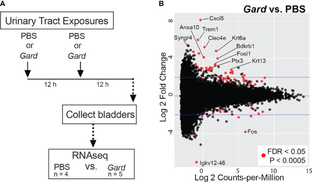
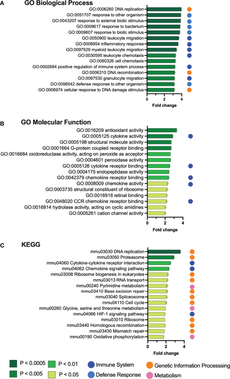
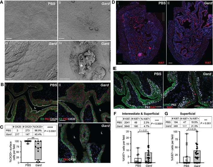
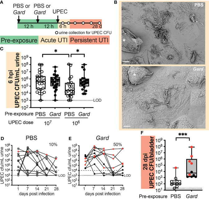
The anaerobic actinobacterium Gardnerella was first isolated from the bladder by suprapubic aspiration more than 50 years ago. Since then, Gardnerella has been increasingly recognized as a common and often abundant member of the female urinary microbiome (urobiome). Some studies even suggest that the presence of Gardnerella is associated with urological disorders in women. We recently reported that inoculation of Gardnerella into the bladders of mice results in urothelial exfoliation. Here, we performed whole bladder RNA-seq in our mouse model to identify additional host pathways involved in the response to Gardnerella bladder exposure. The transcriptional response to Gardnerella reflected the urothelial turnover that is a consequence of exfoliation while also illustrating the activation of pathways involved in inflammation and immunity. Additional timed exposure experiments in mice provided further evidence of a potentially clinically relevant consequence of bladder exposure to Gardnerella—increased susceptibility to subsequent UTI caused by uropathogenic Escherichia coli. Together, these data provide a broader picture of the bladder’s response to Gardnerella and lay the groundwork for future studies examining the impact of Gardnerella on bladder health.
Overview
The claims and statistics of this paper haven’t been checked yet.
Run the check — free, no account needed. It reads the open-access text and checks the sources it cites.
Integrity signals
Registry
Cites
Statistics
Figures
Verification
Claims the paper makes
The claims and statistics of this paper haven’t been checked yet.
Run the check — free, no account needed. It reads the open-access text and checks the sources it cites.
Claims other papers make about this one
Each is checked against this paper’s own text: does it say what the citing paper says it does?
The source backs part of the claim, not all of it — or backs it for a different population, condition or comparison.
In the citing paper
“In addition to these classical uropathogens, several vaginal species, such as Gardnerella vaginalis , Aerococcus , and Ureaplasma are increasingly recognized as contributors to urinary tract pathology, either through direct infection or modulation of host susceptibility [11].”
From Vaginal microbiota and urinary tract infections in women: the protective role of vaginal lactobacilli in counteracting urinary tract infections occurrence and recurrence (2026) · cites this paper as [11]
Experimental — accuracy not yet measured
Self-check
Percentages, sums and reported differences re-computed from the paper’s own numbers.
The self-check needs the paper’s full text.
Citations
Cites
The Etiology and Management of Recurrent Urinary Tract Infections in Postmenopausal Women
Jung, C., & Brubaker, L. (2019) · Climacteric · doi:10.1080/13697137.2018.1551871 · Cited by 169
Research works printed but not linked: 65
- Birch, 1981, Ureaplasma Urealyticum in the Upper Urinary Tracts of Renal Allograft Recipients, J. Infect. Dis., 144, 123, 10.1093/infdis/144.2.123 · DOI
- Borsodi, 2021, Signaling Pathways Mediating Bradykinin-Induced Contraction in Murine and Human Detrusor Muscle, Front. Med. (Lausanne), 8, 10.3389/fmed.2021.745638 · DOI
- Briselden, 1990, Longitudinal Study of the Biotypes of Gardnerella Vaginalis, J. Clin. Microbiol., 28, 2761, 10.1128/jcm.28.12.2761-2764.1990 · DOI
- Brown, 2021, Assessing the Concordance Between Urogenital and Vaginal Microbiota: Can Urine Specimens Be Used as a Proxy for Vaginal Samples, Front. Cell Infect. Microbiol., 11, 10.3389/fcimb.2021.671413 · DOI
- Cao, 2010, Keratin 6 Expression Correlates to Areas of Squamous Differentiation in Multiple Independent Isolates of As(+3)-Induced Bladder Cancer, J. Appl. Toxicol., 30, 416, 10.1002/jat.1513 · DOI
- Cole, 2021, The Pediatric Urobiome in Genitourinary Conditions: A Narrative Review, Pediatr. Nephrol, 10.1007/s00467-021-05274-7 · DOI
- Conover, 2015, Establishment and Characterization of UTI and CAUTI in a Mouse Model, J. Vis. Exp., 100, 10.3791/52892 · DOI
- Dalghi, 2020, The Urothelium: Life in a Liquid Environment, Physiol. Rev., 100, 1621, 10.1152/physrev.00041.2019 · DOI
- Dong, 2015, The Actions of Neurotensin in Rat Bladder Detrusor Contractility, Sci. Rep., 5, 11192, 10.1038/srep11192 · DOI
- Fairley, 1983, Unconventional Bacteria in Urinary Tract Disease: Gardnerella Vaginalis, Kidney Int., 23, 862, 10.1038/ki.1983.107 · DOI
- Foxman, 2014, Urinary Tract Infection Syndromes: Occurrence, Recurrence, Bacteriology, Risk Factors, and Disease Burden, Infect. Dis. Clin. North Am., 28, 1, 10.1016/j.idc.2013.09.003 · DOI
- Gardner, 1954, New Etiologic Agent in Nonspecific Bacterial Vaginitis, Science, 120, 853, 10.1126/science.120.3125.853 · DOI
- Gardner, 1955, Haemophilus Vaginalis Vaginitis: A Newly Defined Specific Infection Previously Classified Non-Specific Vaginitis, Am. J. Obstet. Gynecol., 69, 962, 10.1016/0002-9378(55)90095-8 · DOI
- Garofalo, 2022, Whole-Person, Urobiome-Centric Therapy for Uncomplicated Urinary Tract Infection, Antibiot. (Basel), 11, 10.3390/antibiotics11020218 · DOI · Paper page
- Gatta, 2019, Hyper-Activation of STAT3 Sustains Progression of Non-Papillary Basal-Type Bladder Cancer via FOSL1 Regulome, Cancers (Basel), 11, 10.3390/cancers11091219 · DOI
- Gilbert, 1986, Bacteriuria Due to Ureaplasmas and Other Fastidious Organisms During Pregnancy: Prevalence and Significance, Pediatr. Infect. Dis., 5, S239, 10.1097/00006454-198611010-00007 · DOI
- Gilbert, 2019, Covert Pathogenesis: Transient Exposures to Microbes as Triggers of Disease, PLoS Pathog., 15, 10.1371/journal.ppat.1007586 · DOI
- Gilbert, 2017, Transient Microbiota Exposures Activate Dormant Escherichia Coli Infection in the Bladder and Drive Severe Outcomes of Recurrent Disease, PLoS Pathog., 13, 10.1371/journal.ppat.1006238 · DOI
- Gottschick, 2017, The Urinary Microbiota of Men and Women and its Changes in Women During Bacterial Vaginosis and Antibiotic Treatment, Microbiome, 5, 99, 10.1186/s40168-017-0305-3 · DOI
- Hannan, 2010, Early Severe Inflammatory Responses to Uropathogenic E. Coli Predispose to Chronic and Recurrent Urinary Tract Infection, PLoS Pathog., 6, 10.1371/journal.ppat.1001042 · DOI
- Hillebrand, 2002, Urinary Tract Infections in Pregnant Women With Bacterial Vaginosis, Am. J. Obstet. Gynecol., 186, 916, 10.1067/mob.2002.123987 · DOI
- Hilt, 2014, Urine Is Not Sterile: Use of Enhanced Urine Culture Techniques to Detect Resident Bacterial Flora in the Adult Female Bladder, J. Clin. Microbiol., 52, 871, 10.1128/JCM.02876-13 · DOI
- Hooton, 1989, Association Between Bacterial Vaginosis and Acute Cystitis in Women Using Diaphragms, Arch. Intern. Med., 149, 1932, 10.1001/archinte.1989.00390090014003 · DOI
- Hugenholtz, 2022, Urine and Vaginal Microbiota Compositions of Postmenopausal and Premenopausal Women Differ Regardless of Recurrent Urinary Tract Infection and Renal Transplant Status, Sci. Rep., 12, 2698, 10.1038/s41598-022-06646-1 · DOI
- Jacobs, 2017, Microorganisms Identified in the Maternal Bladder: Discovery of the Maternal Bladder Microbiota, AJP Rep., 7, e188, 10.1055/s-0037-1606860 · DOI
- Jones, 2021, The Urobiome, Urinary Tract Infections, and the Need for Alternative Therapeutics, Microb. Pathog., 161, 105295, 10.1016/j.micpath.2021.105295 · DOI
- Klein, 2020, Urinary Tract Infections: Microbial Pathogenesis, Host-Pathogen Interactions and New Treatment Strategies, Nat. Rev. Microbiol., 18, 211, 10.1038/s41579-020-0324-0 · DOI
- Kline, 2016, Gram-Positive Uropathogens, Polymicrobial Urinary Tract Infection, and the Emerging Microbiota of the Urinary Tract, Microbiol. Spectr., 4, 10.1128/microbiolspec.UTI-0012-2012 · DOI
- Komesu, 2019, Defining the Relationship Between Vaginal and Urinary Microbiomes, Am. J. Obstet. Gynecol, 222, 154.e1, 10.1016/j.ajog.2019.08.011 · DOI
- Krohn, 1989, Comparison of Methods for Diagnosing Bacterial Vaginosis Among Pregnant Women, J. Clin. Microbiol., 27, 1266, 10.1128/jcm.27.6.1266-1271.1989 · DOI
- Kudelski, 2021, Enhanced Expression But Decreased Specific Activity of Matrix Metalloproteinase 10 (MMP-10) in Comparison With Matrix Metalloproteinase 3 (MMP-3) in Human Urinary Bladder Carcinoma, J. Clin. Med., 10, 10.3390/jcm10163683 · DOI
- Lacerda Mariano, 2020, The Immune Response to Infection in the Bladder, Nat. Rev. Urol, 17, 439, 10.1038/s41585-020-0350-8 · DOI
- Lee, 2021, Trefoil Factor 1 Suppresses Epithelial-Mesenchymal Transition Through Inhibition of TGF-Beta Signaling in Gastric Cancer Cells, J. Cancer Prev., 26, 137, 10.15430/JCP.2021.26.2.137 · DOI
- Leopold, 1953, Heretofore Undescribed Organism Isolated From the Genitourinary System, U S Armed Forces Med. J., 4, 263
- Lewis, 2013, Degradation, Foraging, and Depletion of Mucus Sialoglycans by the Vagina-Adapted Actinobacterium Gardnerella Vaginalis, J. Biol. Chem., 288, 12067, 10.1074/jbc.M113.453654 · DOI
- Lin, 2015, Role of Hypoxia Inducible Factor-1alpha (HIF-1alpha) in Innate Defense Against Uropathogenic Escherichia Coli Infection, PloS Pathog., 11, 10.1371/journal.ppat.1004818 · DOI
- Lu, 2021, N6-Methyladenosine-Related Non-Coding RNAs Are Potential Prognostic and Immunotherapeutic Responsiveness Biomarkers for Bladder Cancer, EPMA J., 12, 589, 10.1007/s13167-021-00259-w · DOI
- Marceau, 1997, The Kinin B1 Receptor: An Inducible G Protein Coupled Receptor, Can. J. Physiol. Pharmacol., 75, 725, 10.1139/y97-049 · DOI
- McDonald, 1982, Ureaplasma Urealyticum in Patients With Acute Symptoms of Urinary Tract Infection, J. Urol, 128, 517, 10.1016/S0022-5347(17)53025-6 · DOI
- McDowall, 1981, Anaerobic and Other Fastidious Microorganisms in Asymptomatic Bacteriuria in Pregnant Women, J. Infect. Dis., 144, 114, 10.1093/infdis/144.2.114 · DOI
- Mcfadyen, 1968, Suprapubic Aspiration of Urine in Pregnancy, Lancet, 1, 1112, 10.1016/S0140-6736(68)90185-2 · DOI
- Morrill, 2020, Gardnerella Vaginalis as a Cause of Bacterial Vaginosis: Appraisal of the Evidence From In Vivo Models, Front. Cell Infect. Microbiol., 10, 10.3389/fcimb.2020.00168 · DOI
- Mulvey, 2001, Establishment of a Persistent Escherichia Coli Reservoir During the Acute Phase of a Bladder Infection, Infect. Immun., 69, 4572, 10.1128/IAI.69.7.4572-4579.2001 · DOI
- Mulvey, 2000, Bad Bugs and Beleaguered Bladders: Interplay Between Uropathogenic Escherichia Coli and Innate Host Defenses, Proc. Natl. Acad. Sci. U.S.A., 97, 8829, 10.1073/pnas.97.16.8829 · DOI
- Mysorekar, 2006, Mechanisms of Uropathogenic Escherichia Coli Persistence and Eradication From the Urinary Tract, Proc. Natl. Acad. Sci. U.S.A., 103, 14170, 10.1073/pnas.0602136103 · DOI
- Nicolle, 1982, The Association of Urinary Tract Infection With Sexual Intercourse, J. Infect. Dis., 146, 579, 10.1093/infdis/146.5.579 · DOI
- O'Brien, 2020, Recurrent Escherichia Coli Urinary Tract Infection Triggered by Gardnerella Vaginalis Bladder Exposure in Mice, J. Vis. Exp, 2020, 10.3791/61967 · DOI
- Pearce, 2014, The Female Urinary Microbiome: A Comparison of Women With and Without Urgency Urinary Incontinence, MBio, 5, e01283, 10.1128/mBio.01283-14 · DOI
- Pearce, 2015, The Female Urinary Microbiome in Urgency Urinary Incontinence, Am. J. Obstet. Gynecol., 213, 10.1016/j.ajog.2015.07.009 · DOI
- Price, 2020, The Urobiome of Continent Adult Women: A Cross-Sectional Study, BJOG, 127, 193, 10.1111/1471-0528.15920 · DOI
- Putonti, 2021, Genome Investigation of Urinary Gardnerella Strains and Their Relationship to Isolates of the Vaginal Microbiota, mSphere, 6, 10.1128/mSphere.00154-21 · DOI
- Ravel, 2011, Vaginal Microbiome of Reproductive-Age Women, Proc. Natl. Acad. Sci. U.S.A., 4680, 10.1073/pnas.1002611107 · DOI · Paper page
- Schwartz, 2015, Uropathogenic Escherichia Coli Superinfection Enhances the Severity of Mouse Bladder Infection, PLoS Pathog., 11, 10.1371/journal.ppat.1004599 · DOI
- Schwebke, 2014, Role of Gardnerella Vaginalis in the Pathogenesis of Bacterial Vaginosis: A Conceptual Model, J. Infect. Dis., 210, 338, 10.1093/infdis/jiu089 · DOI
- Somji, 2011, Comparison of Expression Patterns of Keratin 6, 7, 16, 17, and 19 Within Multiple Independent Isolates of As(+3)- and Cd (+2)-Induced Bladder Cancer : Keratin 6, 7, 16, 17, and 19 in Bladder Cancer, Cell Biol. Toxicol., 27, 381, 10.1007/s10565-010-9169-z · DOI
- Sumati, 2009, Association of Urinary Tract Infection in Women With Bacterial Vaginosis, J. Glob. Infect. Dis., 1, 151, 10.4103/0974-777X.56254 · DOI
- Tate, 2021, Pparg Signaling Controls Bladder Cancer Subtype and Immune Exclusion, Nat. Commun., 12, 6160, 10.1038/s41467-021-26421-6 · DOI
- Tessarz, 2008, The TREM-1/DAP12 Pathway, Immunol. Lett., 116, 111, 10.1016/j.imlet.2007.11.021 · DOI
- Vaneechoutte, 2019, Emended Description of Gardnerella Vaginalis and Description of Gardnerella Leopoldii Sp. Nov., Gardnerella Piotii Sp. Nov. And Gardnerella Swidsinskii Sp. Nov., With Delineation of 13 Genomic Species Within the Genus Gardnerella, Int. J. Syst. Evol. Microbiol., 69, 679, 10.1099/ijsem.0.003200 · DOI
- Veranic, 2004, What Determines Differentiation of Urothelial Umbrella Cells, Eur. J. Cell Biol., 83, 27, 10.1078/0171-9335-00351 · DOI
- Wright, 2005, Uropathogenic Escherichia Coli Flagella Aid in Efficient Urinary Tract Colonization, Infect. Immun., 73, 7657, 10.1128/IAI.73.11.7657-7668.2005 · DOI
- Wu, 2021, Analysis of the Expression and Prognostic Value of Annexin Family Proteins in Bladder Cancer, Front. Genet., 12, 10.3389/fgene.2021.731625 · DOI
- Xu, 1999, Multiorgan Autonomic Dysfunction in Mice Lacking the Beta2 and the Beta4 Subunits of Neuronal Nicotinic Acetylcholine Receptors, J. Neurosci., 19, 9298, 10.1523/JNEUROSCI.19-21-09298.1999 · DOI
- Zhang, 2021, Gene Regulatory Networks Analysis of Muscle-Invasive Bladder Cancer Subtypes Using Differential Graphical Model, BMC Genomics, 22, 863, 10.1186/s12864-021-08113-z · DOI
- Zinnemann, 1963, The Taxonomic Position of “Haemophilus vaginalis” (Corynebacterium vaginale), J Pathol Bacteriol, 85, 213, 10.1002/path.1700850120 · DOI
Cited by
Metadata
- Authors
- Gilbert, N. M., O’Brien, V. P., Waller, C., Batourina, E., Mendelsohn, C. L., & Lewis, A. L.
- Affiliations
- Washington University in St. Louis (US)
- Columbia University Irving Medical Center (US)
- University of California San Diego (US)
- Corresponding author
- Gilbert, Nicole M.
- Venue
- Frontiers in Cellular and Infection Microbiology
- Publisher
- Frontiers Media SA
- Published
- 2022-06-16
- Volume
- 12
- Pages
- 909799-909799
- ISSN
- 2235-2988 (ISSN-L)
- PMC
- PMC9245024
- OpenAlex
- W4283027576
- Cited by
- 23
- References
- 1
- Citations by year
- 2026: 7 · 2025: 6 · 2024: 5 · 2023: 4 · 2022: 1
- FWCI
- 2.82
- Type
- journal article
- Language
- en
- Open access
- gold
- doi.org
- Licence
- Locations
- Frontiers in Cellular and Infection Microbiology (publishedVersion, cc-by)
- PubMed Central (publishedVersion, cc-by)
- PubMed (publishedVersion)
- Digital Commons@Becker (Washington University School of Medicine) (submittedVersion, cc-by)
- DOAJ (DOAJ: Directory of Open Access Journals) (submittedVersion, cc-by-sa)
- Funders
- National Science Foundation (1143954)
- American Heart Association
- National Institutes of Health (DGE-1143954, CA013696, DK104309)
- Washington University School of Medicine in St. Louis
- Center for Cellular Imaging, Washington University
- National Cancer Institute (P30CA013696)
- National Institute of Allergy and Infectious Diseases (R21AI152049, AI152049, AI114635, R01AI114635)
- National Institute of Diabetes and Digestive and Kidney Diseases (DK092586, R03DK132442, K01DK110225, R21DK092586, DK110225, U54DK104309)
- National Center for Research Resources (C06-RR-015502)
- Topics
- Health Sciences › Medicine › Urology › Urinary Tract Infections Management
- Life Sciences › Immunology and Microbiology › Microbiology › Reproductive tract infections research
- Health Sciences › Medicine › Urology › Urinary Bladder and Prostate Research
- Keywords
- Gardnerella, uropathogenic Escherichia coli, urinary tract infection, urinary microbiome, inflammation, host immune response, mouse model
- MeSH terms
- Animals, Urinary Bladder, Escherichia coli Infections, Female, Humans, Urinary Tract Infections, Gene Expression, Gardnerella, Mice, Uropathogenic Escherichia coli
About these checks
Registry, retraction, Cites and statistics results are computed, not judged, with one exception: a printed reference that no index lists is read once by a model before it counts as a citation error. Claims are extracted by a model and kept only when their quote appears verbatim in the text; each cited source is then checked, with a model’s help, for whether it says what the claim relies on. Those verdicts can be wrong — read the claim’s quote and the cited source before relying on one.
Statistics and arithmetic are recomputed, not judged.
Confidence bands are shown only for fields calibrated on our annotated test set. None is calibrated yet, so no band is shown.
What counts as a claim ›How we measure confidence ›How we check this ›
Are you an author, or do you see a problem with this page? Request a review
Data sources
Bibliographic data: Semantic Scholar, OpenAlex, Crossref.
Contains information from the Semantic Scholar Academic Graph Datasets, made available under the ODC Attribution License (ODC-BY 1.0).
Semantic Scholar’s data is described in: Kinney, R., et al. (2023). The Semantic Scholar Open Data Platform. arXiv:2301.10140.
