

The DOI matches its Crossref record; 81 of the 81 references it cites were found in a registry or an index.
P2X7 on Mouse T Cells: One Channel, Many Functions
Open accessAbstract
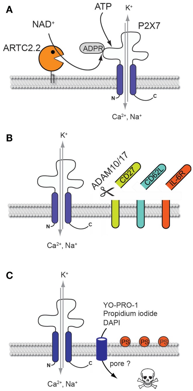
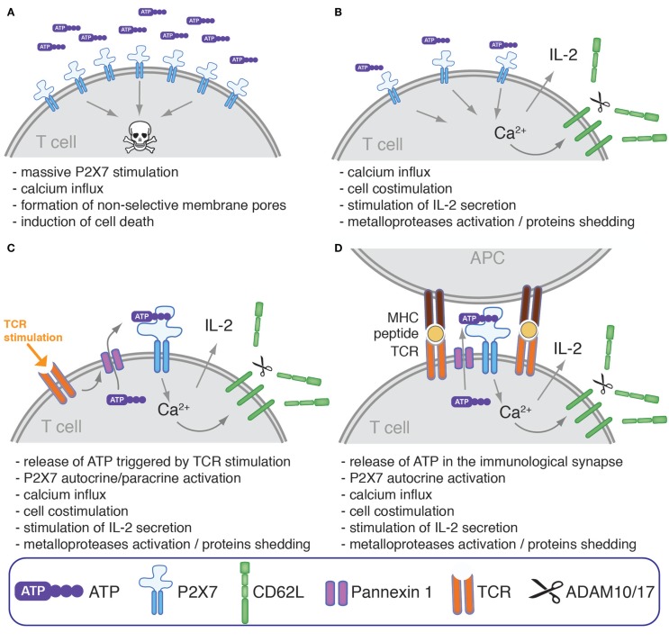
The P2X7 receptor is an adenosine triphosphate (ATP)-gated cation channel that is expressed by several cells of the immune system. P2X7 is best known for its proinflammatory role in promoting inflammasome formation and release of mature interleukin (IL)-1β by innate immune cells. Mounting evidence indicates that P2X7 is also an important regulatory receptor of murine and human T cell functions. Murine T cells express a sensitive splice variant of P2X7 that can be activated either by non-covalent binding of ATP or, in the presence of nicotinamide adenine dinucleotide, by its covalent ADP-ribosylation catalyzed by the ecto-ADP-ribosyltransferase ARTC2.2. Prolonged activation of P2X7 by either one of these pathways triggers the induction of T cell death. Conversely, lower concentrations of ATP can activate P2X7 to enhance T cell proliferation and production of IL-2. In this review, we will highlight the molecular and cellular consequences of P2X7 activation on mouse T cells and its versatile role in T cell homeostasis and activation. Further, we will discuss important differences in the function of P2X7 on human and murine T cells.
Overview
The claims and statistics of this paper haven’t been checked yet.
Run the check — free, no account needed. It reads the open-access text and checks the sources it cites.
Integrity signals
Registry
Cites
Statistics
Figures
Verification
Claims the paper makes
The claims and statistics of this paper haven’t been checked yet.
Run the check — free, no account needed. It reads the open-access text and checks the sources it cites.
Claims other papers make about this one
Each is checked against this paper’s own text: does it say what the citing paper says it does?
The source backs part of the claim, not all of it — or backs it for a different population, condition or comparison.
In the citing paper
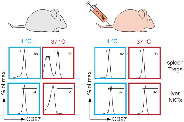
“For example, activation of the danger receptor P2RX7 by extracellular NAD or ATP, released by dying cells during processing, can lead to shedding of surface molecules such as CD62L and CD27, the masking of CD8β and the induction of cell death in susceptible cells like Treg, NKT, and Trm cells [286-288].”
From Guidelines for the use of flow cytometry and cell sorting in immunological studies (third edition) (2021) · cites this paper as [286]
Experimental — accuracy not yet measured
Self-check
Percentages, sums and reported differences re-computed from the paper’s own numbers.
The self-check needs the paper’s full text.
Citations
Cites
Coombes, J. L., Siddiqui, K. R. R., Arancibia-Cárcamo, C. V., Hall, J. A., Sun, C., Belkaid, Y., & Powrie, F. M. (2007) · The Journal of Experimental Medicine · doi:10.1084/jem.20070590 · Cited by 2,708
Deaglio, S., Dwyer, K. M., Gao, W., Friedman, D. J., Usheva, A., Erat, A., Chen, J., Enjyoji, K., Linden, J. M., Oukka, M., et al. (2007) · The Journal of Experimental Medicine · doi:10.1084/jem.20062512 · Cited by 2,613
Ghiringhelli, F., Apetoh, L., Tesniere, A., Aymeric, L., Ma, Y., Ortiz, C., Vermaelen, K., Panaretakis, T., Mignot, G., Ullrich, E., et al. (2009) · Nature Medicine · doi:10.1038/nm.2028 · Cited by 1,898
Retinoic acid imprints gut-homing specificity on T cells
Iwata, M., Hirakiyama, A., Eshima, Y., Kagechika, H., Kato, C., & Song, S.-Y. (2004) · Immunity · doi:10.1016/j.immuni.2004.08.011 · Cited by 1,541
Altered Cytokine Production in Mice Lacking P2X7Receptors
Solle, M., Labasi, J. M., Perregaux, D. G., Stam, E. J., Petrushova, N., Koller, B. H., Griffiths, R., & Gabel, C. A. (2001) · Journal of Biological Chemistry · doi:10.1074/jbc.m006781200 · Cited by 924
Immune cell regulation by autocrine purinergic signalling
JUNGER, W. G. (2011) · Nature reviews. Immunology · doi:10.1038/nri2938 · Cited by 855
Pannexin membrane channels are mechanosensitive conduits for ATP
Bao, L., Locovei, S., & Dahl, G. (2004) · FEBS Letters · doi:10.1016/j.febslet.2004.07.009 · Cited by 837
Disruption of the P2X7 purinoceptor gene abolishes chronic inflammatory and neuropathic pain
Chessell, I. P., Hatcher, J. P., Bountra, C., Michel, A. D., Hughes, J. P., Green, P., Egerton, J., Murfin, M., Richardson, J. C., Peck, W. L., et al. (2005) · Pain · doi:10.1016/j.pain.2005.01.002 · Cited by 807
A novel pathway combining calreticulin exposure and ATP secretion in immunogenic cancer cell death
Garg, A. D., Krysko, D. V., Verfaillie, T., Kaczmarek, A., Ferreira, G. B., Marysael, T., Rubio, N., Firczuk, M., Mathieu, C., Roebroek, A. J. M., et al. (2012) · The EMBO Journal · doi:10.1038/emboj.2011.497 · Cited by 800
Critical contribution of liver natural killer T cells to a murine model of hepatitis
Takeda, K., Hayakawa, Y., Kaer, L. V., Matsuda, H., Yagita∥, H., & Okumura, K. (2000) · Proceedings of the National Academy of Sciences · doi:10.1073/pnas.040566697 · Cited by 538
Research works printed but not linked: 1
- Freedman, 1999, ATP-evoked Ca2+ transients and currents in murine thymocytes: possible role for P2X receptors in death by neglect, Eur J Immunol, 29, 1635, 10.1002/(SICI)1521-4141(199905)29:051635::AID-IMMU16353.3.CO;2-2 · DOI
Cited by
Sorted by citations
Items 101–116 of 116
The Role of Microglial P2X7 Receptor Phosphorylation in Neuropathic Pain
Pilapil, A. (2016) · Libraries and Cultural Resources (University of Calgary) · doi:10.11575/prism/28333
Efeitos da sinalização purinérgica durante a infecção aguda e crônica pelo Plasmodium chabaudi AS.
de Salles, É. M. (2017) · Digital Library of Theses and Dissertations (Universidade de São Paulo) · doi:10.11606/t.42.2017.tde-12042017-141823
Amaral, E. P. (2016) · Digital Library of Theses and Dissertations (Universidade de São Paulo) · doi:10.11606/t.42.2016.tde-14042016-104430
Caracterizção imunofuncional de um inibidor de linfócitos presente na saliva de Aedes aegypti.
Bizzarro, B. (2017) · Digital Library of Theses and Dissertations (Universidade de São Paulo) · doi:10.11606/t.42.2017.tde-09032017-091514
How lipid specific T cells become effectors
Wang, H. (2019) · University of Minnesota Digital Conservancy (University of Minnesota)
Siqueira, R. (2019) · Digital Library of Theses and Dissertations (Universidade de São Paulo) · doi:10.11606/t.42.2019.tde-02102019-143339
Novel therapeutic targets in uveitis
Tempest-Roe, S. (2017) · Spiral (Imperial College London) · doi:10.25560/75500
Er-Lukowiak, M., Duan, Y., Rassendren, F., Ulmann, L., Nicke, A., Ufer, F., Friese, M., Koch-Nolte, F., Magnus, T., & Rissiek, B. (2020) · bioRxiv · doi:10.1101/2020.06.10.139410 · Cited by 1
Self-citationIdentification of Follicular T Cells in the Gut
Georgiev, H., Papadogianni, G., & Bernhardt, G. (2021) · Methods in molecular biology · doi:10.1007/978-1-0716-1736-6_8
Progress in undestanding of relationship between P2X7R and pancreatic ductal adenocarcinoma
Chi, Z.-C. (2025) · World Chinese Journal of Digestology · doi:10.11569/wcjd.v33.i8.599
Wilmes, M., Pinto-Espinoza, C., Ludewig, P., Stabernack, J., Liesz, A., Nicke, A., Gelderblom, M., Gerloff, C. P., Falzoni, S., Tolosa, E., et al. (2022) · Figshare · doi:10.6084/m9.figshare.21321416
Self-citationAbad, C., Demeules, M., Guillou, C., Gondé, H., Zoubaïri, R., Tan, Y.-V., Pinto-Espinoza, C., Schäfer, W., Mann, A. M., Vouret‐Craviari, V., et al. (2024) · Figshare · doi:10.6084/m9.figshare.26666614
Self-citationXénobiotiques hépatotoxiques : études de métabolisme et mécanismes d’action
Daré, B. L. (2021) · theses.fr (ABES) · doi:10.70675/c23bbc49z8149z4490zaba3z80027345e9cc
Bergero, G., Mazzocco, Y. L., Gallardo, Z. M. C., Rivarola, W., Rosso, S. D., & Aoki, M. P. (2026) · Frontiers in Immunology · doi:10.3389/fimmu.2026.1770276
Kamel, A. S. E. D., Shalaby, H. N., El-Said, Y. A. M., & Nawwar, D. A. (2026) · Drug Development Research · doi:10.1002/ddr.70318
Metadata
- Authors
- Rissiek, B., Haag, F., Boyer, O., Koch-Nolte, F., & Adriouch, S.
- Affiliations
- Universität Hamburg (DE)
- Inserm (FR)
- Corresponding author
- Adriouch, Sahil
- Venue
- Frontiers in Immunology
- Publisher
- Frontiers Media SA
- Published
- 2015-05-19
- Volume
- 6
- Pages
- 204-204
- ISSN
- 1664-3224 (ISSN-L)
- PMC
- PMC4436801
- OpenAlex
- W1564355150
- Cited by
- 116
- References
- 81
- Citations by year
- 2026: 4 · 2025: 7 · 2024: 7 · 2023: 10 · 2022: 8 · 2021: 15 · 2020: 13 · 2019: 14 · 2018: 15 · 2017: 11 · 2016: 12
- FWCI
- 1.62
- Type
- journal article
- Language
- en
- Open access
- gold
- doi.org
- Licence
- cc-by
- Locations
- Frontiers in Immunology (publishedVersion, cc-by)
- Europe PMC (PubMed Central) (publishedVersion, cc-by)
- PubMed Central (publishedVersion, cc-by)
- PubMed (publishedVersion)
- HAL (submittedVersion, other-oa)
- HAL (submittedVersion)
- DOAJ (DOAJ: Directory of Open Access Journals) (submittedVersion)
- Funders
- Deutsche Forschungsgemeinschaft (SFB 877, HA2369/5)
- Institut National de la Santé et de la Recherche Médicale
- Normandie Université
- Topics
- Life Sciences › Biochemistry, Genetics and Molecular Biology › Physiology › Adenosine and Purinergic Signaling
- Life Sciences › Biochemistry, Genetics and Molecular Biology › Physiology › Calcium signaling and nucleotide metabolism
- Life Sciences › Biochemistry, Genetics and Molecular Biology › Molecular Biology › Biochemical and Molecular Research
- Keywords
- P2X7 receptor, T cells, ATP-gated cation channels, ADP-ribosylation, T cell death, T cell proliferation, interleukin-2, T cell homeostasis, T cell activation, splice variants
About these checks
Registry, retraction, Cites and statistics results are computed, not judged, with one exception: a printed reference that no index lists is read once by a model before it counts as a citation error. Claims are extracted by a model and kept only when their quote appears verbatim in the text; each cited source is then checked, with a model’s help, for whether it says what the claim relies on. Those verdicts can be wrong — read the claim’s quote and the cited source before relying on one.
Statistics and arithmetic are recomputed, not judged.
Confidence bands are shown only for fields calibrated on our annotated test set. None is calibrated yet, so no band is shown.
What counts as a claim ›How we measure confidence ›How we check this ›
Are you an author, or do you see a problem with this page? Request a review
