Cited by 29

The DOI matches its Crossref record; 1 of the 1 references it cites were found in a registry or an index.

Safeguarding of Fetal Growth by Mast Cells and Natural Killer Cells: Deficiency of One Is Counterbalanced by the Other

Meyer, Nicole, Woidacki, Katja, Maurer, Marcus, Zenclussen, Ana Claudia·2017·Frontiers in Immunology

Overview

The claims and statistics of this paper haven’t been checked yet.

Run the check — free, no account needed. It reads the open-access text and checks the sources it cites.

A few free checks a day · papers already verified open instantly · results are public

Integrity signals

Registry

Crossref · 10.3389/fimmu.2017.00711

Cites

1 of 1 references verified0 citation errors · 0 not checkable

Statistics

Figures

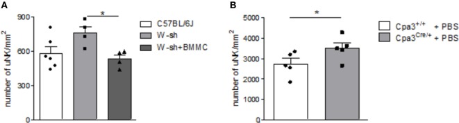
Figure 1 Mast cell (MC)-deficient mice show increased numbers of uterine natural killer cells (uNKs) in contrast to wild type (WT). Bone marrow-derived MCs (BMMC) reconstitution restored this phenotype. Numbers of uNKs cells per 1 mm2 were quantified in Dolichos biflorus agglutinin lectin-stained 5 µm paraffin cross sections of implantations of WT C57BL/6J (n = 6), MC-deficient W-sh (n = 4), or BMMC-reconstituted W-sh (n = 4) mice (A) as well as in Cpa3+/+ control mice (n = 5) vs. MC-deficient Cpa3Cre/+ (n = 5) mice (B) at gd10. Results are presented as means with SEM. Also individual values are shown by dots, squares and triangles. Statistical differences were obtained using Mann–Whitney U-test (*P < 0.05). MCs, mast cells; NKs, natural killer cells; u, uterine; WT, wild type; BMMC, bone marrow-derived mast cells; W-sh, C57BL/6J-KitW-sh/W-sh.
Figure 1 Mast cell (MC)-deficient mice show increased numbers of uterine natural killer cells (uNKs) in contrast to wild type (WT). Bone marrow-derived MCs (BMMC) reconstitution restored this phenotype. Numbers of uNKs cells per 1 mm2 were quantified in Dolichos biflorus agglutinin lectin-stained 5 µm paraffin cross sections of implantations of WT C57BL/6J (n = 6), MC-deficient W-sh (n = 4), or BMMC-reconstituted W-sh (n = 4) mice (A) as well as in Cpa3+/+ control mice (n = 5) vs. MC-deficient Cpa3Cre/+ (n = 5) mice (B) at gd10. Results are presented as means with SEM. Also individual values are shown by dots, squares and triangles. Statistical differences were obtained using Mann–Whitney U-test (*P < 0.05). MCs, mast cells; NKs, natural killer cells; u, uterine; WT, wild type; BMMC, bone marrow-derived mast cells; W-sh, C57BL/6J-KitW-sh/W-sh.CC BY · PubMed Central
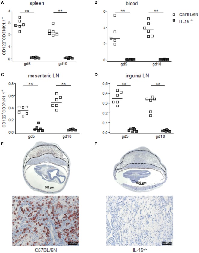
Figure 2 Interleukin (IL)-15-deficient mice are characterized by very low levels of peripheral (p) and the absence of uterine natural killer cells (uNKs) in contrast to wild-type mice. Percentages of CD122+CD3−NK1.1+ pNKs in spleen (A), blood (B), mesenteric lymph nodes (LN) (C), and inguinal LNs (D) at gd5 and gd10 were analyzed by flow cytometry in samples from C57BL/6N (n = 5–6) or IL-15−/− (n = 6) mice. Results are presented with squares showing the individual values for each mouse and medians are indicated. Statistical analysis was performed by the Mann–Whitney U-test (**P < 0.001). Representative Dolichos biflorus agglutinin (DBA) lectin-stained cross sections of implantations from C57BL/6N (E) or IL-15−/− (F) mice at gd10 are depicted (scale bar = 500 μm). uNKs can be identified as DBA lectin-positive, brown-stained cells within the decidua basalis (dashed lines marked area). Lower pictures show cutouts from the decidua basalis with higher magnification (scale bar = 100 μm) (F) confirms the absolute NK absence at gd10. IL, interleukin; p, peripheral; u, uterine; LN, lymph nodes; DBA, dolichos biflorus agglutinin.
Figure 2 Interleukin (IL)-15-deficient mice are characterized by very low levels of peripheral (p) and the absence of uterine natural killer cells (uNKs) in contrast to wild-type mice. Percentages of CD122+CD3−NK1.1+ pNKs in spleen (A), blood (B), mesenteric lymph nodes (LN) (C), and inguinal LNs (D) at gd5 and gd10 were analyzed by flow cytometry in samples from C57BL/6N (n = 5–6) or IL-15−/− (n = 6) mice. Results are presented with squares showing the individual values for each mouse and medians are indicated. Statistical analysis was performed by the Mann–Whitney U-test (**P < 0.001). Representative Dolichos biflorus agglutinin (DBA) lectin-stained cross sections of implantations from C57BL/6N (E) or IL-15−/− (F) mice at gd10 are depicted (scale bar = 500 μm). uNKs can be identified as DBA lectin-positive, brown-stained cells within the decidua basalis (dashed lines marked area). Lower pictures show cutouts from the decidua basalis with higher magnification (scale bar = 100 μm) (F) confirms the absolute NK absence at gd10. IL, interleukin; p, peripheral; u, uterine; LN, lymph nodes; DBA, dolichos biflorus agglutinin.CC BY · PubMed Central
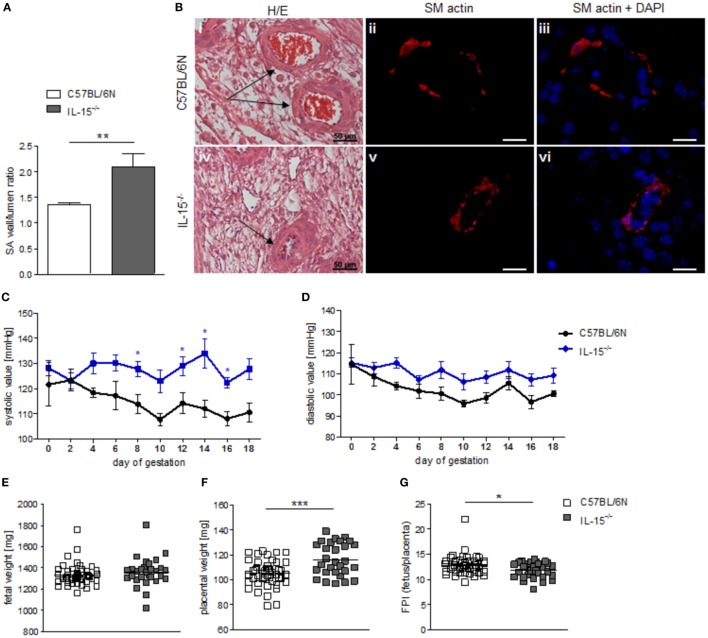
Figure 3 Interleukin (IL)-15−/− mice showed impaired remodeling of spiral arteries (SAs), but no hypertension throughout pregnancy. While natural killer cell (NK) absence had no impact on fetal growth, it was associated with higher placental weight and lower fetoplacental-index (FPI). (A) SA wall and lumen perimeters of 3–10 SAs per animal of BL/6N (n = 6) and IL-15−/− (n = 5) mice were measured and SA wall-to-lumen ratio were calculated. Results are presented as means with SEM. Statistical differences were obtained using Mann–Whitney U-test (**P < 0.01). (B) Representative images of H/E-stained sections (scale bar = 50 μm) and immunofluorescence for smooth muscle (SM) actin and SM actin + DAPI (scale bar = 20 μm) of SAs (indicated by arrows) of BL/6N and IL-15−/− mice are shown. Systolic (C) and diastolic blood (D) pressure values for pregnant C57BL/6N (n = 5) or IL-15−/− (n = 5) mice from gd0 to gd18. Data were recorded every second day. For (C,D), results are presented as mean ± SEM. Statistical differences were obtained using Mann–Whitney U-test (*P < 0.05,). Fetal weights (E), placental weights (F), and FPI (G) from C57BL/6N (mothers: n = 5, fetus/placenta: n = 46) or IL-15−/− (mothers: n = 5, fetus/placenta: n = 29) females at gd18. For (E–G), results are presented with squares showing individual values for each fetus/placenta and means are indicated. Statistical differences were obtained using unpaired t-test (*P < 0.05, ***P < 0.001). IL, interleukin; SA, spiral arteries, NK, natural killer cells; FPI, fetoplacental-index; SM, smooth muscle.
Figure 3 Interleukin (IL)-15−/− mice showed impaired remodeling of spiral arteries (SAs), but no hypertension throughout pregnancy. While natural killer cell (NK) absence had no impact on fetal growth, it was associated with higher placental weight and lower fetoplacental-index (FPI). (A) SA wall and lumen perimeters of 3–10 SAs per animal of BL/6N (n = 6) and IL-15−/− (n = 5) mice were measured and SA wall-to-lumen ratio were calculated. Results are presented as means with SEM. Statistical differences were obtained using Mann–Whitney U-test (**P < 0.01). (B) Representative images of H/E-stained sections (scale bar = 50 μm) and immunofluorescence for smooth muscle (SM) actin and SM actin + DAPI (scale bar = 20 μm) of SAs (indicated by arrows) of BL/6N and IL-15−/− mice are shown. Systolic (C) and diastolic blood (D) pressure values for pregnant C57BL/6N (n = 5) or IL-15−/− (n = 5) mice from gd0 to gd18. Data were recorded every second day. For (C,D), results are presented as mean ± SEM. Statistical differences were obtained using Mann–Whitney U-test (*P < 0.05,). Fetal weights (E), placental weights (F), and FPI (G) from C57BL/6N (mothers: n = 5, fetus/placenta: n = 46) or IL-15−/− (mothers: n = 5, fetus/placenta: n = 29) females at gd18. For (E–G), results are presented with squares showing individual values for each fetus/placenta and means are indicated. Statistical differences were obtained using unpaired t-test (*P < 0.05, ***P < 0.001). IL, interleukin; SA, spiral arteries, NK, natural killer cells; FPI, fetoplacental-index; SM, smooth muscle.CC BY · PubMed Central
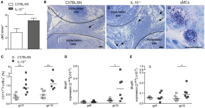
Figure 4 Interleukin (IL)-15−/− mice exhibit increased numbers of uterine mast cells (uMCs). (A) Numbers of uMCs per 1 mm2 were quantified in tolouidine blue-stained 5 µm paraffin sections from uteri of C57BL/6N (n = 6) and IL-15−/− mice (n = 6) at gd5. Results are presented as mean with SEM. Statistical differences were obtained using Mann–Whitney U-test (*P < 0.05). (B) Representative pictures for (A) are shown for C57BL/6N (i) and IL-15−/− (ii) mice (scale bar = 100 μm). uMCs (iii) are localized between the implantation sites and are indicated by arrows (scale bar = 20 μm) in (i) and (ii). (C) Percentages of CD117+FcεRα+ MCs in uterus (gd 5) or decidua (gd 10) samples were analyzed by flow cytometry. mRNA levels of MC-related proteases (Mcpt)-1 (D) and Mcpt-6 (E) in uterine (gd 5) and decidual (gd 10) tissue of BL/6N and IL-15−/− mice were measured. For (C–E), results are presented with squares showing individual values. Additionally, the medians are indicated. Statistical differences were obtained using non-parametric Mann–Whitney U-test (*P < 0.05, **P < 0.005). IL, interleukin; uMC, uterine mast cell; Mcpt, MC-related protease.
Figure 4 Interleukin (IL)-15−/− mice exhibit increased numbers of uterine mast cells (uMCs). (A) Numbers of uMCs per 1 mm2 were quantified in tolouidine blue-stained 5 µm paraffin sections from uteri of C57BL/6N (n = 6) and IL-15−/− mice (n = 6) at gd5. Results are presented as mean with SEM. Statistical differences were obtained using Mann–Whitney U-test (*P < 0.05). (B) Representative pictures for (A) are shown for C57BL/6N (i) and IL-15−/− (ii) mice (scale bar = 100 μm). uMCs (iii) are localized between the implantation sites and are indicated by arrows (scale bar = 20 μm) in (i) and (ii). (C) Percentages of CD117+FcεRα+ MCs in uterus (gd 5) or decidua (gd 10) samples were analyzed by flow cytometry. mRNA levels of MC-related proteases (Mcpt)-1 (D) and Mcpt-6 (E) in uterine (gd 5) and decidual (gd 10) tissue of BL/6N and IL-15−/− mice were measured. For (C–E), results are presented with squares showing individual values. Additionally, the medians are indicated. Statistical differences were obtained using non-parametric Mann–Whitney U-test (*P < 0.05, **P < 0.005). IL, interleukin; uMC, uterine mast cell; Mcpt, MC-related protease.CC BY · PubMed Central

Verification

Claims the paper makes

The claims and statistics of this paper haven’t been checked yet.

Run the check — free, no account needed. It reads the open-access text and checks the sources it cites.

A few free checks a day · papers already verified open instantly · results are public

Claims other papers make about this one

Each is checked against this paper’s own text: does it say what the citing paper says it does?

Self-check

Percentages, sums and reported differences re-computed from the paper’s own numbers.

The self-check needs the paper’s full text.

Citations

Cites

Show all 1 references

Research works printed but not linked: 31

  1. Moll, 1975, Hemodynamic implications of hemochorial placentation, Eur J Obstet Gynecol Reprod Biol, 5, 67, 10.1016/0028-2243(75)90131-8 · DOI
  2. Pijnenborg, 2006, The uterine spiral arteries in human pregnancy: facts and controversies, Placenta, 27, 939, 10.1016/j.placenta.2005.12.006 · DOI
  3. Bulmer, 1991, Granulated lymphocytes in human endometrium: histochemical and immunohistochemical studies, Hum Reprod, 6, 791, 10.1093/oxfordjournals.humrep.a137430 · DOI
  4. Kennedy, 2000, Reversible defects in natural killer and memory CD8 T cell lineages in interleukin 15-deficient mice, J Exp Med, 191, 771, 10.1084/jem.191.5.771 · DOI
  5. Ashkar, 2003, Assessment of requirements for IL-15 and IFN regulatory factors in uterine NK cell differentiation and function during pregnancy, J Immunol, 171, 2937, 10.4049/jimmunol.171.6.2937 · DOI
  6. Barber, 2003, The uterine NK cell population requires IL-15 but these cells are not required for pregnancy nor the resolution of a Listeria monocytogenes infection, J Immunol, 171, 37, 10.4049/jimmunol.171.1.37 · DOI
  7. Woidacki, 2013, Mast cells rescue implantation defects caused by c-kit deficiency, Cell Death Dis, 4, e462, 10.1038/cddis.2012.214 · DOI · Paper page
  8. Grimbaldeston, 2005, Mast cell-deficient W-sash c-kit mutant Kit W-sh/W-sh mice as a model for investigating mast cell biology in vivo, Am J Pathol, 167, 835, 10.1016/S0002-9440(10)62055-X · DOI · Paper page
  9. Meyer, 2017, Chymase-producing cells of the innate immune system are required for decidual vascular remodeling and fetal growth, Sci Rep, 7, 45106, 10.1038/srep45106 · DOI · Paper page
  10. Brosens, 1967, The physiological response of the vessels of the placental bed to normal pregnancy, J Pathol Bacteriol, 93, 569, 10.1002/path.1700930218 · DOI
  11. Khong, 1986, Inadequate maternal vascular response to placentation in pregnancies complicated by pre-eclampsia and by small-for-gestational age infants, Br J Obstet Gynaecol, 93, 1049, 10.1111/j.1471-0528.1986.tb07830.x · DOI · Paper page
  12. Zenclussen, 2015, Cellular regulation of the uterine microenvironment that enables embryo implantation, Front Immunol, 6, 321, 10.3389/fimmu.2015.00321 · DOI
  13. Ashkar, 2000, Interferon gamma contributes to initiation of uterine vascular modification, decidual integrity, and uterine natural killer cell maturation during normal murine pregnancy, J Exp Med, 192, 259, 10.1084/jem.192.2.259 · DOI
  14. Peel, 1989, Granulated metrial gland cells, Adv Anat Embryol Cell Biol, 115, 1, 10.1007/978-3-642-74170-8_1 · DOI
  15. Croy, 1993, Granulated metrial gland cells: a natural killer cell subset of the pregnant murine uterus, Microsc Res Tech, 25, 189, 10.1002/jemt.1070250302 · DOI
  16. Brosens, 1972, The role of the spiral arteries in the pathogenesis of preeclampsia, Obstet Gynecol Annu, 1, 177
  17. Croy, 2011, Identification of the primary outcomes that result from deficient spiral arterial modification in pregnant mice, Pregnancy Hypertens, 1, 87, 10.1016/j.preghy.2010.10.002 · DOI
  18. Yamaleyeva, 2015, Uterine artery dysfunction in pregnant ACE2 knockout mice is associated with placental hypoxia and reduced umbilical blood flow velocity, Am J Physiol Endocrinol Metab, 309, E84, 10.1152/ajpendo.00596.2014 · DOI
  19. Greenwood, 2000, Ultrastructural studies of implantation sites from mice deficient in uterine natural killer cells, Placenta, 21, 693, 10.1053/plac.2000.0556 · DOI
  20. Chakraborty, 2011, Natural killer cells direct hemochorial placentation by regulating hypoxia-inducible factor dependent trophoblast lineage decisions, Proc Natl Acad Sci U S A, 108, 16295, 10.1073/pnas.1109478108 · DOI
  21. Woidacki, 2015, Transfer of regulatory T cells into abortion-prone mice promotes the expansion of uterine mast cells and normalizes early pregnancy angiogenesis, Sci Rep, 5, 13938, 10.1038/srep13938 · DOI
  22. Welle, 1997, Development, significance, and heterogeneity of mast cells with particular regard to the mast cell-specific proteases chymase and tryptase, J Leukoc Biol, 61, 233, 10.1002/jlb.61.3.233 · DOI
  23. McNeil, 1991, Molecular cloning of the mouse mast cell protease-5 gene. A novel secretory granule protease expressed early in the differentiation of serosal mast cells, J Biol Chem, 266, 20316, 10.1016/S0021-9258(18)54925-5 · DOI
  24. Beaven, 2009, Our perception of the mast cell from Paul Ehrlich to now, Eur J Immunol, 39, 11, 10.1002/eji.200838899 · DOI
  25. Croy, 2003, Update on pathways regulating the activation of uterine natural killer cells, their interactions with decidual spiral arteries and homing of their precursors to the uterus, J Reprod Immunol, 59, 175, 10.1016/S0165-0378(03)00046-9 · DOI
  26. Galli, 2005, Mast cells as “tunable” effector and immunoregulatory cells: recent advances, Annu Rev Immunol, 23, 749, 10.1146/annurev.immunol.21.120601.141025 · DOI · Paper page
  27. Lash, 2011, Do uterine natural killer (uNK) cells contribute to female reproductive disorders?, J Reprod Immunol, 88, 156, 10.1016/j.jri.2011.01.003 · DOI
  28. Zhang, 2011, Natural killer cell-triggered vascular transformation: maternal care before birth?, Cell Mol Immunol, 8, 1, 10.1038/cmi.2010.38 · DOI
  29. Tirado-González, 2012, Uterine NK cells are critical in shaping DC immunogenic functions compatible with pregnancy progression, PLoS One, 7, e46755, 10.1371/journal.pone.0046755 · DOI
  30. Brown, 2014, M1/M2 macrophage polarity in normal and complicated pregnancy, Front Immunol, 5, 606, 10.3389/fimmu.2014.00606 · DOI
  31. Vargas-Rojas, 2016, Th1, Th2, Th17 and Treg levels in umbilical cord blood in preeclampsia, J Matern Fetal Neonatal Med, 29, 1642, 10.3109/14767058.2015.1057811 · DOI

Cited by

Sorted by citations

See all 29

Metadata

Authors
Meyer, N., Woidacki, K., Maurer, M., & Zenclussen, A. C.
Venue
Frontiers in Immunology
Published
2017-06-16
Volume
8
ISSN
1664-3224
Cited by
29
References
1
Funders
Deutsche Forschungsgemeinschaft (ZE 526/6-2)

About these checks

Registry, retraction, Cites and statistics results are computed, not judged, with one exception: a printed reference that no index lists is read once by a model before it counts as a citation error. Claims are extracted by a model and kept only when their quote appears verbatim in the text; each cited source is then checked, with a model’s help, for whether it says what the claim relies on. Those verdicts can be wrong — read the claim’s quote and the cited source before relying on one.

Statistics and arithmetic are recomputed, not judged.

Confidence bands are shown only for fields calibrated on our annotated test set. None is calibrated yet, so no band is shown.

What counts as a claim ›How we measure confidence ›How we check this ›

Are you an author, or do you see a problem with this page? Request a review

How to cite

Cite CoChat for the checks on this page, and the paper for its own findings.

This page
CoChat. (2026). Citation check: Safeguarding of Fetal Growth by Mast Cells and Natural Killer Cells: Deficiency of One Is Counterbalanced by the Other. https://app.cochat.ai/papers/10.3389/fimmu.2017.00711
The paper
Meyer, N., Woidacki, K., Maurer, M., & Zenclussen, A. C. (2017). Safeguarding of Fetal Growth by Mast Cells and Natural Killer Cells: Deficiency of One Is Counterbalanced by the Other. Frontiers in Immunology. https://doi.org/10.3389/fimmu.2017.00711