

The DOI matches its Crossref record; 2 of the 2 references it cites were found in a registry or an index.
Mast cells rescue implantation defects caused by c‐kit deficiency
Overview
The claims and statistics of this paper haven’t been checked yet.
Run the check — free, no account needed. It reads the open-access text and checks the sources it cites.
Integrity signals
Registry
Cites
Statistics
Figures
Verification
Claims the paper makes
The claims and statistics of this paper haven’t been checked yet.
Run the check — free, no account needed. It reads the open-access text and checks the sources it cites.
Claims other papers make about this one
Each is checked against this paper’s own text: does it say what the citing paper says it does?
In the citing paper
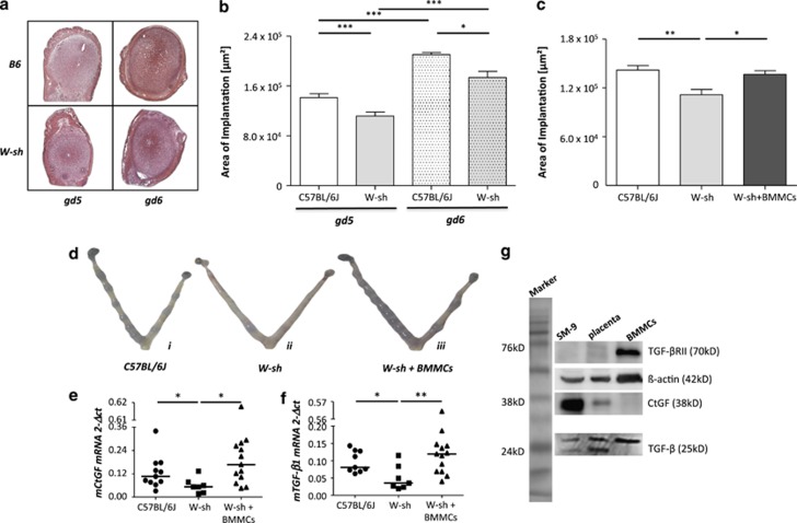
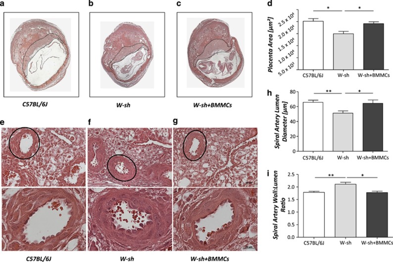
“Mast cell‐deficient female mice display defective spiral artery remodeling and impaired implantation, collectively resulting in fetal growth restriction. 122 , 124 , 125 Interestingly, mast cells appear to mediate these processes cooperatively with uterine NK cells, since both can (partially) compensate in the absence of the other. 125”
From Mast cell ontogeny: From fetal development to life‐long health and disease (2023) · cites this paper as [122]
In the citing paper
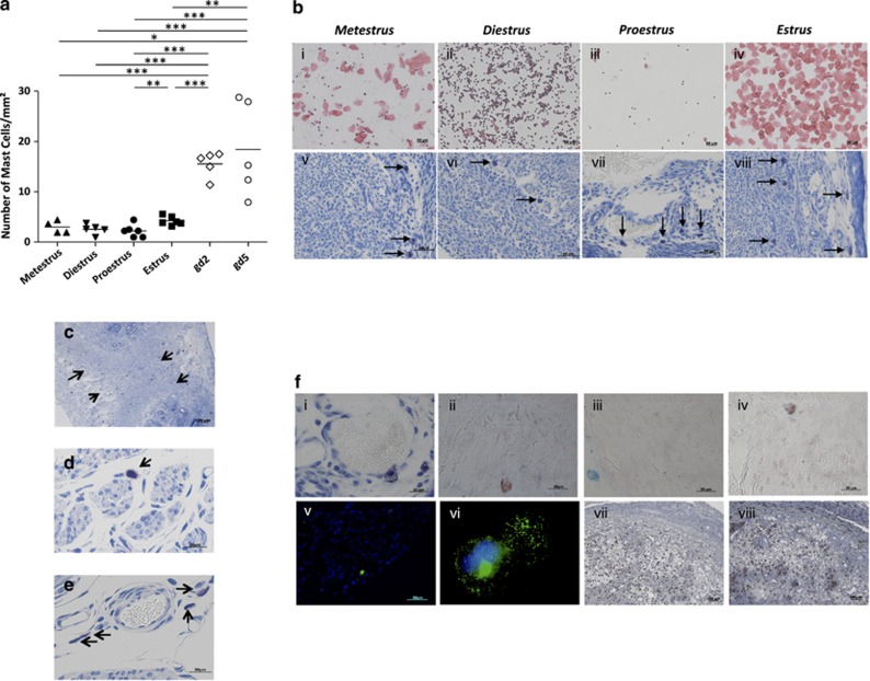
“In the uterus, mast cell numbers increase during the receptive stage of the estrous cycle and early pregnancy, when they are in close association with the blood vessels at the implantation sites. 122 , 123”
From Mast cell ontogeny: From fetal development to life‐long health and disease (2023) · cites this paper as [122]
Self-check
Percentages, sums and reported differences re-computed from the paper’s own numbers.
The self-check needs the paper’s full text.
Citations
Cites
Jensen, F., Woudwyk, M., Teles, A., Woidacki, K., Taran, F., Costa, S., Malfertheiner, S. F., & Zenclussen, A. C. (2010) · PLoS ONE · doi:10.1371/journal.pone.0014409 · Cited by 145 · cited 2×
“and progesterone promoteMCmigration from the periphery to the uterus.(25) To investigate the role of MCs in pregnancy, we used C57BL/6J-Kit mice.”
BackgroundMethodSelf-citationZhao, X. J. (2001) · Thorax · doi:10.1136/thorax.56.3.205 · Cited by 120 · cited 1×
“This appears to be regulated through endocrine mechanisms, which is not surprising as they express estrogen and progesterone receptors.(37) We recently found that estradiol”
Background
Research works printed but not linked: 49
- Ogawa M, Matsuzaki Y, Nishikawa S, Hayashi S, Kunisada T, Sudo T et al. Expression and function of c-kit in hemopoietic progenitor cells. J Exp Med 1991; 174: 63–71. · DOI
- Dastych J, Metcalfe DD . Stem cell factor induces mast cell adhesion to fibronectin. J Immunol 1994; 152: 213–219. · DOI
- Ohta H, Yomogida K, Dohmae K, Nishimune Y . Regulation of proliferation and differentiation in spermatogonial stem cells: the role of c-kit and its ligand SCF. Development 2000; 127: 2125–2131. · DOI
- Hutt KJ, McLaughlin EA, Holland MK . KIT/KIT ligand in mammalian oogenesis and folliculogenesis: roles in rabbit and murine ovarian follicle activation and oocyte growth. Biol Reprod 2006; 75: 421–433. · DOI
- Moore PA, Belvedere O, Orr A, Pieri K, LaFleur DW, Feng P et al. BLyS: member of the tumor necrosis factor family and B lymphocyte stimulator. Science 1999; 285: 260–263. · DOI
- Kehrl JH, Wakefield LM, Roberts AB, Jakowlew S, Alvarez-Mon M, Derynck R et al. Production of transforming growth factor beta by human T lymphocytes and its potential role in the regulation of T cell growth. J Exp Med 1986; 163: 1037–1050. · DOI
- Besmer P . The kit ligand encoded at the murine Steel locus: a pleiotropic growth and differentiation factor. Curr Opin Cell Biol 1991; 3: 939–946. · DOI
- Sharkey AM, Jokhi PP, King A, Loke YW, Brown KD, Smith SK . Expression of c-kit and kit ligand at the human maternofetal interface. Cytokine 1994; 6: 195–205. · DOI
- Horie K, Fujita J, Takakura K, Kanzaki H, Kaneko Y, Iwai M et al. Expression of c-kit protein during placental development. Biol Reprod 1992; 47: 614–620. · DOI
- Nocka K, Tan JC, Chiu E, Chu TY, Ray P, Traktman P et al. Molecular bases of dominant negative and loss of function mutations at the murine c-kit/white spotting locus: W37, Wv, W41 and W. EMBO J 1990; 9: 1805–1813. · DOI
- Lyon MF, Glenister PH . A new allele sash (Wsh) at the W-locus and a spontaneous recessive lethal in mice. Genet Res 1982; 39: 315–322. · DOI
- Nagle DL, Kozak CA, Mano H, Chapman VM, Bućan M . Physical mapping of the Tec and Gabrb1 loci reveals that the Wsh mutation on mouse chromosome 5 is associated with an inversion. Hum Mol Genet 1995; 4: 2073–2079. · DOI
- Wolters PJ, Mallen-St Clair J, Lewis CC, Villalta SA, Baluk P, Erle DJ et al. Tissue-selective mast cell reconstitution and differential lung gene expression in mast cell-deficient Kit(W-sh)/Kit(W-sh) sash mice. Clin Exp Allergy 2005; 35: 82–88. · DOI · Paper page
- Grimbaldeston MA, Chen CC, Piliponsky AM, Tsai M, Tam SY, Galli SJ . Mast cell-deficient W-sash c-kit mutant Kit W-sh/W-sh mice as a model for investigating mast cell biology in vivo. Am J Pathol 2005; 167: 835–848. · DOI · Paper page
- Berrozpe G, Timokhina I, Yukl S, Tajima Y, Ono M, Zelenetz AD et al. The W(sh), W(57), and Ph Kit expression mutations define tissue-specific control elements located between -23 and -154 kb upstream of Kit. Blood 1999; 94: 2658–2666. · DOI
- Galli SJ, Nakae S, Tsai M . Mast cells in the development of adaptive immune responses. Nat Immunol 2005; 6: 135–142. · DOI
- Horie K, Fujita J, Takakura K, Kanzaki H, Suginami H, Iwai M et al. The expression of c-kit protein in human adult and fetal tissues. Hum Reprod 1993; 8: 1955–1962. · DOI
- Mori A, Zhai YL, Toki T, Nikaido T, Fujii S . Distribution and heterogeneity of mast cells in the human uterus. Hum Reprod 1997; 12: 368–372. · DOI
- Varayoud J, Ramos JG, Bosquiazzo VL, Muñoz-de-Toro M, Luque EH . Mast cells degranulation affects angiogenesis in the rat uterine cervix during pregnancy. Reproduction 2004; 127: 379–387. · DOI · Paper page
- Bosquiazzo VL, Ramos JG, Varayoud J, Muñoz-de-Toro M, Luque EH . Mast cell degranulation in rat uterine cervix during pregnancy correlates with expression of vascular endothelial growth factor mRNA and angiogenesis. Reproduction 2007; 133: 1045–1055. · DOI · Paper page
- Bytautiene E, Vedernikov YP, Saade GR, Romero R, Garfield RE . IgE-independent mast cell activation augments contractility of nonpregnant and pregnant guinea pig myometrium. Int Arch Allergy Immunol 2008; 147: 140–146. · DOI
- Norrby K . Mast cells and angiogenesis. APMIS 2002; 110: 355–371. · DOI
- Shelesnyak MC . Antihistamines and the ovum. Br Med J 1963; 16: 1268. · DOI
- Murphy VE, Clifton VL, Gibson PG . Asthma exacerbations during pregnancy: incidence and association with adverse pregnancy outcomes. Thorax 2006; 61: 169–176. · DOI
- Menzies FM, Higgins CA, Shepherd MC, Nibbs RJ, Nelson SM . Mast cells reside in myometrium and cervix, but are dispensable in mice for successful pregnancy and labor. Immunol Cell Biol 2011; 90: 321–329. · DOI
- Michaloudi HC, Papadopoulos GC . Mast cells in the sheep, hedgehog and rat forebrain. J Anat 1999; 195: 269–276. · DOI
- Scholten J, Hartmann K, Gerbaulet A, Krieg T, Müller W, Testa G et al. Mast cell- specific Cre/loxP-mediated recombination in vivo. Transgenic Res 2008; 17: 307–315. · DOI
- Song H, Lim H, Paria BC, Matsumoto H, Swift LL, Morrow J et al. Cytosolic phospholipase A2 alpha is crucial for “on-time” embryo implantation that directs subsequent development. Development 2002; 129: 2879–2889. · DOI
- Valent P, Sillaber C, Baghestanian M, Bankl HC, Kiener HP, Lechner K et al. What have mast cells to do with edema formation, the consecutive repair and fibrinolysis? Int Arch Allergy Immunol 1998; 115: 2–8. · DOI
- Metz M, Maurer M . Mast cells-key effector cells in immune responses. Trends Immunol 2007; 28: 234–241. · DOI
- Galli SJ . Mast cells and basophils. Curr Opin Hematol 2000; 7: 32–39. · DOI
- Rabinovich GA, Croci DO . Regulatory circuits mediated by lectin-glycan interactions in autoimmunity and cancer. Immunity 2012; 36: 322–335. · DOI
- von Wolff M, Wang X, Gabius HJ, Strowitzki T . Galectin fingerprinting in human endometrium and decidua during the menstrual cycle and in early gestation. Mol Hum Reprod 2005; 11: 189–194. · DOI
- Fischer I, Redel S, Hofmann S, Kuhn C, Friese K, Walzel H et al. Stimulation of syncytium formation in vitro in human trophoblast cells by galectin-1. Placenta 2010; 31: 825–832. · DOI
- Dudeck A, Dudeck J, Scholten J, Petzold A, Surianarayanan S, Köhler A et al. Mast cells are key promoters of contact allergy that mediate the adjuvant effects of haptens. Immunity 2011; 34: 973–984. · DOI
- Müsch W, Wege AK, Männel DN, Hehlgans T . Generation and characterization of alpha-chymase-Cre transgenic mice. Genesis 2008; 46: 163–166. · DOI
- Feyerabend TB, Weiser A, Tietz A, Stassen M, Harris N, Kopf M et al. Cre-mediated cell ablation contests mast cell contribution in models of antibody- and T cell-mediated autoimmunity. Immunity 2011; 35: 832–844. · DOI
- Aflalo ED, Sod-Moriah UA, Potashnik G, Har-Vardi I . Expression of plasminogen activators in preimplantation rat embryos developed in vivo and in vitro. Reprod Biol Endocrinol 2005; 3: 7. · DOI
- Oh CK . Mast cell mediators in airway remodeling. Chem Immunol Allergy 2005; 87: 85–100. · DOI
- Carmeliet P, Schoonjans L, Kieckens L, Ream B, Degen J, Bronson R et al. Physiological consequences of loss of plasminogen activator gene function in mice. Nature 1994; 368: 419–424. · DOI
- Labied S, Blacher S, Carmeliet P, Noël A, Frankenne F, Foidart JM et al. Transient reduction of placental angiogenesis in PAI-1-deficient mice. Physiol Genomics 2011; 43: 188–198. · DOI
- Uzumcu M, Homsi MF, Ball DK, Coskun S, Jaroudi K, Hollanders JM et al. Localization of connective tissue growth factor in human uterine tissues. Mol Hum Reprod 2000; 6: 1093–1098. · DOI
- Surveyor GA, Wilson AK, Brigstock DR . Localization of connective tissue growth factor during the period of embryo implantation in the mouse. Biol Reprod 1998; 59: 1207–1213. · DOI
- Frazier K, Williams S, Kothapalli D, Klapper H, Grotendorst GR . Stimulation of fibroblast cell growth, matrix production, and granulation tissue formation by connective tissue growth factor. J Invest Dermatol 1996; 107: 404–411. · DOI
- Miller DA, Lee A, Pelton RW, Chen EY, Moses HL, Derynck R . Murine transforming growth factor-beta 2 cDNA sequence and expression in adult tissues and embryos. Mol Endocrinol 1989; 3: 1108–1114. · DOI
- Shull MM, Ormsby I, Kier AB, Pawlowski S, Diebold RJ, Yin M et al. Targeted disruption of the mouse transforming growth factor-β1 gene results in multifocal inflammatory disease. Nature 1992; 359: 693–699. · DOI · Paper page
- Yamazaki M, Tsujimura T, Morii E, Isozaki K, Onoue H, Nomura S et al. C-kit gene is expressed by skin mast cells in embryos but not in puppies of Wsh/Wsh mice: age-dependent abolishment of c-kit gene expression. Blood 1994; 83: 3509–3516. · DOI
- Zenclussen AC, Gerlof K, Zenclussen ML, Sollwedel A, Bertoja AZ, Ritter T et al. Abnormal T cell reactivity against paternal antigens in spontaneous abortion: adoptive transfer of pregnancy-induced CD4+CD25+ regulatory T cells prevents fetal rejection in a murine abortion model. Am J Pathol 2005; 166: 811–822. · DOI
- Croy BA, Zhang J, Tayade C, Colucci F, Yadi H, Yamada AT . Analysis of uterine natural killer cells in mice. Methods Mol Biol 2011; 612: 465–503. · DOI
Cited by
Sorted by citations
Mast cell ontogeny: From fetal development to life‐long health and disease
Chia, S. L., Kapoor, S., Carvalho, C., Bajénoff, M., & Gentek, R. (2023) · Immunological Reviews · doi:10.1111/imr.13191 · Cited by 60
Metadata
- Authors
- Woidacki, K., Popovic, M., Metz, M., Schumacher, A., Linzke, N., Teles, A., Poirier, F., Fest, S., Jensen, F., Rabinovich, G. A., Maurer, M., & Zenclussen, A. C.
- Venue
- Cell Death & Disease
- Published
- 2013-01-17
- Volume
- 4
- Issue
- 1
- Pages
- e462-e462
- ISSN
- 2041-4889
- PMC
- PMC3564001
- Cited by
- 88
- References
- 2
- Language
- en
About these checks
Registry, retraction, Cites and statistics results are computed, not judged, with one exception: a printed reference that no index lists is read once by a model before it counts as a citation error. Claims are extracted by a model and kept only when their quote appears verbatim in the text; each cited source is then checked, with a model’s help, for whether it says what the claim relies on. Those verdicts can be wrong — read the claim’s quote and the cited source before relying on one.
Statistics and arithmetic are recomputed, not judged.
Confidence bands are shown only for fields calibrated on our annotated test set. None is calibrated yet, so no band is shown.
What counts as a claim ›How we measure confidence ›How we check this ›
Are you an author, or do you see a problem with this page? Request a review
How to cite
Cite CoChat for the checks on this page, and the paper for its own findings.
- This page
- CoChat. (2026). Citation check: Mast cells rescue implantation defects caused by c‐kit deficiency. https://app.cochat.ai/papers/10.1038/cddis.2012.214
- The paper
- Woidacki, K., Popovic, M., Metz, M., Schumacher, A., Linzke, N., Teles, A., Poirier, F., Fest, S., Jensen, F., Rabinovich, G. A., Maurer, M., & Zenclussen, A. C. (2013). Mast cells rescue implantation defects caused by c‐kit deficiency. Cell Death & Disease. https://doi.org/10.1038/cddis.2012.214
