

The DOI matches its Crossref record; 134 of the 134 references it cites were found in a registry or an index.
Nanobodies and Nanobody-Based Human Heavy Chain Antibodies As Antitumor Therapeutics
Open accessAbstract
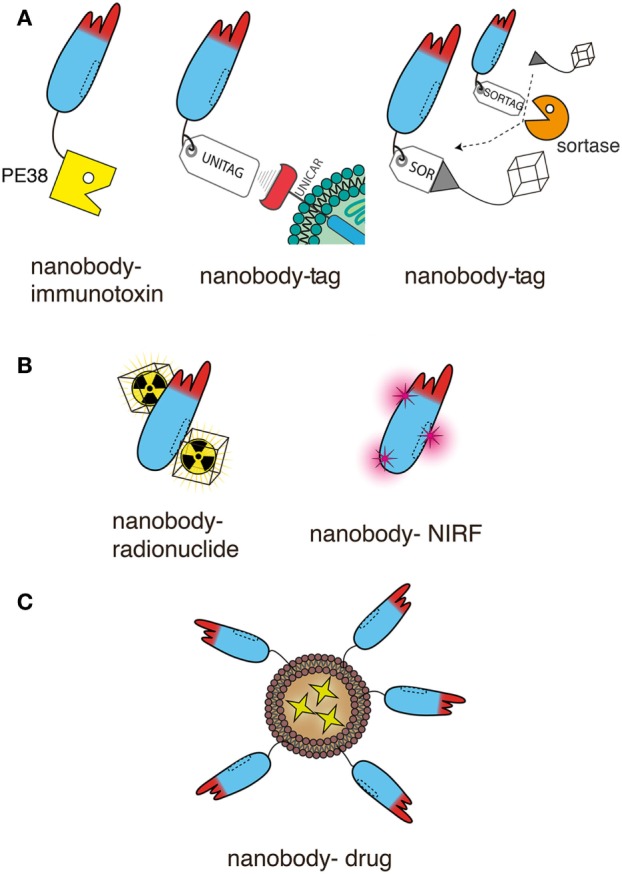
Monoclonal antibodies have revolutionized cancer therapy. However, delivery to tumor cells in vivo is hampered by the large size (150 kDa) of conventional antibodies. The minimal target recognition module of a conventional antibody is composed of two non-covalently associated variable domains (VH and VL). The proper orientation of these domains is mediated by their hydrophobic interface and is stabilized by their linkage to disulfide-linked constant domains (CH1 and CL). VH and VL domains can be fused via a genetic linker into a single chain variable fragment (scFv). scFv modules in turn can be fused to one another, e.g. to generate a bispecific T cell engager (BiTE), or they can be fused in various orientations to antibody hinge and Fc domains to generate bi- and multispecific antibodies. However, the inherent hydrophobic interaction of VH and VL domains limits the stability and solubility of engineered antibodies, often causing aggregation and/or mispairing of V-domains. Nanobodies (15 kDa) and nanobody-based human heavy chain antibodies (75 kDa) can overcome these limitations. Camelids naturally produce antibodies composed only of heavy chains in which the target recognition module is composed of a single variable domain (VHH or nanobody). Advantageous features of nanobodies include their small size, high solubility, high stability, and excellent tissue penetration in vivo. Nanobodies can readily be linked genetically to Fc-domains, other nanobodies, peptide tags, or toxins and can be conjugated chemically at a specific site to drugs, radionuclides, photosensitizers, and nanoparticles. These properties make them particularly suited for specific and efficient targeting of tumors in vivo. Chimeric nanobody-heavy chain antibodies combine advantageous features of nanobodies and human Fc domains in about half the size of a conventional antibody. In this review, we discuss recent developments and perspectives for applications of nanobodies and nanobody-based human heavy chain antibodies as anti-tumor therapeutics.
Overview
The claims and statistics of this paper haven’t been checked yet.
Run the check — free, no account needed. It reads the open-access text and checks the sources it cites.
Integrity signals
Registry
Cites
Statistics
Figures
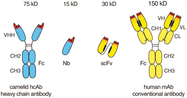
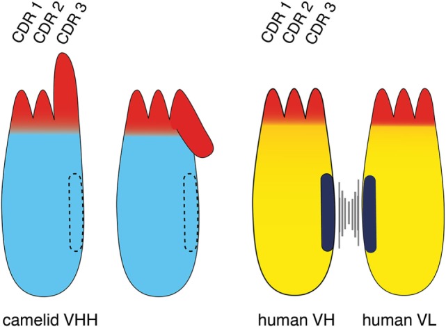
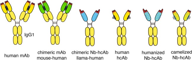
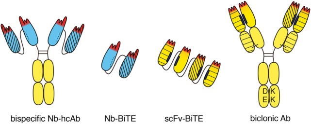

Verification
Claims the paper makes
The claims and statistics of this paper haven’t been checked yet.
Run the check — free, no account needed. It reads the open-access text and checks the sources it cites.
Claims other papers make about this one
Each is checked against this paper’s own text: does it say what the citing paper says it does?
The source backs part of the claim, not all of it — or backs it for a different population, condition or comparison.
In the citing paper
“One of the most promising is the H chain only antibody, or nanobody that are naturally occurring in camelids (llamas, alpacas and camels) [27] and sharks [228, 229] (reviewed by Muyldermans [230] and Bannas et al. [27]).”
From Computational approaches to therapeutic antibody design: established methods and emerging trends (2020) · cites this paper as [27]
Experimental — accuracy not yet measuredIn the citing paper
“The accumulated methodology of computational antibody protocols could potentially be applied to novel antibody formats with intrinsically better biophysical properties, such as nanobodies [27].”
From Computational approaches to therapeutic antibody design: established methods and emerging trends (2020) · cites this paper as [27]
In the citing paper
“Single domain antibodies have attracted attention because of their smaller size and better biophysical properties with respect to antibodies (higher stability and lower suspected immunogenicity) [27].”
From Computational approaches to therapeutic antibody design: established methods and emerging trends (2020) · cites this paper as [27]
Self-check
Percentages, sums and reported differences re-computed from the paper’s own numbers.
The self-check needs the paper’s full text.
Citations
Cites
Delivering nanomedicine to solid tumors
Jain, R. K., & Stylianopoulos, T. (2010) · Nature Reviews Clinical Oncology · doi:10.1038/nrclinonc.2010.139 · Cited by 3,265
Naturally occurring antibodies devoid of light chains
Hamers-Casterman, C., Atarhouch, T., Muyldermans, S., Robinson, G., Hammers, C., Songa, E. B., Bendahman, N., & Hammers, R. (1993) · Nature · doi:10.1038/363446a0 · Cited by 3,241
Rapid generation of functional human IgG antibodies derived from Fab-on-phage display libraries
Jostock, T., Vanhove, M., Brepoels, E., van Gool, R., Daukandt, M., Wehnert, A., van Hegelsom, R., Dransfield, D., Sexton, D., Devlin, M., et al. (2004) · Journal of Immunological Methods · doi:10.1016/j.jim.2004.03.014 · Cited by 2,572
Nanobodies: natural single-domain antibodies
Muyldermans, S. (2013) · Annual Review of Biochemistry · doi:10.1146/annurev-biochem-063011-092449 · Cited by 2,304
Scott, A. M., Wolchok, J. D., & Old, L. J. (2012) · Nature reviews. Cancer · doi:10.1038/nrc3236 · Cited by 2,244
Blinatumomab versus Chemotherapy for Advanced Acute Lymphoblastic Leukemia
Kantarjian, H. M., Stein, A. S., Gökbuget, N., Fielding, A. K., Schuh, A. C., Ribera, J., Wei, A. H., Dombret, H., Foà, R., Bassan, R., et al. (2017) · New England Journal of Medicine · doi:10.1056/nejmoa1609783 · Cited by 2,204
Reshaping human antibodies for therapy
Riechmann, L., Clark, M., Waldmann, H., & Winter, G. P. (1988) · Nature · doi:10.1038/332323a0 · Cited by 1,565
The therapeutic monoclonal antibody market
Ecker, D. M., Jones, S. D., & Levine, H. L. (2014) · mAbs · doi:10.4161/19420862.2015.989042 · Cited by 1,369
Adeno-Associated Virus (AAV) as a Vector for Gene Therapy
Naso, M. F., Tomkowicz, B., Perry, W. L., & Strohl, W. R. (2017) · BioDrugs · doi:10.1007/s40259-017-0234-5 · Cited by 1,362
Drug targeting to tumors: Principles, pitfalls and (pre-) clinical progress
Lammers, T., Kiessling, F., Hennink, W. E., & Storm, G. (2012) · Journal of Controlled Release · doi:10.1016/j.jconrel.2011.09.063 · Cited by 1,324
Cited by
Sorted by citations
Items 201–300 of 572
Ruiz‐Vega, G., Soler, M., Estévez, M. ‐., Ramirez‐Priego, P., Pazos, M. D., Noriega, M. A., Margolles, Y., Francés‐Gómez, C., Geller, R., Matusali, G., et al. (2022) · Sensors & Diagnostics · doi:10.1039/d2sd00082b · Cited by 26
Principles and current clinical landscape of NK cell engaging bispecific antibody against cancer
Tian, H., Guan, B., Li, H., Tu, X., Zhang, C., & Tang, B. (2023) · Human Vaccines & Immunotherapeutics · doi:10.1080/21645515.2023.2256904 · Cited by 25
Zheng, L., Wang, H., Zhong, X., Lin, J. H., Shi, G., Bai, C., Yang, R., Huang, Z., Jiang, Y., Wei, J., et al. (2024) · Biomaterials · doi:10.1016/j.biomaterials.2024.122809 · Cited by 22
Fan, X., Zhou, Y., Yu, J., Li, F., Chen, Q., Song, H., Feng, D., Liu, W., Nian, R., & Xian, M. (2019) · Separation and Purification Technology · doi:10.1016/j.seppur.2019.05.068 · Cited by 22
Diagnostics and Therapeutics in Targeting HER2 Breast Cancer: A Novel Approach
Vi, C., Mandarano, G., & Shigdar, S. L. (2021) · International Journal of Molecular Sciences · doi:10.3390/ijms22116163 · Cited by 25
Hu, X., Fan, J., Ma, Q., Han, L., Cao, Z., Xu, C., Luan, J., Jing, G., Nan, Y., Wu, T., et al. (2022) · Journal of Nanobiotechnology · doi:10.1186/s12951-022-01456-z · Cited by 22
Expanding and improving nanobody repertoires using a yeast display method: Targeting SARS-CoV-2
Cross, F. R., Fridy, P. C., Ketaren, N. E., Mast, F. D., Li, S., Olivier, J. P., Pecani, K., Chait, B. T., Aitchison, J. D., & Rout, M. P. (2023) · Journal of Biological Chemistry · doi:10.1016/j.jbc.2023.102954 · Cited by 21
Hofland, T., Endstra, S., Gomes, C. K. P., de Boer, R., de Weerdt, I., Bobkov, V., Riedl, J., Heukers, R., Smit, M. J., Eldering, E., et al. (2019) · HemaSphere · doi:10.1097/hs9.0000000000000308 · Cited by 22
Zhang, N., Zheng, Q., Wang, Y., Lin, J., Wang, H., Liu, R., Yan, M., Chen, X., Yang, J., & Chen, X. (2021) · Frontiers in Pharmacology · doi:10.3389/fphar.2021.681424 · Cited by 22
Verma, V., Sinha, N., & Raja, A. (2025) · mAbs · doi:10.1080/19420862.2025.2486390 · Cited by 21
Nanobody-based bispecific antibody engagers targeting CTLA-4 or PD-L1 for cancer immunotherapy
Liu, X., Gall, C. M. L., Alexander, R., Borgman, E., Balligand, T., & Ploegh, H. L. (2025) · Nature Biomedical Engineering · doi:10.1038/s41551-025-01447-z · Cited by 21
Campenhout, R. V., Muyldermans, S., Vinken, M., Devoogdt, N., & Groof, T. W. M. D. (2021) · Biomolecules · doi:10.3390/biom11010063 · Cited by 22
Silva-Pilipich, N., Smerdou, C., & Vanrell, L. (2021) · Microorganisms · doi:10.3390/microorganisms9091956 · Cited by 22
Emerging applications of nanobodies in cancer therapy
Awad, R. M., Meeus, F., Ceuppens, H., Ertveldt, T., Hanssens, H., Lecocq, Q., Mateusiak, Ł., Zeven, K., Valenta, H., Groof, T. W. M. D., et al. (2022) · International review of cytology · doi:10.1016/bs.ircmb.2022.03.010 · Cited by 21
Camels’ biological fluids contained nanobodies: promising avenue in cancer therapy
Al‐Numair, N. S., Theyab, A., Alzahrani, F. A., Shams, A. M., Alanazi, I. O., Oyouni, A. A. A., Al‐Amer, O. M., Mavromatis, C., Saadeldin, I. M., Abdali, W. A., et al. (2022) · Cancer Cell International · doi:10.1186/s12935-022-02696-7 · Cited by 24
Bannas, P., & Koch‐Nolte, F. (2018) · Frontiers in Immunology · doi:10.3389/fimmu.2018.02559 · Cited by 21
Next‐Generation Polymeric Nanomedicines for Oncology: Perspectives and Future Directions
Fletcher, N. L., Kempe, K., & Thurecht, K. J. (2020) · Macromolecular Rapid Communications · doi:10.1002/marc.202000319 · Cited by 21
Nanobodies—Useful Tools for Allergy Treatment?
Flicker, S., Zettl, I., & Tillib, S. V. (2020) · Frontiers in Immunology · doi:10.3389/fimmu.2020.576255 · Cited by 21
Identification of nanobodies against hepatocellular carcinoma marker glypican-3
Wang, W., Xu, C., Wang, H., & Jiang, C. (2021) · Molecular Immunology · doi:10.1016/j.molimm.2021.01.010 · Cited by 20
Wen, J., Moloney, E. B., Canning, A., Donohoe, E., Ritter, T., Wang, J., Xiang, D., Wu, J., & Li, Y. (2021) · Pharmacological Research · doi:10.1016/j.phrs.2021.105833 · Cited by 22
Construction of Synthetic Nanobody Library in Mammalian Cells by dsDNA‐Based Strategies**
Zhao, Y., Wang, Y., Su, W., & Li, S. (2021) · ChemBioChem · doi:10.1002/cbic.202100286 · Cited by 21
Zettl, I., Ivanova, T. I., Zghaebi, M., Rutovskaya, M. V., Ellinger, I., Goryainova, O. S., Kollárová, J., Villazala‐Merino, S., Lupinek, C., Weichwald, C., et al. (2022) · Frontiers in Immunology · doi:10.3389/fimmu.2022.1022418 · Cited by 21
Song, W., Wei, W., Lan, X., & Cai, W. (2023) · European Journal of Nuclear Medicine and Molecular Imaging · doi:10.1007/s00259-023-06266-5 · Cited by 20
Koerselman, M., Morshuis, L., & Karperien, M. (2023) · Acta Biomaterialia · doi:10.1016/j.actbio.2023.07.045 · Cited by 20
Fernández‐Quintero, M. L., Guarnera, E., Müsil, D., Pekar, L., Sellmann, C., Freire, F., Sousa, R. L., Santos, S. P., Freitas, M. C., Bandeiras, T. M., et al. (2024) · Protein Science · doi:10.1002/pro.5176 · Cited by 21
Nanotherapeutics for multiple myeloma
Zheleznyak, A., Shokeen, M., & Achilefu, S. (2018) · Wiley Interdisciplinary Reviews Nanomedicine and Nanobiotechnology · doi:10.1002/wnan.1526 · Cited by 21
Al‐Seragi, M., Chen, Y., & Duong, F. (2025) · Biochemical Society Transactions · doi:10.1042/bst20241419 · Cited by 19
Antibody-oligonucleotide conjugates in cancer therapy: Potential and Promise
Meng, Q., Yang, M., Xing, F., Xie, Z., Hao, Y., Jiang, P. X., & Xiao, B. (2025) · Critical Reviews in Oncology/Hematology · doi:10.1016/j.critrevonc.2025.104858 · Cited by 19
Advances in Nanobody-Based Platforms for Precision Cancer Diagnosis and Therapy
Yuan, Y., Zhuoga, C., Zeng, C., Zhou, W., Chen, W., Xu, Z. C., & Jiang, X. (2025) · Polymer science & technology. · doi:10.1021/polymscitech.5c00080 · Cited by 20
McInnes, I. B., Coates, L. C., Mease, P. J., Ogdie, A., Kavanaugh, A., Eder, L., Schett, G. A., Kivitz, A., McGonagle, D., Brennan, N., et al. (2025) · Nature Medicine · doi:10.1038/s41591-025-03971-6 · Cited by 19
Engineered Bifunctional Proteins for Targeted Cancer Therapy: Prospects and Challenges
Du, Y., & Xu, J. (2021) · Advanced Materials · doi:10.1002/adma.202103114 · Cited by 19
A review on emerging targeted therapies for the management of metastatic colorectal cancers
Deshmukh, R., Prajapati, M. K., & Harwansh, R. K. (2023) · Medical Oncology · doi:10.1007/s12032-023-02020-x · Cited by 19
Arras, P., Yoo, H. B., Pekar, L., Schröter, C. J., Clarke, T., Krah, S., Klewinghaus, D., Siegmund, V., Evers, A., & Zielonka, S. (2023) · mAbs · doi:10.1080/19420862.2023.2261149 · Cited by 19
BCMA/CD47-directed universal CAR-T cells exhibit excellent antitumor activity in multiple myeloma
Lu, Q., Li, H., Wu, Z., Zhu, Z., Zhang, Z., Yang, D., & Tong, A. (2024) · Journal of Nanobiotechnology · doi:10.1186/s12951-024-02512-6 · Cited by 19
Rapid LC–MS Method for Accurate Molecular Weight Determination of Membrane and Hydrophobic Proteins
Lippens, J. L., Egea, P. F., Spahr, C. S., Vaish, A., Keener, J. E., Marty, M. T., Loo, J. A., & Campuzano, I. D. G. (2018) · Analytical Chemistry · doi:10.1021/acs.analchem.8b03843 · Cited by 19
VHH characterization.Recombinant VHHs: Production, characterization and affinity
Chabrol, E., Stojko, J., Nicolas, A., Botzanowski, T., Fould, B., Antoine, M., Cianférani, S., Ferry, G., & Boutin, J. A. (2019) · Analytical Biochemistry · doi:10.1016/j.ab.2019.113491 · Cited by 18
Evaluation of P2X7 Receptor Function in Tumor Contexts Using rAAV Vector and Nanobodies (AAVnano)
Demeules, M., Scarpitta, A., Abad, C., Gondé, H., Hardet, R., Pinto-Espinoza, C., Eichhoff, A. M., Schäfer, W., Haag, F., Koch‐Nolte, F., et al. (2020) · Frontiers in Oncology · doi:10.3389/fonc.2020.01699 · Cited by 23
Abadi, B., Ahmadi‐Zeidabadi, M., Dini, L., & Vergallo, C. (2020) · Hematology/Oncology and Stem Cell Therapy · doi:10.1016/j.hemonc.2020.08.001 · Cited by 22
Peyron, I., Kizlik‐Masson, C., Dubois, M., Atsou, S., Ferrière, S., Denis, C. V., Lenting, P. J., Casari, C., & Christophe, O. D. (2020) · Research and Practice in Thrombosis and Haemostasis · doi:10.1002/rth2.12420 · Cited by 19
The role of single-domain antibodies (or nanobodies) in SARS-CoV-2 neutralization
Zebardast, A., Hosseini, P., Hasanzadeh, A., & Latifi, T. (2021) · Molecular Biology Reports · doi:10.1007/s11033-021-06819-7 · Cited by 19
Lam, K. H., Perry, K., Shoemaker, C. B., & Jin, R. (2020) · Toxins · doi:10.3390/toxins12100616 · Cited by 18
Baum, N., Fliegert, R., Bauche, A., Hambach, J., Menzel, S., Haag, F., Bannas, P., & Koch‐Nolte, F. (2020) · Cancers · doi:10.3390/cancers13010076 · Cited by 18
Anti-drug antibodies in the current management of cancer
Borregón, M., Martínez, K., Ramos, A., Ramos, I., Berzal, B., Mazariegos, M., Martínez, E., Hernández, T., Doger, B., & Moreno, V. (2022) · Cancer Chemotherapy and Pharmacology · doi:10.1007/s00280-022-04418-2 · Cited by 19
Modern Advances in CARs Therapy and Creating a New Approach to Future Treatment
Sadowski, K. A., Olejarz, W., & Basak, G. W. (2022) · International Journal of Molecular Sciences · doi:10.3390/ijms232315006 · Cited by 19
Leading Edge: Intratumor Delivery of Monoclonal Antibodies for the Treatment of Solid Tumors
Blanco, E., Chocarro, L., Fernández, L., Bocanegra, A., Arasanz, H., Echaide, M., Garnica, M., Piñeiro‐Hermida, S., Kochan, G. T., & Escors, D. (2023) · International Journal of Molecular Sciences · doi:10.3390/ijms24032676 · Cited by 18
Park, C.-Y., Kim, K., Kim, Y., Zhu, R., Hain, L., Seferovic, H., Kim, M., Woo, H. J., Hwang, H., Lee, S. H., et al. (2024) · ACS Nano · doi:10.1021/acsnano.4c00360 · Cited by 18
Unlocking the Gates: Therapeutic Agents for Noninvasive Drug Delivery Across the Blood-Brain Barrier
Culkins, C., Adomanis, R., Phan, N., Robinson, B., Slaton, E. W., Lothrop, E., Chen, Y., & Kimmel, B. R. (2024) · Molecular Pharmaceutics · doi:10.1021/acs.molpharmaceut.4c00604 · Cited by 22
Yang, S., Li, L., Yin, S., YouJun, S., Khan, M. U. Z., He, X., Li, Y., Gao, X. M., Liu, X., & Cai, J. (2018) · Applied Microbiology and Biotechnology · doi:10.1007/s00253-018-9324-7 · Cited by 18
Development of tilapia collagen and chitosan composite hydrogels for nanobody delivery
Fan, X., Liang, Y., Cui, Y., Li, F., Sun, Y., Yang, J., Song, H., Bao, Z., & Nian, R. (2020) · Colloids and Surfaces B Biointerfaces · doi:10.1016/j.colsurfb.2020.111261 · Cited by 18
Arafiles, J. V. V., Hirose, H., Hirai, Y., Kuriyama, M., Sakyiamah, M. M., Nomura, W., Sonomura, K., Imanishi, M., Otaka, A., Tamamura, H., et al. (2021) · Angewandte Chemie International Edition · doi:10.1002/anie.202016754 · Cited by 21
Nanobody Paratope Ensembles in Solution Characterized by MD Simulations and NMR
Fernández‐Quintero, M. L., DeRose, E. F., Gabel, S. A., Mueller, G. A., & Liedl, K. R. (2022) · International Journal of Molecular Sciences · doi:10.3390/ijms23105419 · Cited by 16
Site-Specific Radioactive Labeling of Nanobodies
Crauwels, M., Massa, S., Martin, C., Betti, C., Ballet, S., Devoogdt, N., Xavier, C., & Muyldermans, S. (2018) · Methods in molecular biology · doi:10.1007/978-1-4939-8648-4_26 · Cited by 18
Lamtha, T., Tabtimmai, L., Bangphoomi, K., Kiriwan, D., Malik, A. A., Chaicumpa, W., Henegouwen, P. M. P. V. B. E., & Choowongkomon, K. (2021) · Protein Engineering Design and Selection · doi:10.1093/protein/gzab030 · Cited by 17
Puthenveetil, R., Christenson, E. T., & Vinogradova, O. (2022) · Membranes · doi:10.3390/membranes12020227 · Cited by 18
Imaging and therapeutic targeting of the tumor immune microenvironment with biologics
Arnouk, S. M., Groof, T. W. M. D., & Ginderachter, J. A. V. (2022) · Advanced Drug Delivery Reviews · doi:10.1016/j.addr.2022.114239 · Cited by 17
Synthetic nanobodies as tools to distinguish IgG Fc glycoforms
Kao, K. S., Gupta, A., Zong, G., Li, C., Kerschbaumer, I., Borghi, S., Achkar, J. M., Bournazos, S., Wang, L., & Ravetch, J. V. (2022) · Proceedings of the National Academy of Sciences · doi:10.1073/pnas.2212658119 · Cited by 17
Emerging Landscape of Nanobodies and Their Neutralizing Applications against SARS-CoV-2 Virus
Feng, X., & Wang, H. (2023) · ACS Pharmacology & Translational Science · doi:10.1021/acsptsci.3c00042 · Cited by 19
TEMPRO: nanobody melting temperature estimation model using protein embeddings
Alvarez, J. A. E., & Dean, S. N. (2024) · Scientific Reports · doi:10.1038/s41598-024-70101-6 · Cited by 17
Anti-EGFR Binding Nanobody Delivery System to Improve the Diagnosis and Treatment of Solid Tumours
Wang, L., Zhang, G., Qin, L., Ye, H., Wang, Y., Long, B., & Jiao, Z. (2020) · Recent Patents on Anti-Cancer Drug Discovery · doi:10.2174/1574892815666200904111728 · Cited by 17
Nikooharf, A., Arezumand, R., Mansouri, K., Khoshi, A., & Ahmadabad, H. N. (2020) · Molecular Biotechnology · doi:10.1007/s12033-020-00275-7 · Cited by 17
Prospective Applications of Single-Domain Antibodies in Biomedicine
Tillib, S. V. (2020) · Molecular Biology · doi:10.1134/s0026893320030164 · Cited by 17
Research trends in the use of nanobodies for cancer therapy
Hou, J., Du, K., Li, J., Li, Z., Cao, S., Zhang, S., Huang, W., Liu, H., Yang, X., Sun, S., et al. (2025) · Journal of Controlled Release · doi:10.1016/j.jconrel.2025.01.045 · Cited by 18
Yan, Y., Cheng, X., Li, L., Zhang, R., Zhu, Y., Wu, Z., & Ding, K. (2021) · Frontiers in Oncology · doi:10.3389/fonc.2021.669393 · Cited by 17
Nanobody-based pannexin1 channel inhibitors reduce inflammation in acute liver injury
Campenhout, R. V., Groof, T. W. M. D., Kadam, P. H., Kwak, B. R., Muyldermans, S., Devoogdt, N., & Vinken, M. (2023) · Journal of Nanobiotechnology · doi:10.1186/s12951-023-02137-1 · Cited by 16
Yang, J., Lin, S., Chen, Z., Yang, F., Guo, L., Wang, L., Duan, Y., Zhang, X., Dai, Y., Yin, K., et al. (2023) · PLoS Pathogens · doi:10.1371/journal.ppat.1011804 · Cited by 16
Li, H., & Ye, B. (2024) · Synthetic and Systems Biotechnology · doi:10.1016/j.synbio.2024.09.001 · Cited by 16
Antibody–antigen complex modelling in the era of immunoglobulin repertoire sequencing
Raybould, M. I. J., Wong, W. K., & Deane, C. M. (2019) · Molecular Systems Design & Engineering · doi:10.1039/c9me00034h · Cited by 14
Hacisuleyman, A., & Erman, B. (2020) · Journal of Biological Physics · doi:10.1007/s10867-020-09548-3 · Cited by 16
Isolation of nanobodies with potential to reduce patients' IgE binding to Bet v 1
Zettl, I., Ivanova, T. I., Strobl, M. R., Weichwald, C., Goryainova, O. S., Khan, E., Rutovskaya, M. V., Focke‐Tejkl, M., Drescher, A., Bohle, B., et al. (2021) · Allergy · doi:10.1111/all.15191 · Cited by 16
Flexible on-demand cell-free protein synthesis platform based on a tube-in-tube reactor
Zhou, C., Lin, X., Lu, Y., & Zhang, J. (2019) · Reaction Chemistry & Engineering · doi:10.1039/c9re00394k · Cited by 17
Hambach, J., Mann, A. M., Bannas, P., & Koch‐Nolte, F. (2022) · Frontiers in Immunology · doi:10.3389/fimmu.2022.1005800 · Cited by 14
Stahl, P., Kollenda, S., Sager, J., Schmidt, L., Schroer, M. A., Stauber, R. H., Epple, M., & Knauer, S. K. (2023) · Small · doi:10.1002/smll.202300871 · Cited by 15
Lee, H. E., Cho, A. H., Hwang, J., Kim, J. W., Yang, H. R., Ryu, T., Jung, Y., & Lee, S. (2024) · International Journal of Molecular Sciences · doi:10.3390/ijms25094791 · Cited by 13
Wang, Q., Cheng, Q., Yao, G., Wang, Z., Zhu, L., Zeng, Z., Jia, L., Du, Y., Xue, J., & Gao, C. (2024) · Nano Today · doi:10.1016/j.nantod.2024.102507 · Cited by 15
Перспективы использования однодоменных антител в биомедицине
Tillib, S. V. (2020) · Molecular Biology · doi:10.31857/s0026898420030167 · Cited by 15
Beyond Allotypes: The Influence of Allelic Diversity in Antibody Constant Domains
Warrender, A. K., & Kelton, W. (2020) · Frontiers in Immunology · doi:10.3389/fimmu.2020.02016 · Cited by 16
Nanotechnology-based approaches in the fight against SARS-CoV-2
Albaz, A. A., Rafeeq, M. M., Sain, Z. M., Almutairi, W. A., Alamri, A. S., Aloufi, A. H., Almalki, W. H., & Tarique, M. (2021) · AIMS Microbiology · doi:10.3934/microbiol.2021023 · Cited by 15
Novel Ion Channel Targets and Drug Delivery Tools for Controlling Glioblastoma Cell Invasiveness
Varricchio, A., Ramesh, S. A., & Yool, A. J. (2021) · International Journal of Molecular Sciences · doi:10.3390/ijms222111909 · Cited by 17
Structure of the Pf12 and Pf41 heterodimeric complex of Plasmodium falciparum 6-cysteine proteins
Dietrich, M. H., Chan, L., Adair, A., Boulet, C., O’Neill, M. T., Tan, L. L., Keremane, S., Mok, Y.-F., Lo, A. W. H., Gilson, P. R., et al. (2022) · FEMS Microbes · doi:10.1093/femsmc/xtac005 · Cited by 14
Impact of tissue penetration and albumin binding on design of T cell targeted bispecific agents
Kopp, A., Kwon, H., Johnston, C. M., Vance, S., Legg, J. W., Galson-Holt, L., & Thurber, G. M. (2024) · Neoplasia · doi:10.1016/j.neo.2023.100962 · Cited by 15
Zheng, L., Wang, H., Zhou, J., Shi, G., Ma, J., Jiang, Y., Dong, Z., Li, J., He, Y., Wu, D., et al. (2024) · Journal for ImmunoTherapy of Cancer · doi:10.1136/jitc-2024-008888 · Cited by 14
Structure-guided mutations in CDRs for enhancing the affinity of neutralizing SARS-CoV-2 nanobody
Singh, V., Bhutkar, M., Choudhary, S., Nehul, S., Kumar, R., Singla, J., Kumar, P., & Tomar, S. (2024) · Biochemical and Biophysical Research Communications · doi:10.1016/j.bbrc.2024.150746 · Cited by 14
Huang, K., Li, Z., Su, T., Shen, D., Hu, S., & Cheng, K. (2019) · Advanced Therapeutics · doi:10.1002/adtp.201900009 · Cited by 14
Purification of animal immunoglobulin G ( IgG ) using peptoid affinity ligands
Reese, H. R., Bordelon, T., Odeh, F., Broussard, A., Kormos, C. M., Murphy, A., Shanahan, C. C., & Menegatti, S. (2020) · Biotechnology Progress · doi:10.1002/btpr.2994 · Cited by 14
Beta Cell Imaging—From Pre-Clinical Validation to First in Man Testing
Demine, S., Schulte, M. I., Territo, P. R., & Eizirik, D. L. (2020) · International Journal of Molecular Sciences · doi:10.3390/ijms21197274 · Cited by 15
Binding Mechanism of Neutralizing Nanobodies Targeting SARS-CoV-2 Spike Glycoprotein
Gölcük, M., Hacisuleyman, A., Erman, B., Yıldız, A., & Gür, M. (2021) · Journal of Chemical Information and Modeling · doi:10.1021/acs.jcim.1c00695 · Cited by 15
Pan, J., Li, N., Renn, A., Zhu, H., Chen, L., Shen, M., Hall, M. D., Qian, M., Pastan, I. H., & Ho, M. (2022) · Molecular Cancer Therapeutics · doi:10.1158/1535-7163.mct-21-0778 · Cited by 14
Phage Displayed Domain Antibodies (dAb) for Detection of Allergenic Pistachio Proteins in Foods
Madrid, R., García-García, A., González, I., Martı́n, R., & Garcı́a, T. (2020) · Foods · doi:10.3390/foods9091230 · Cited by 13
PLAbDab-nano: a database of camelid and shark nanobodies from patents and literature
Gordon, G. L., Greenshields‐Watson, A., Agarwal, P., Wong, A., Boyles, F., Hummer, A. M., Hernandez, A. G. L., & Deane, C. M. (2024) · Nucleic Acids Research · doi:10.1093/nar/gkae881 · Cited by 13
van Dijk, B., van Duyvenbode, J. F. F. H., de Vor, L., Nurmohamed, F. R. H. A., Lam, M. G. E. H., Poot, A. J., Ramakers, R. M., Koustoulidou, S., Beekman, F. J., van Strijp, J. A. G., et al. (2023) · International Journal of Molecular Sciences · doi:10.3390/ijms24054374 · Cited by 13
AGuIX nanoparticle-nanobody bioconjugates to target immune checkpoint receptors
Carmès, L., Bort, G., Lux, F., Seban, L., Rocchi, P., Muradova, Z., Hagège, A., Heinrich, L., Delolme, F., Gueguen‐Chaignon, V., et al. (2023) · Nanoscale · doi:10.1039/d3nr04777f · Cited by 12
Nanobody-Based Drug Delivery: Emerging Strategies for Targeted Cancer Therapy
Ghosh, N., Sepay, N., Paul, M., & Im, J. (2026) · International Journal of Nanomedicine · doi:10.2147/ijn.s584604 · Cited by 14
Percipalle, M., Hunashal, Y., Steyaert, J., Fogolari, F., & Esposito, G. (2021) · Molecules · doi:10.3390/molecules26123567 · Cited by 12
Designing of nanobodies against Dengue virus Capsid: a computational affinity maturation approach
Chaudhuri, D., Majumder, S., Datta, J., & Giri, K. (2022) · Journal of Biomolecular Structure and Dynamics · doi:10.1080/07391102.2022.2029773 · Cited by 12
Wu, S., Luo, Q., Li, F., Zhang, S., Zhang, C., Liu, J., Shao, B., Yang, H., Tan, T., Dong, X., et al. (2024) · British Journal of Haematology · doi:10.1111/bjh.19631 · Cited by 13
Zheng, L., Ding, Y., Xu, X., Wang, H., Shi, G., Li, Y., He, Y., Gong, Y., Zhang, X., Wei, J., et al. (2025) · Acta Pharmaceutica Sinica B · doi:10.1016/j.apsb.2025.03.039 · Cited by 12
Unique Benefits of Tumor-Specific Nanobodies for Fluorescence Guided Surgery
Lwin, T. M., Hoffman, R. M., & Bouvet, M. (2021) · Biomolecules · doi:10.3390/biom11020311 · Cited by 12
Shajari, S., Farajollahi, M. M., Behdani, M., & Tarighi, P. (2022) · Molecular Biotechnology · doi:10.1007/s12033-022-00485-1 · Cited by 12
Mirror-Image Single-Domain Antibody for a Novel Nonimmunogenic Drug Scaffold
Aoki, K., Manabe, A., Kimura, H., Katoh, Y., Inuki, S., Ohno, H., Nonaka, M., & Oishi, S. (2023) · Bioconjugate Chemistry · doi:10.1021/acs.bioconjchem.3c00372 · Cited by 11
Boutin, L., Barjon, C., Chauvet, M., Lafrance, L., Senechal, E., Bourges, D., Vigne, E., & Scotet, E. (2024) · European Journal of Immunology · doi:10.1002/eji.202350773 · Cited by 14
Metadata
- Authors
- Bannas, P., Hambach, J., & Koch-Nolte, F.
- Affiliations
- University Medical Center Hamburg-Eppendorf (DE)
- Venue
- Frontiers in Immunology
- Publisher
- Frontiers Media SA
- Published
- 2017-11-22
- Volume
- 8
- Pages
- 1603-1603
- ISSN
- 1664-3224 (ISSN-L)
- PMC
- PMC5702627
- OpenAlex
- W2769445535
- Cited by
- 572
- References
- 134
- Citations by year
- 2026: 51 · 2025: 68 · 2024: 93 · 2023: 62 · 2022: 70 · 2021: 85 · 2020: 68 · 2019: 48 · 2018: 27
- FWCI
- 5.92
- Type
- journal article
- Language
- en
- Open access
- gold
- doi.org
- Licence
- cc-by
- Locations
- Frontiers in Immunology (publishedVersion, cc-by)
- Europe PMC (PubMed Central) (publishedVersion, cc-by)
- PubMed Central (publishedVersion, cc-by)
- PubMed (publishedVersion)
- DOAJ (DOAJ: Directory of Open Access Journals) (submittedVersion)
- Funders
- Wilhelm Sander-Stiftung (2012.113.2)
- Topics
- Life Sciences › Immunology and Microbiology › Immunology › Monoclonal and Polyclonal Antibodies Research
- Life Sciences › Immunology and Microbiology › Immunology › Toxin Mechanisms and Immunotoxins
- Health Sciences › Medicine › Radiology, Nuclear Medicine and Imaging › Radiopharmaceutical Chemistry and Applications
- Keywords
- nanobodies, VHH, camelid antibodies, antibody engineering, tumor targeting, antibody aggregation, bispecific T-cell engagers, antibody-drug conjugates, radionuclide conjugation, photosensitizer conjugation, nanoparticle conjugation, cancer immunotherapy
About these checks
Registry, retraction, Cites and statistics results are computed, not judged, with one exception: a printed reference that no index lists is read once by a model before it counts as a citation error. Claims are extracted by a model and kept only when their quote appears verbatim in the text; each cited source is then checked, with a model’s help, for whether it says what the claim relies on. Those verdicts can be wrong — read the claim’s quote and the cited source before relying on one.
Statistics and arithmetic are recomputed, not judged.
Confidence bands are shown only for fields calibrated on our annotated test set. None is calibrated yet, so no band is shown.
What counts as a claim ›How we measure confidence ›How we check this ›
Are you an author, or do you see a problem with this page? Request a review
