

The DOI matches its Crossref record; 3 of the 3 references it cites were found in a registry or an index.
Breaching the Bacterial Envelope: The Pivotal Role of Perforin-2 (MPEG1) Within Phagocytes
Abstract
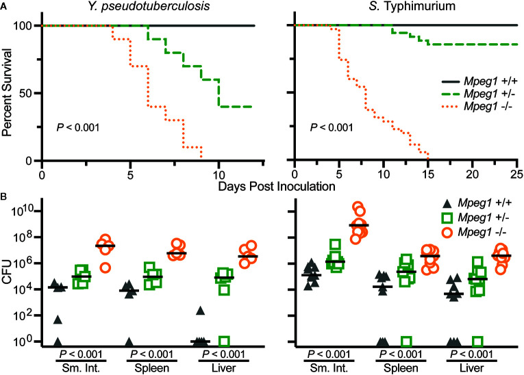
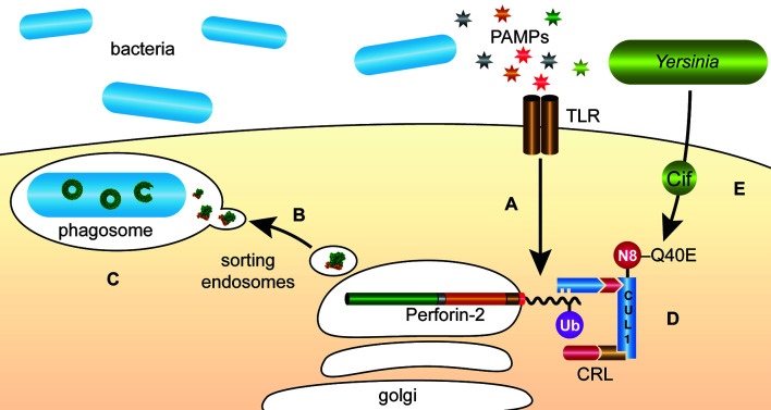
The membrane attack complex (MAC) of the complement system and Perforin-1 are well characterized innate immune effectors. MAC is composed of C9 and other complement proteins that target the envelope of gram-negative bacteria. Perforin-1 is deployed when killer lymphocytes degranulate to destroy virally infected or cancerous cells. These molecules polymerize with MAC-perforin/cholesterol-dependent cytolysin (MACPF/CDC) domains of each monomer deploying amphipathic β-strands to form pores through target lipid bilayers. In this review we discuss one of the most recently discovered members of this family; Perforin-2, the product of the Mpeg1 gene. Since their initial description more than 100 years ago, innumerable studies have made macrophages and other phagocytes some of the best understood cells of the immune system. Yet remarkably it was only recently revealed that Perforin-2 underpins a pivotal function of phagocytes; the destruction of phagocytosed microbes. Several studies have established that phagocytosed bacteria persist and in some cases flourish within phagocytes that lack Perforin-2. When challenged with either gram-negative or gram-positive pathogens Mpeg1 knockout mice succumb to infectious doses that the majority of wild-type mice survive. As expected by their immunocompromised phenotype, bacterial pathogens replicate and disseminate to deeper tissues of Mpeg1 knockout mice. Thus, this evolutionarily ancient gene endows phagocytes with potent bactericidal capability across taxa spanning sponges to humans. The recently elucidated structures of mammalian Perforin-2 reveal it to be a homopolymer that depends upon low pH, such as within phagosomes, to transition to its membrane-spanning pore conformation. Clinical manifestations of Mpeg1 missense mutations further highlight the pivotal role of Perforin-2 within phagocytes. Controversies and gaps within the field of Perforin-2 research are also discussed as well as animal models that may be used to resolve the outstanding issues. Our review concludes with a discussion of bacterial counter measures against Perforin-2.
Overview
The claims and statistics of this paper haven’t been checked yet.
Run the check — free, no account needed. It reads the open-access text and checks the sources it cites.
Integrity signals
Registry
Cites
Statistics
Figures
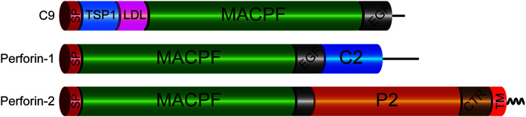
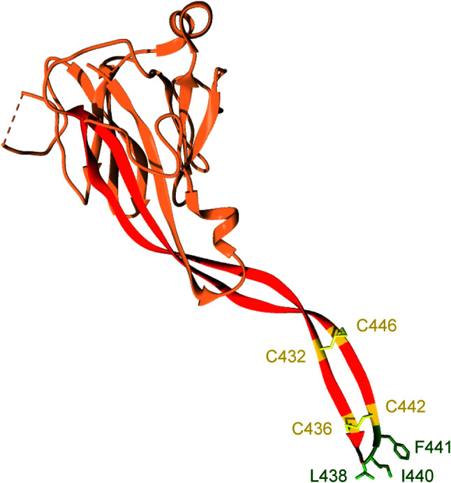
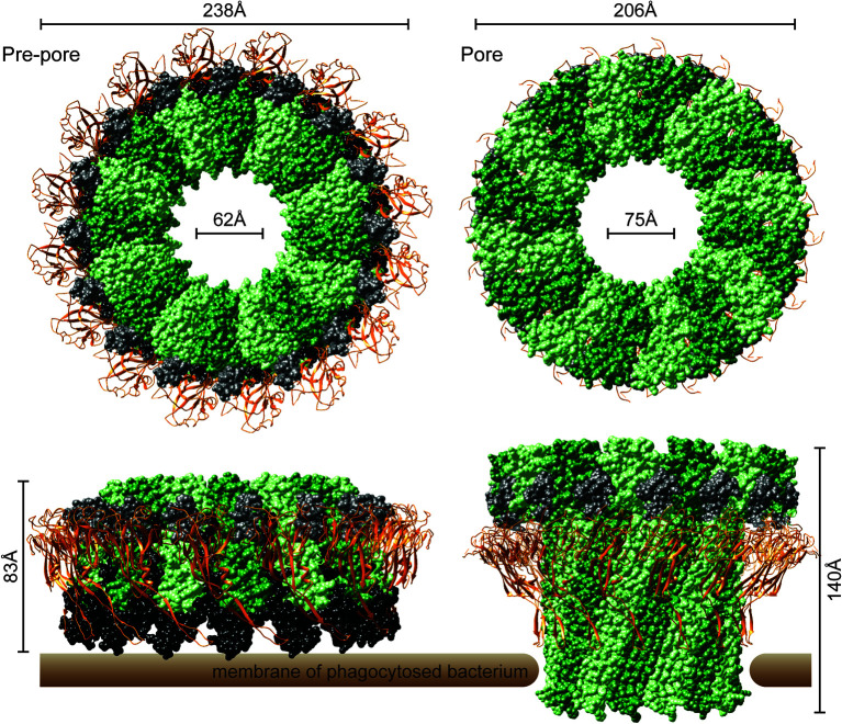
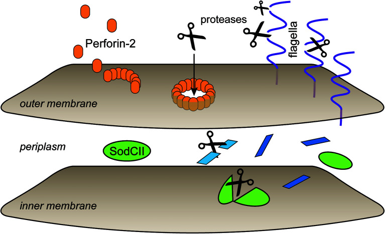
Verification
Claims the paper makes
The claims and statistics of this paper haven’t been checked yet.
Run the check — free, no account needed. It reads the open-access text and checks the sources it cites.
Claims other papers make about this one
Each is checked against this paper’s own text: does it say what the citing paper says it does?
In the citing paper
“MPEG1 (macrophage expressed gene 1) inhibits the acidification of phagocytic vacuole (44).”
From Loss of Dnmt3a impairs hematopoietic homeostasis and myeloid cell skewing via the PI3Kinase pathway (2023) · cites this paper as [44]
Self-check
Percentages, sums and reported differences re-computed from the paper’s own numbers.
The self-check needs the paper’s full text.
Citations
Cites
Perforin and granzymes: function, dysfunction and human pathology
Voskoboinik, I., Whisstock, J. C., & Trapani, J. A. (2015) · Nature Reviews Immunology · doi:10.1038/nri3839 · Cited by 1,327
The SCF ubiquitin ligase: insights into a molecular machine
Cardozo, T., & Pagano, M. (2004) · Nature Reviews Molecular Cell Biology · doi:10.1038/nrm1471 · Cited by 1,114
Endosomes are specialized platforms for bacterial sensing and NOD2 signalling
Nakamura, N., Lill, J. R., Phung, Q., Jiang, Z., Bakalarski, C., de Mazière, A., Klumperman, J., Schlatter, M., Delamarre, L., & Mellman, I. (2014) · Nature · doi:10.1038/nature13133 · Cited by 313
Research works printed but not linked: 102
- Lukoyanova, 2016, The membrane attack complex, perforin and cholesterol-dependent cytolysin superfamily of pore-forming proteins, J Cell Sci, 129, 10.1242/jcs.182741 · DOI
- Ni, 2017, Repurposing a pore: Highly conserved perforin-like proteins with alternative mechanisms, Philos Trans R Soc B Biol Sci, 372, 10.1098/rstb.2016.0212 · DOI
- Gilbert, 2002, Pore-forming toxins, Cell Mol Life Sci, 59, 10.1007/s00018-002-8471-1 · DOI
- Anderluh, 2014, Distribution of MACPF/CDC proteins, Subcell Biochem, 80, 7, 10.1007/978-94-017-8881-6_2 · DOI
- Yoshida, 1989, Study of cell kinetics in anaplastic thyroid carcinoma transplanted to nude mice, J Surg Oncol, 41, 1, 10.1002/jso.2930410104 · DOI
- Gilbert, 1999, Two structural transitions in membrane pore formation by pneumolysin, the pore-forming toxin of Streptococcus pneumoniae, Cell, 97, 10.1016/S0092-8674(00)80775-8 · DOI
- Podack, 1982, Polymerization of the ninth component of complement (C9): Formation of poly(C9) with a tubular ultrastructure resembling the membrane attack complex of complement, Proc Natl Acad Sci USA, 79, 10.1073/pnas.79.2.574 · DOI
- Morgan, 2016, Terminal complexes of the complement system: new structural insights and their relevance to function, Immunol Rev, 274, 10.1111/imr.12461 · DOI · Paper page
- Law, 2010, The structural basis for membrane binding and pore formation by lymphocyte perforin, Nature, 468, 10.1038/nature09518 · DOI
- Dudkina, 2016, Structure of the poly-C9 component of the complement membrane attack complex, Nat Commun, 7, 10588, 10.1038/ncomms10588 · DOI
- Podack, 1982, Circular polymerization of the ninth component of complement. Ring closure of the tubular complex confers resistance to detergent dissociation and to proteolytic degradation, J Biol Chem, 10.1073/pnas.79.2.574 · DOI
- Spilsbury, 1995, Isolation of a novel macrophage-specific gene by differential cDNA analysis, Blood, 10.1182/blood.v85.6.1620.bloodjournal8561620 · DOI
- Dangelo, 2012, Perforin evolved from a gene duplication of MPEG1, followed by a complex pattern of gene gain and loss within Euteleostomi, BMC Evol Biol, 12, 59, 10.1186/1471-2148-12-59 · DOI
- McCormack, 2015, Perforin-2 is essential for intracellular defense of parenchymal cells and phagocytes against pathogenic bacteria, eLife, 4, 1, 10.7554/eLife.06508 · DOI
- Garin, 2001, The phagosome proteome: Insight into phagosome functions, J Cell Biol, 4, e06508, 10.1083/jcb.152.1.165 · DOI
- Duclos, 2011, The endosomal proteome of macrophage and dendritic cells, Proteomics, 11, 10.1002/pmic.201000577 · DOI
- McCormack, 2015, Enteric pathogens deploy cell cycle inhibiting factors to block the bactericidal activity of Perforin-2, eLife, 4, e06505, 10.7554/eLife.06505 · DOI
- Ni, 2020, Structure and mechanism of bactericidal mammalian perforin-2, an ancient agent of innate immunity, Sci Adv, 6, eaax8286, 10.1126/sciadv.aax8286 · DOI
- Pang, 2019, The cryo-EM structure of the acid activatable pore-forming immune effector Macrophage-expressed gene 1, Nat Commun, 10.1038/s41467-019-12279-2 · DOI
- Christie, 2018, Cholesterol-dependent cytolysins: from water-soluble state to membrane pore, Biophys Rev, 10.1007/s12551-018-0448-x · DOI
- Tweten, 2005, Cholesterol-dependent cytolysins, a family of versatile pore-forming toxins, Infect Immun, 10.1128/IAI.73.10.6199-6209.2005 · DOI
- Hackam, 1997, Regulation of phagosomal acidification. Differential targeting of Na +/H + exchangers, Na +/K +-ATPases, and vacuolar-type H +-ATPases, J Biol Chem, 10.1074/jbc.272.47.29810 · DOI
- Beyenbach, 2006, The V-type H+ ATPase: Molecular structure and function, physiological roles and regulation, J Exp Biol, 10.1242/jeb.02014 · DOI
- Bai, 2018, Perforin-2 Breaches the Envelope of Phagocytosed Bacteria Allowing Antimicrobial Effectors Access to Intracellular Targets, J Immunol, 201, 10.4049/jimmunol.1800365 · DOI
- Wiens, 2005, Innate immune defense of the sponge Suberites domuncula against bacteria involves a MyD88-dependent signaling pathway: Induction of a perforin-like molecule, J Biol Chem, 280, 10.1074/jbc.M504049200 · DOI
- Walters, 2020, The Complicated Evolutionary Diversification of the Mpeg-1/Perforin-2 Family in Cnidarians, Front Immunol, 11, 10.3389/fimmu.2020.01690 · DOI
- Choi, 2020, Functional characterization and gene expression profile of perforin-2 in starry flounder (Platichthys stellatus), Fish Shellfish Immunol, 107, 10.1016/j.fsi.2020.11.011 · DOI
- Estévez-Calvar, 2011, Involvement of pore-forming molecules in immune defense and development of the Mediterranean mussel (Mytilus galloprovincialis), Dev Comp Immunol, 35, 10.1016/j.dci.2011.03.023 · DOI
- Bathige, 2014, Antibacterial activity and immune responses of a molluscan macrophage expressed gene-1 from disk abalone, Haliotis discus discus, Fish Shellfish Immunol, 39, 10.1016/j.fsi.2014.05.012 · DOI
- Modak, 2020, Contrasting immunomodulatory effects of probiotic and pathogenic bacteria on eastern oyster, crassostrea virginica, larvae, Vaccines, 8, 588, 10.3390/vaccines8040588 · DOI
- Kemp, 2011, Identification and characterisation of the Mpeg1 homologue in the South African abalone, Haliotis midae, Fish Shellfish Immunol, 31, 10.1016/j.fsi.2011.07.010 · DOI
- Ni, 2019, Grouper (Epinephelus coioides) Mpeg1s : Molecular identification, expression analysis, and antimicrobial activity, Fish Shellfish Immunol, 92, 10.1016/j.fsi.2019.06.060 · DOI
- Jagannathan-Bogdan, 2013, Hematopoiesis, Dev, 140, 10.1242/dev.083147 · DOI
- Mo, 2016, Comparative transcriptional profile of the fish parasite Cryptocaryon irritans, Parasites Vectors, 9, 1, 10.1186/s13071-016-1919-1 · DOI
- Ni, 2020, Distribution of Mpeg1+ cells in healthy grouper (Epinephelus coioides) and after Cryptocaryon irritans infection, Fish Shellfish Immunol, 104, 10.1016/j.fsi.2020.06.018 · DOI
- Le Pennec, 2019, The challenge of the sponge suberites domuncula (Olivi, 1792) in the presence of a symbiotic bacterium and a pathogen bacterium, Genes (Basel), 10.3390/genes10070485 · DOI
- Libro, 2013, RNA-seq profiles of immune related genes in the staghorn coral Acropora cervicornis Infected with white band disease, PLoS One, 10.1371/journal.pone.0081821 · DOI
- Gignoux-Wolfsohn, 2015, Identification of candidate coral pathogens on white band disease-infected staghorn coral, PLoS One, 10.1371/journal.pone.0134416 · DOI
- Duke, 1989, Purified perforin induces target cell lysis but not DNA fragmentation, J Exp Med, 10.1084/jem.170.4.1451 · DOI
- Masson, 1985, Isolation of a lytic, pore-forming protein (perforin) from cytolytic T-lymphocytes, J Biol Chem, 260, 10.1016/S0021-9258(17)39328-6 · DOI
- He, 2011, An Mpeg (macrophage expressed gene) from the Pacific oyster Crassostrea gigas: Molecular characterization and gene expression, Fish Shellfish Immunol, 30, 10.1016/j.fsi.2011.01.009 · DOI
- Biesecker, 1980, The ninth component of human complement: Purification and physicochemical characterization, J Immunol, 124, 10.4049/jimmunol.124.3.1291 · DOI
- Biesecker, 1982, An amphiphilic structure of the ninth component of human complement. Evidence from analysis of fragments produced by α-thrombin, J Biol Chem, 10.1016/S0021-9258(18)34964-0 · DOI
- Kontermann, 1989, N-Deglycosylation of human complement component C9 reduces its hemolytic activity, Mol Immunol, 10.1016/0161-5890(89)90056-4 · DOI
- Franc, 2017, Proteoform Profile Mapping of the Human Serum Complement Component C9 Revealing Unexpected New Features of N-, O-, and C-Glycosylation, Anal Chem, 10.1021/acs.analchem.6b04527 · DOI
- Spicer, 2018, The first transmembrane region of complement component-9 acts as a brake on its self-assembly, Nat Commun, 10.1038/s41467-018-05717-0 · DOI
- Chen, 2009, Glycoproteomics analysis of human liver tissue by combination of multiple enzyme digestion and hydrazide chemistry, J Proteome Res, 10.1021/pr8008012 · DOI
- Shental-Bechor, 2008, Effect of glycosylation on protein folding: A close look at thermodynamic stabilization, Proc Natl Acad Sci U S A, 10.1073/pnas.0801340105 · DOI
- Lee, 2015, Effects of N-glycosylation on protein conformation and dynamics: Protein Data Bank analysis and molecular dynamics simulation study, Sci Rep, 5, 8926, 10.1038/srep08926 · DOI
- McCormack, 2016, Perforin-2 Protects Host Cells and Mice by Restricting the Vacuole to Cytosol Transitioning of a Bacterial Pathogen, Infect Immun, 84, 10.1128/iai.01434-15 · DOI
- McCormack, 2013, Inhibition of intracellular bacterial replication in fibroblasts is dependent on the perforin-like protein (Perforin-2) encoded by macrophage-expressed gene 1, J Innate Immun, 5, 10.1159/000345249 · DOI
- Strbo, 2019, Single cell analyses reveal specific distribution of anti-bacterial molecule Perforin-2 in human skin and its modulation by wounding and Staphylococcus aureus infection, Exp Dermatol, 28, 10.1111/exd.13870 · DOI
- Ellett, 2011, Phagocytes, Granulocytes, and Myelopoiesis mpeg1 promoter transgenes direct macrophage-lineage expression in zebrafish, Blood, 27, 10.1182/blood-2010-10-314120 · DOI
- Zakrzewska, 2010, Macrophage-specific gene functions in Spi1-directed innate immunity, Blood, 116, 1, 10.1182/blood-2010-01-262873 · DOI
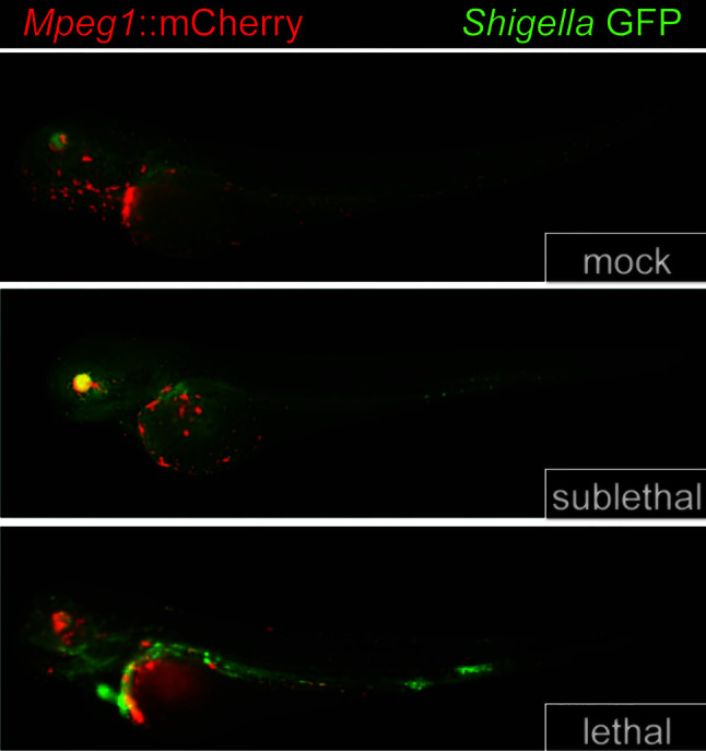
- Benard, 2015, Macrophage-expressed perforins Mpeg1 and Mpeg1.2 have an anti-bacterial function in zebrafish, J Innate Immun, 7, 10.1159/000366103 · DOI
- Mostowy, 2013, The Zebrafish as a New Model for the In Vivo Study of Shigella flexneri Interaction with Phagocytes and Bacterial Autophagy, PLoS Pathog, 9, e81003588, 10.1371/journal.ppat.1003588 · DOI
- McCormack, 2015, Perforin-2/Mpeg1 and other pore-forming proteins throughout evolution, J Leukoc Biol, 98, 10.1189/jlb.4MR1114-523RR · DOI
- Podack, 2016, Killing of microbes and cancer by the immune system with three mammalian pore-forming killer proteins, Front Immunol, 7, 10.3389/fimmu.2016.00464 · DOI
- Podack, 1977, The SC5b-7 complex: Formation, isolation, properties, and subunit composition, J Immunol, 10.4049/jimmunol.119.6.2024 · DOI
- Podack, 1980, Membrane attack complex of complement. A structural analysis of its assembly, J Exp Med, 10.1084/jem.151.2.301 · DOI
- Tschopp, 1981, Membranolysis by the ninth component of human complement, Biochem Biophys Res Commun, 10.1016/0006-291X(81)91981-1 · DOI
- Tschopp, 1982, Formation of transmembrane tubules by spontaneous polymerization of the hydrophilic complement protein C9, Nature, 10.1038/298534a0 · DOI
- Podack, 1983, Assembly of two types of tubules with putative cytolytic function by cloned natural killer cells, Nature, 10.1038/302442a0 · DOI
- Podack, 1985, Cytolytic T-cell granules: biochemical properties and functional specificity, Adv Exp Med Biol, 10.1007/978-1-4684-8326-0_8 · DOI
- Podack, 1985, Isolation and biochemical and functional characterization of perforin 1 from cytolytic T-cell granules, Proc Natl Acad Sci U S A, 10.1073/pnas.82.24.8629 · DOI
- Lichtenheld, 1988, Structure and function of human perforin, Nature, 10.1038/335448a0 · DOI
- Podack, 1989, Structure of perforin and its role in cytolysis, Year Immunol
- McCormack, 2013, Killing machines: Three pore-forming proteins of the immune system, Immunol Res, 57, 10.1007/s12026-013-8469-9 · DOI
- Fields, 2013, Perforin-2 Restricts Growth of Chlamydia trachomatis in Macrophages, Infect Immun, 81, 10.1128/iai.00497-13 · DOI
- Darville, 2010, Pathogenesis of genital tract disease due to Chlamydia trachomatis, J Infect Dis, 201, 10.1086/652397 · DOI
- Keb, 2020, An Ancient Molecular Arms Race: Chlamydia vs. Membrane Attack Complex/Perforin (MACPF) Domain Proteins, Front Immunol, 10.3389/fimmu.2020.01490 · DOI
- Pastar, 2020, Staphylococcus epidermidis Boosts Innate Immune Response by Activation of Gamma Delta T Cells and Induction of Perforin-2 in Human Skin, Front Immunol, 11, 10.3389/fimmu.2020.550946 · DOI
- Gayle, 2020, Maternal- and Fetal-Encoded Perforin-2 Limits Placental Infection by a Bloodborne Pathogen, J Immunol, 205, 10.4049/jimmunol.2000615 · DOI
- Cheers, 1984, Listeriosis in beige mice and their heterozygous littermates, Immunology, 51
- Portnoy, 2002, The cell biology of Listeria monocytogenes infection: The intersection of bacterial pathogenesis and cell-mediated immunity, J Cell Biol, 10.1083/jcb.200205009 · DOI
- Dramsi, 2002, Listeriolysin O: A genuine cytolysin optimized for an intracellular parasite, J Cell Biol, 10.1083/jcb.200202121 · DOI
- McCormack, 2020, An Essential Role for Perforin-2 in Type I IFN Signaling, J Immunol, 10.4049/jimmunol.1901013 · DOI
- Kayagaki, 2011, Non-canonical inflammasome activation targets caspase-11, Nature, 10.1038/nature10558 · DOI · Paper page
- Kenneth, 2012, An inactivating caspase 11 passenger mutation originating from the 129 murine strain in mice targeted for c-IAP1, Biochem J, 10.1042/BJ20120249 · DOI
- Thomas, 2011, Structural linkage between ligand discrimination and receptor activation by Type I interferons, Cell, 10.1016/j.cell.2011.06.048 · DOI
- Weng, 2019, HawkDock: a web server to predict and analyze the protein-protein complex based on computational docking and MM/GBSA, Nucleic Acids Res, 10.1093/nar/gkz397 · DOI
- Ringwald, 2000, GXD: A gene expression database for the laboratory mouse: Current status and recent enhancements, Nucleic Acids Res, 10.1093/nar/28.1.115 · DOI
- Hemberger, 2000, cDNA subtraction cloning reveals novel genes whose temporal and spatial expression indicates association with trophoblast invasion, Dev Biol, 10.1006/dbio.2000.9705 · DOI
- Karczewski, 2020, The mutational constraint spectrum quantified from variation in 141,456 humans, Nature, 581, 10.1038/s41586-020-2308-7 · DOI · Paper page
- McCormack, 2017, MPEG1/perforin-2 mutations in human pulmonary nontuberculous mycobacterial infections, JCI Insight, 2, 1, 10.1172/jci.insight.89635 · DOI
- Merselis, 2020, MPEG1/Perforin-2 Haploinsufficiency Associated Polymicrobial Skin Infections and Considerations for Interferon-γ Therapy, Front Immunol, 11, 10.3389/fimmu.2020.601584 · DOI
- Bosu, 2008, Cullin-RING ubiquitin ligases: Global regulation and activation cycles, Cell Div, 10.1186/1747-1028-3-7 · DOI
- Charpentier, 2004, Identification of the secretion and translocation domain of the enteropathogenic and enterohemorrhagic Escherichia coli effector Cif, using TEM-1 β-lactamase as a new fluorescence-based reporter, J Bacteriol, 10.1128/JB.186.16.5486-5495.2004 · DOI
- Crow, 2012, The molecular basis of ubiquitin-like protein NEDD8 deamidation by the bacterial effector protein Cif, Proc Natl Acad Sci U S A, 109, 10.1073/pnas.1112107109 · DOI
- Cui, 2010, Glutamine deamidation and dysfunction of ubiquitin/NEDD8 induced by a bacterial effector family, Science, 80, 10.1126/science.1193844 · DOI
- Reddick, 2014, Bacteria fighting back: How pathogens target and subvert the host innate immune system, Mol Cell, 54, 10.1016/j.molcel.2014.03.010 · DOI
- Wilkinson, 1984, Marine sponges discriminate between food bacteria and bacterial symbionts: Electron microscope radioautography and in situ evidence, Proc R Soc London - Biol Sci, 10.1098/rspb.1984.0018 · DOI
- Wehrl, 2007, Bacterial uptake by the marine sponge Aplysina aerophoba, Microb Ecol, 10.1007/s00248-006-9090-4 · DOI
- Olano, 2000, Phagocytic activities of the gorgonian coral Swiftia exserta, J Invertebr Pathol, 10.1006/jipa.2000.4974 · DOI
- Adema, 1991, Molluscan Hemocyte-Mediated Cytotoxicity - the Role of Reactive Oxygen Intermediates, Rev Aquat Sci
- Mohandas, 1985, Mechanism of lysosomal enzyme release from Mercenaria mercenaria granulocytes: A scanning electron microscope study, J Invertebr Pathol, 10.1016/0022-2011(85)90148-X · DOI
- Willenz, 1984, Ultrastructural localization of lysosomal digestion in the fresh water sponge Ephydatia fluviatilis, J Ultrasructure Res, 10.1016/S0022-5320(84)90112-6 · DOI
- Krawczyk, 2020, To Kill But Not Be Killed: Controlling the Activity of Mammalian Pore-Forming Proteins, Front Immunol, 10.3389/fimmu.2020.601405 · DOI
- Bayly-Jones, 2020, Ancient but Not Forgotten: New Insights Into MPEG1, a Macrophage Perforin-Like Immune Effector, Front Immunol, 10.3389/fimmu.2020.581906 · DOI
- Hung, 2020, Cellular context of IL-33 expression dictates impact on anti-helminth immunity, Sci Immunol, 10.1126/sciimmunol.abc6259 · DOI
- Pettersen, 2004, UCSF Chimera - A visualization system for exploratory research and analysis, J Comput Chem, 10.1002/jcc.20084 · DOI
- Pettersen, 2020, UCSF ChimeraX: Structure visualization for researchers, educators, and developers, Protein Sci, 30, 70, 10.1002/pro.3943 · DOI
Cited by
Loss of Dnmt3a impairs hematopoietic homeostasis and myeloid cell skewing via the PI3Kinase pathway
Palam, L. R., Ramdas, B., Pickerell, K., Pasupuleti, S. K., Kanumuri, R., Cesarano, A., Szymanski, M., Selman, B., Dave, U. P., Sandusky, G., et al. (2023) · JCI Insight · doi:10.1172/jci.insight.163864 · Cited by 13
Metadata
- Authors
- Merselis, L. C., Rivas, Z. P., & Munson, G. P.
- Venue
- Frontiers in Immunology
- Publisher
- Frontiers Media SA
- Published
- 2021-02-22
- Volume
- 12
- ISSN
- 1664-3224
- PMC
- PMC7937864
- Cited by
- 18
- References
- 3
- Funders
- National Institutes of Health (R01AI110810, R21AI156567)
About these checks
Registry, retraction, Cites and statistics results are computed, not judged, with one exception: a printed reference that no index lists is read once by a model before it counts as a citation error. Claims are extracted by a model and kept only when their quote appears verbatim in the text; each cited source is then checked, with a model’s help, for whether it says what the claim relies on. Those verdicts can be wrong — read the claim’s quote and the cited source before relying on one.
Statistics and arithmetic are recomputed, not judged.
Confidence bands are shown only for fields calibrated on our annotated test set. None is calibrated yet, so no band is shown.
What counts as a claim ›How we measure confidence ›How we check this ›
Are you an author, or do you see a problem with this page? Request a review
