

The DOI matches its Crossref record; 108 of the 108 references it cites were found in a registry or an index.
Translational AI and Deep Learning in Diagnostic Pathology
Open accessAbstract
There has been an exponential growth in the application of AI in health and in pathology. This is resulting in the innovation of deep learning technologies that are specifically aimed at cellular imaging and practical applications that could transform diagnostic pathology. This paper reviews the different approaches to deep learning in pathology, the public grand challenges that have driven this innovation and a range of emerging applications in pathology. The translation of AI into clinical practice will require applications to be embedded seamlessly within digital pathology workflows, driving an integrated approach to diagnostics and providing pathologists with new tools that accelerate workflow and improve diagnostic consistency and reduce errors. The clearance of digital pathology for primary diagnosis in the US by some manufacturers provides the platform on which to deliver practical AI. AI and computational pathology will continue to mature as researchers, clinicians, industry, regulatory organizations and patient advocacy groups work together to innovate and deliver new technologies to health care providers: technologies which are better, faster, cheaper, more precise, and safe.
Overview
The claims and statistics of this paper haven’t been checked yet.
Run the check — free, no account needed. It reads the open-access text and checks the sources it cites.
Integrity signals
Registry
Cites
Statistics
Figures
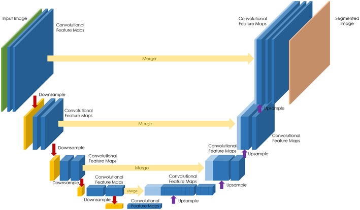
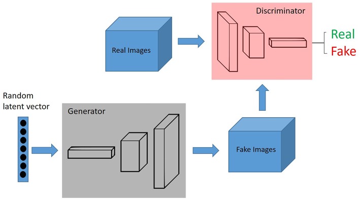
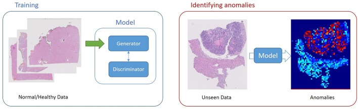
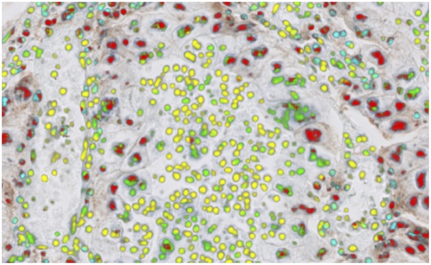
Verification
Claims the paper makes
The claims and statistics of this paper haven’t been checked yet.
Run the check — free, no account needed. It reads the open-access text and checks the sources it cites.
Claims other papers make about this one
Each is checked against this paper’s own text: does it say what the citing paper says it does?
In the citing paper
“On the other hand, the recent advancement in AI-based deep learning technologies focusing on cellular images can transform education in diagnostic pathology [24].”
From Artificial Intelligence Revolutionizing the Field of Medical Education (2023) · cites this paper as [24]
Self-check
Percentages, sums and reported differences re-computed from the paper’s own numbers.
The self-check needs the paper’s full text.
Citations
Cites
PD-1 and PD-L1 Immune Checkpoint Blockade to Treat Breast Cancer
Hartkopf, A. D., Taran, F., Wallwiener, M., Walter, C. B., Krämer, B., Grischke, E., & Brucker, S. Y. (2016) · Breast Care · doi:10.1159/000453569 · Cited by 30,989
Unpaired image-to-image translation using cycle-consistent adversarial networks
Zhu, J.-Y., Park, T., Isola, P., & Efros, A. A. (2017) · 2017 IEEE International Conference on Computer Vision (ICCV) · doi:10.1109/iccv.2017.244 · Cited by 23,602
Nishimura, R., Osako, T., Nishiyama, Y., Tashima, R., Nakano, M., Fujisue, M., Toyozumi, Y., & Arima, N. (2013) · Oncology · doi:10.1159/000353099 · Cited by 13,239
Comprehensive molecular portraits of human breast tumours
The Cancer Genome Atlas Network (2012) · Nature · doi:10.1038/nature11412 · Cited by 12,129
The Ki-67 protein: From the known and the unknown
Scholzen, T. E., & Gerdes, J. (2000) · Journal of Cellular Physiology · doi:10.1002/(sici)1097-4652(200003)182:3 · Cited by 4,707
Generative Adversarial Networks
Goodfellow, I. J., Pouget-Abadie, J., Mirza, M., Xu, B., Warde-Farley, D., Ozair, S., Courville, A., & Bengio, Y. (2014) · arXiv · doi:10.48550/arxiv.1406.2661 · Cited by 4,477
Deep Residual Learning for Image Recognition
He, K., Zhang, X., Ren, S., & Sun, J. (2015) · arXiv · doi:10.48550/arxiv.1512.03385 · Cited by 4,196
Ehteshami Bejnordi, B., Veta, M., Johannes van Diest, P., van Ginneken, B., Karssemeijer, N., Litjens, G., van der Laak, J. A. W. M., and the CAMELYON16 Consortium, Hermsen, M., Manson, Q. F., et al. (2017) · JAMA · doi:10.1001/jama.2017.14585 · Cited by 3,293
Epstein, J. I., Egevad, L., Amin, M. B., Delahunt, B., Srigley, J. R., & Humphrey, P. A. (2015) · The American Journal of Surgical Pathology · doi:10.1097/pas.0000000000000530 · Cited by 3,246
Coudray, N., Ocampo, P. S., Sakellaropoulos, T., Narula, N., Snuderl, M., Fenyö, D., Moreira, A. L., Razavian, N., & Tsirigos, A. (2018) · Nature Medicine · doi:10.1038/s41591-018-0177-5 · Cited by 2,930
Research works printed but not linked: 6
- Press Announcements - FDA Allows Marketing of First Whole Slide Imaging System for Digital Pathology2017
- Detection of mitosis and evaluation of nuclear atypia score in breast cancer histological images RouxL 22nd International Conference on Pattern Recognition 2014. MITOS-ATYPIA Contest.2014
- Mitosis detection in breast cancer histology images via deep cascaded networks11606 ChenH DouQ WangX QinJ HengP-A AAAI PressProceedings of the Thirtieth AAAI Conference on Artificial Intelligence2016
- Automated segmentation of epithelial tissue in prostatectomy slides using deep learning BultenW LitjensGJS Hulsbergen-van de KaaCA van der LaakJ Proceedings Medical Imaging 2018: Digital Pathology.2018
- Towards grading gleason score using generically trained deep convolutional neural networks11637 KallenH MolinJ HeydenA LundstromC AstromK In: 2016 IEEE 13th Int Symp on Biomed Im (ISBI).2016
- Automatic lymphocyte detection on gastric cancer ihc images using deep learning2004 GarciaE HermozaR CastanonCB CanoL CastilloM CastannedaC IEEE 30th International Symposium on Computer Based Medical Systems (CBMS)2017
Cited by
Sorted by citations
Digital pathology and artificial intelligence in translational medicine and clinical practice
Baxi, V., Edwards, R., Montalto, M., & Saha, S. (2022) · Modern Pathology · doi:10.1038/s41379-021-00919-2 · Cited by 592
Artificial intelligence and computational pathology
Cui, M., & Zhang, D. Y. (2021) · Laboratory Investigation · doi:10.1038/s41374-020-00514-0 · Cited by 544
Multimodal data fusion for cancer biomarker discovery with deep learning
Steyaert, S., Pizurica, M., Nagaraj, D., Khandelwal, P., Hernandez-Boussard, T., Gentles, A. J., & Gevaert, O. (2023) · Nature Machine Intelligence · doi:10.1038/s42256-023-00633-5 · Cited by 402
Artificial intelligence (AI) and big data in cancer and precision oncology
Dlamini, Z., Francies, F. Z., Hull, R., & Marima, R. M. (2020) · Computational and Structural Biotechnology Journal · doi:10.1016/j.csbj.2020.08.019 · Cited by 419
Medical image analysis based on deep learning approach
Puttagunta, M. K., & Ravi, S. (2021) · Multimedia Tools and Applications · doi:10.1007/s11042-021-10707-4 · Cited by 349
Korngiebel, D. M., & Mooney, S. D. (2021) · npj Digital Medicine · doi:10.1038/s41746-021-00464-x · Cited by 259
Application of Artificial Intelligence in Medicine: An Overview
Liu, P.-R., Lu, L., Zhang, J.-Y., Huo, T.-T., Liu, S.-X., & Ye, Z.-W. (2021) · Current Medical Science · doi:10.1007/s11596-021-2474-3 · Cited by 258
Arya, S. S., Dias, S. B., Jelinek, H. F., Hadjileontiadis, L. J., & Pappa, A. (2023) · Biosensors and Bioelectronics · doi:10.1016/j.bios.2023.115387 · Cited by 161
Artificial Intelligence Revolutionizing the Field of Medical Education
Narayanan, S., Ramakrishnan, R., Durairaj, E., & Das, A. (2023) · Cureus · doi:10.7759/cureus.49604 · Cited by 164
The Xception model: A potential feature extractor in breast cancer histology images classification
Sharma, S., & Kumar, S. (2021) · ICT Express · doi:10.1016/j.icte.2021.11.010 · Cited by 137
An Overview of Artificial Intelligence in Oncology
Farina, E. M. J. D. M., Nabhen, J. J., Dacoregio, M. I., Batalini, F., & de Moraes, F. Y. (2022) · Future Science OA · doi:10.2144/fsoa-2021-0074 · Cited by 145
A bird's-eye view of deep learning in bioimage analysis
Meijering, E. (2020) · Computational and Structural Biotechnology Journal · doi:10.1016/j.csbj.2020.08.003 · Cited by 126
Introduction to digital pathology and computer-aided pathology
Nam, S., Chong, Y., Jung, C. K., Kwak, T.-Y., Lee, J. Y., Park, J., Rho, M. J., & Go, H. (2020) · Journal of Pathology and Translational Medicine · doi:10.4132/jptm.2019.12.31 · Cited by 126
Computational pathology: A survey review and the way forward
Hosseini, M. S., Bejnordi, B. E., Trinh, V. Q., Chan, L., Hasan, D., Li, X., Yang, S. C., Kim, T., Zhang, H., Wu, T., et al. (2024) · Journal of Pathology Informatics · doi:10.1016/j.jpi.2023.100357 · Cited by 142
AI empowering traditional Chinese medicine?
Song, Z., Chen, G., & Chen, C. Y. (2024) · Chemical Science · doi:10.1039/d4sc04107k · Cited by 122
Höhn, J., Krieghoff-Henning, E., Jutzi, T. B., von Kalle, C., Utikal, J. S., Meier, F., Gellrich, F. F., Hobelsberger, S., Hauschild, A., Schlager, J. G., et al. (2021) · European Journal of Cancer · doi:10.1016/j.ejca.2021.02.032 · Cited by 106
Machine Learning and Big Data Provide Crucial Insight for Future Biomaterials Discovery and Research
Kerner, J., Dogan, A. B., & von Recum, H. A. (2020) · Acta Biomaterialia · doi:10.1016/j.actbio.2021.05.053 · Cited by 106
The inclusion of augmented intelligence in medicine: A framework for successful implementation
Bazoukis, G., Hall, J. L., Loscalzo, J., Antman, E. M., Fuster, V., & Armoundas, A. A. (2022) · Cell Reports Medicine · doi:10.1016/j.xcrm.2021.100485 · Cited by 111
Automated feature detection in dental periapical radiographs by using deep learning
Khan, H. A., Haider, M. A., Ansari, H. A., Ishaq, H., Kiyani, A., Sohail, K., Muhammad, M., & Khurram, S. A. (2020) · Oral Surgery Oral Medicine Oral Pathology and Oral Radiology · doi:10.1016/j.oooo.2020.08.024 · Cited by 100
AlQuraishi, M., & Sorger, P. K. (2021) · Nature Methods · doi:10.1038/s41592-021-01283-4 · Cited by 96
The Value of Artificial Intelligence in Laboratory Medicine
Paranjape, K., Schinkel, M., Hammer, R., Schouten, B., Panday, R. S. N., Elbers, P., Kramer, M. H. H., & Nanayakkara, P. W. B. (2020) · American Journal of Clinical Pathology · doi:10.1093/ajcp/aqaa170 · Cited by 102
Bazoukis, G., Stavrakis, S., Zhou, J., Bollepalli, S. C., Tse, G., Zhang, Q., Singh, J. P., & Armoundas, A. A. (2020) · Heart Failure Reviews · doi:10.1007/s10741-020-10007-3 · Cited by 91
Automated quality assessment of large digitised histology cohorts by artificial intelligence
Haghighat, M., Browning, L., Sirinukunwattana, K., Malacrino, S., Alham, N. K., Colling, R. T., Cui, Y., Rakha, E. A., Hamdy, F. C., Verrill, C. L., et al. (2022) · Scientific Reports · doi:10.1038/s41598-022-08351-5 · Cited by 89
Artificial intelligence in healthcare: a comprehensive review of its ethical concerns
Kooli, C., & Muftah, H. A. A. (2022) · Technological Sustainability · doi:10.1108/techs-12-2021-0029 · Cited by 97
Algorithms for Ethical Decision-Making in the Clinic: A Proof of Concept
Meier, L. J., Hein, A., Diepold, K. J., & Buyx, A. (2022) · The American Journal of Bioethics · doi:10.1080/15265161.2022.2040647 · Cited by 98
Homeyer, A., Geißler, C., Schwen, L. O., Zakrzewski, F., Evans, T., Strohmenger, K., Westphal, M., Bülow, R. D., Kargl, M., Karjauv, A., et al. (2022) · Modern Pathology · doi:10.1038/s41379-022-01147-y · Cited by 88
Introduction to Artificial Intelligence and Machine Learning for Pathology
Harrison, J. H., Gilbertson, J. R., Hanna, M. G., Olson, N. H., Seheult, J. N., Sorace, J. M., & Stram, M. N. (2021) · Archives of Pathology & Laboratory Medicine · doi:10.5858/arpa.2020-0541-cp · Cited by 91
Lee, S. H., Song, I. H., & Jang, H. (2021) · International Journal of Cancer · doi:10.1002/ijc.33599 · Cited by 82
Evaluating Cancer-Related Biomarkers Based on Pathological Images: A Systematic Review
Xie, X. S., Wang, X., Liang, Y., Yang, J., Wu, Y. P., Li, L., Sun, X., Bing, P., He, B., Tian, G., et al. (2021) · Frontiers in Oncology · doi:10.3389/fonc.2021.763527 · Cited by 85
Quantifying Explainers of Graph Neural Networks in Computational Pathology
Jaume, G., Pati, P., Bozorgtabar, B., Foncubierta–Rodríguez, A., Anniciello, A. M., Feroce, F., Rau, T. T., Thiran, J., Gabrani, M., & Göksel, O. (2021) · IEEE Computer Society Conference on Computer Vision and Pattern Recognition/Proceedings - IEEE Computer Society Conference on Computer Vision and Pattern Recognition/Proceedings · doi:10.1109/cvpr46437.2021.00801 · Cited by 89
Current challenges in metastasis research and future innovation for clinical translation
Parker, A. L., Benguigui, M., Fornetti, J., Goddard, E. T., Lucotti, S., Insua‐Rodríguez, J., & Wiegmans, A. P. (2022) · Clinical & Experimental Metastasis · doi:10.1007/s10585-021-10144-5 · Cited by 82
Deep learning‐based model for diagnosing Alzheimer's disease and tauopathies
Koga, S., Ikeda, A., & Dickson, D. W. (2021) · Neuropathology and Applied Neurobiology · doi:10.1111/nan.12759 · Cited by 77
Role of AI and Histopathological Images in Detecting Prostate Cancer: A Survey
Ayyad, S. M., Shehata, M., Shalaby, A. M., El‐Ghar, M. A., Ghazal, M. A., El-Melegy, M., Abdel-Hamid, N. B., Labib, L. M., Ali, H. A., & El-Baz, A. S. (2021) · Sensors · doi:10.3390/s21082586 · Cited by 76
Jang, H., Lee, A., Kang, J., Song, I. H., & Lee, S. H. (2020) · World Journal of Gastroenterology · doi:10.3748/wjg.v26.i40.6207 · Cited by 76
Developing image analysis methods for digital pathology
Bankhead, P. (2022) · The Journal of Pathology · doi:10.1002/path.5921 · Cited by 76
Artificial Intelligence: Review of Current and Future Applications in Medicine
Thomas, L. B. (2021) · Federal Practitioner · doi:10.12788/fp.0174 · Cited by 72
Deep Learning of Histopathology Images at the Single Cell Level
Lee, K., Lockhart, J. H., Xie, M., Chaudhary, R., Slebos, R. J. C., Flores, E. R., Chung, C. H., & Tan, A. C. (2021) · Frontiers in Artificial Intelligence · doi:10.3389/frai.2021.754641 · Cited by 67
Cheng, G., Zhang, F., Xing, Y., Hu, X., Zhang, H., Chen, S., Li, M., Peng, C., Ding, G., Zhang, D., et al. (2022) · Frontiers in Immunology · doi:10.3389/fimmu.2022.893198 · Cited by 65
Farris, A. B., Vizcarra, J. C., Amgad, M. T., Cooper, L., Gutman, D. A., & Hogan, J. (2020) · Histopathology · doi:10.1111/his.14304 · Cited by 61
Abele, N., Tiemann, K., Krech, T., Wellmann, A., Schaaf, C. P., Länger, F. P., Peters, A., Donner, A., Keil, F., Daifalla, K., et al. (2023) · Modern Pathology · doi:10.1016/j.modpat.2022.100033 · Cited by 57
Digitization of Pathology Labs: A Review of Lessons Learned
Schwen, L. O., Kiehl, T., Carvalho, R., Zerbe, N., & Homeyer, A. (2023) · Laboratory Investigation · doi:10.1016/j.labinv.2023.100244 · Cited by 59
Survey of XAI in Digital Pathology
Pocevičiūtė, M., Eilertsen, G., & Lundström, C. (2020) · Lecture notes in computer science · doi:10.1007/978-3-030-50402-1_4 · Cited by 53
Marsh, J. N., Liu, T., Wilson, P. C., Swamidass, S. J., & Gaut, J. P. (2021) · JAMA Network Open · doi:10.1001/jamanetworkopen.2020.30939 · Cited by 55
PyHIST: A Histological Image Segmentation Tool
Muñoz-Aguirre, M., Ntasis, V. F., Rojas, S., & Guigó, R. (2020) · PLoS Computational Biology · doi:10.1371/journal.pcbi.1008349 · Cited by 51
Koga, S., Ghayal, N. B., & Dickson, D. W. (2021) · Journal of Neuropathology & Experimental Neurology · doi:10.1093/jnen/nlab005 · Cited by 51
The in vitro micronucleus assay using imaging flow cytometry and deep learning
Rodrigues, M. A., Probst, C., Zayats, A., Davidson, B., Riedel, M. J., Li, Y., & Vidya Venkatachalam, A. (2021) · npj Systems Biology and Applications · doi:10.1038/s41540-021-00179-5 · Cited by 49
Artificial Intelligence in Pathology: From Prototype to Product
Homeyer, A., Lotz, J., Schwen, L. O., Weiss, N., Romberg, D., Höfener, H., Zerbe, N., & Hufnagl, P. (2021) · Journal of Pathology Informatics · doi:10.4103/jpi.jpi_84_20 · Cited by 52
Alam, R., Abdul‐Ghafar, J., Yim, K., Thakur, N., Lee, S. H., Jang, H., Jung, C. K., & Chong, Y. (2022) · Cancers · doi:10.3390/cancers14112590 · Cited by 46
Mei, T., Wang, T., & Zhou, Q. (2024) · Clinical and Experimental Medicine · doi:10.1007/s10238-024-01324-0 · Cited by 47
Ahmad, F., & Muhmood, T. (2024) · Colloids and Surfaces B Biointerfaces · doi:10.1016/j.colsurfb.2024.114041 · Cited by 46
Abdeltawab, H., Khalifa, F., Ghazal, M. A., Liang Cheng, C., El-Baz, A. S., & Gondim, D. D. (2022) · Journal of Pathology Informatics · doi:10.1016/j.jpi.2022.100093 · Cited by 43
Geaney, A., O’Reilly, P., Maxwell, P., James, J. A., McArt, D., & Salto-Tellez, M. (2023) · Oncogene · doi:10.1038/s41388-023-02857-6 · Cited by 42
Saeidnia, H. R., Firuzpour, F., Kozak, M., & majd, H. S. (2025) · Artificial Intelligence Review · doi:10.1007/s10462-025-11117-w · Cited by 48
Zubair, M., Owais, M., Hassan, T., Bendechache, M., Hussain, M., Hussain, I., & Werghi, N. (2025) · Scientific Reports · doi:10.1038/s41598-025-97256-0 · Cited by 61
Application of digital pathology and machine learning in the liver, kidney and lung diseases
Wu, B., & Moeckel, G. W. (2023) · Journal of Pathology Informatics · doi:10.1016/j.jpi.2022.100184 · Cited by 42
Park, J. H., Kim, E. Y., Luchini, C., Eccher, A., Tizaoui, K., Shin, J. I., & Lim, B. J. (2022) · International Journal of Molecular Sciences · doi:10.3390/ijms23052462 · Cited by 38
Biosensors for healthcare: an artificial intelligence approach
Davé, S., Dave, A., Radhakrishnan, S., Das, J., & Dave, S. (2022) · Elsevier eBooks · doi:10.1016/b978-0-323-88464-8.00008-7 · Cited by 39
Current Trends in Artificial Intelligence Application for Endourology and Robotic Surgery
Chang, T., Seufert, C., Eminağa, O., Shkolyar, E., Hu, J. C., & Liao, J. C. (2020) · Urologic Clinics of North America · doi:10.1016/j.ucl.2020.09.004 · Cited by 37
Petrick, N., Akbar, S., Kenny, H., Nofech‐Mozes, S., Sahiner, B., Gavrielides, M. A., Kalpathy–Cramer, J., Drukker, K., & Martel, A. L. (2021) · Journal of medical imaging · doi:10.1117/1.jmi.8.3.034501 · Cited by 38
Deepfake Histologic Images for Enhancing Digital Pathology
Falahkheirkhah, K., Tiwari, S., Yeh, K. L., Gupta, S., Herrera-Hernandez, L. P., McCarthy, M. R., Jiménez, R. E., Cheville, J. C., & Bhargava, R. (2023) · Laboratory Investigation · doi:10.1016/j.labinv.2022.100006 · Cited by 38
Advanced image generation for cancer using diffusion models
Kidder, B. L. (2024) · Biology Methods and Protocols · doi:10.1093/biomethods/bpae062 · Cited by 38
Deep learning powers cancer diagnosis in digital pathology
He, Y., Zhao, H. P., & Wong, S. T. C. (2020) · Computerized Medical Imaging and Graphics · doi:10.1016/j.compmedimag.2020.101820 · Cited by 37
Krijgsman, D., van Leeuwen, M. B., van der Ven, J., Almeida, V., Vlutters, R., Halter, D., Kuppen, P. J. K., van de Velde, C. J. �., & Wimberger-Friedl, R. (2020) · IEEE Journal of Biomedical and Health Informatics · doi:10.1109/jbhi.2020.3003475 · Cited by 35
Jang, H., Go, J., Kim, Y.-H., & Lee, S. H. (2023) · Cancers · doi:10.3390/cancers15225389 · Cited by 33
Rashidi, H. H., Bowers, K., & Gil, M. R. (2023) · Journal of Thrombosis and Haemostasis · doi:10.1016/j.jtha.2022.12.019 · Cited by 33
Recent Advances in Melanoma Diagnosis and Prognosis Using Machine Learning Methods
Grossarth, S. N., Mosley, D. A., Madden, C., Ike, J., Smith, I. T., Huo, Y., & Wheless, L. (2023) · Current Oncology Reports · doi:10.1007/s11912-023-01407-3 · Cited by 36
Arun, S., Grosheva, M. N., Kosenko, M. G., Robertus, J. L., Blyuss, O. B., Gabe, R., Munblit, D., & Offman, J. (2025) · npj Precision Oncology · doi:10.1038/s41698-025-00940-7 · Cited by 34
Jang, H., Song, I.-H., & Lee, S. H. (2021) · Cancers · doi:10.3390/cancers13153811 · Cited by 34
Joint categorical and ordinal learning for cancer grading in pathology images
Vuong, T. T. L., Kim, K., Song, B., & Kwak, J. T. (2021) · Medical Image Analysis · doi:10.1016/j.media.2021.102206 · Cited by 30
Yao, Z., Jin, T., Mao, B., Lu, B., Zhang, Y., Li, S., & Chen, W. (2022) · Frontiers in Oncology · doi:10.3389/fonc.2022.815951 · Cited by 30
Patch-level Tumor Classification in Digital Histopathology Images with Domain Adapted Deep Learning
Xia, T., Kumar, A., Feng, D. D., & Kim, J. (2018) · Annual International Conference of the IEEE Engineering in Medicine and Biology Society (EMBC) · doi:10.1109/embc.2018.8512353 · Cited by 30
Dudgeon, S. N., Wen, S., Hanna, M. G., Gupta, R., Amgad, M. T., Sheth, M., Marble, H. D., Huang, R., Herrmann, M. D., Szu, C. H., et al. (2021) · Journal of Pathology Informatics · doi:10.4103/jpi.jpi_83_20 · Cited by 29
Deep Learning Identification of Stiffness Markers in Breast Cancer
Sneider, A., Kiemen, A. L., Kim, J. H., Wu, P.-H., Habibi, M., White, M. J., Phillip, J. M., Gu, L., & Wirtz, D. (2020) · Biomaterials · doi:10.1016/j.biomaterials.2022.121540 · Cited by 28
Larey, A., Aknin, E., Daniel, N., Osswald, G. A., Caldwell, J. M., Rochman, M., Wasserman, T., Collins, M. H., Arva, N. C., Yang, G., et al. (2022) · Frontiers in Medicine · doi:10.3389/fmed.2022.950728 · Cited by 28
Artificial Intelligence in Digital Pathology for Bladder Cancer: Hype or Hope? A Systematic Review
Khoraminia, F., Fuster, S., Kanwal, N., Olislagers, M., Engan, K., van Leenders, G. J. L. H., Stubbs, A., Akram, F., & Zuiverloon, T. C. M. (2023) · Cancers · doi:10.3390/cancers15184518 · Cited by 29
Tokarz, D. A., Steinbach, T., Lokhande, A., Srivastava, G., Ugalmugle, R., Co, C. A., Shockley, K. R., Singletary, E., Cesta, M. F., Thomas, H. C., et al. (2020) · Toxicologic Pathology · doi:10.1177/0192623320972614 · Cited by 29
Jang, H., Lee, A., Kang, J. K., Song, I. H., & Lee, S. H. (2021) · World Journal of Gastroenterology · doi:10.3748/wjg.v27.i44.7687 · Cited by 29
The need for measurement science in digital pathology
Romanchikova, M., Thomas, S. A., Dexter, A., Shaw, M., Partarrieau, I., Smith, N. A. S., Venton, J., Adeogun, M., Brettle, D. S., & Turpin, R. J. (2022) · Journal of Pathology Informatics · doi:10.1016/j.jpi.2022.100157 · Cited by 27
Oster, H. S., Crouch, S., Smith, A. G., Yu, G., Shrkihe, B. A., Baruch, S., Kolomansky, A., Ben‐Ezra, J. M., Naor, S., Fenaux, P., et al. (2021) · Blood Advances · doi:10.1182/bloodadvances.2020004055 · Cited by 25
Lee, S. H., Lee, Y., & Jang, H. (2022) · International Journal of Cancer · doi:10.1002/ijc.34251 · Cited by 26
Teacher-Student chain for efficient semi-supervised histology image classificationPreprint
Shaw, S., Pajak, M., Lisowska, A., Tsaftaris, S. A., & O'Neil, A. Q. (2020) · arXiv (Cornell University) · doi:10.48550/arxiv.2003.08797 · Cited by 26
Aoyama, Y., Maruko, I., Kawano, T., Yokoyama, T., Ogawa, Y., Maruko, R., & Iida, T. (2021) · PLoS ONE · doi:10.1371/journal.pone.0244469 · Cited by 23
Guo, H., Diao, L., Zhou, X., Chen, J.-N., Zhou, Y., Fang, Q., He, Y., Dziadziuszko, R., Zhou, C., & Hirsch, F. R. (2021) · Translational Lung Cancer Research · doi:10.21037/tlcr-21-96 · Cited by 23
Kuklyte, J., Fitzgerald, J., Nelissen, S. R., Wei, H., Whelan, A., Power, A., Ahmad, A., Miarka, M., Gregson, M., Maxwell, M., et al. (2021) · Toxicologic Pathology · doi:10.1177/0192623320986423 · Cited by 24
Identifying pathological slices of gastric cancer via deep learning
Tung, C., Chang, H.-C., Yang, B.-Z., Hou, K.-J., Tsai, H.-H., Tsai, C.-Y., & Yu, P.-T. (2022) · Journal of the Formosan Medical Association · doi:10.1016/j.jfma.2022.05.004 · Cited by 22
The present and future role of artificial intelligence and machine learning in anesthesiology
Alexander, J. C., Romito, B. T., & Çobanoğlu, M. C. (2020) · International Anesthesiology Clinics · doi:10.1097/aia.0000000000000294 · Cited by 22
Prognostic markers of inflammation in endometrioid and clear cell ovarian cancer
Gallego, A., Mendiola, M., Hernando, B., Berjón, A., Cádiz, A., Chaves-Urbano, B., Heredia-Soto, V., Spagnolo, E., Hernández, A., Hardisson, D., et al. (2022) · International Journal of Gynecological Cancer · doi:10.1136/ijgc-2022-003353 · Cited by 21
AI in medical diagnosis: A contextualised study of patient motivations and concerns
Hajiheydari, N., Delgosha, M. S., & Saheb, T. (2025) · Social Science & Medicine · doi:10.1016/j.socscimed.2025.117850 · Cited by 21
Multi-omic and quantum machine learning integration for lung subtypes classification
Saggi, M. K., Bhatia, A. S., Mensah, I. K., Gowher, H., & Kais, S. (2025) · Future Generation Computer Systems · doi:10.1016/j.future.2025.107905 · Cited by 27
Deman, F., Broeckx, G., Declercq, S., Degotte, Q., Raymaekers, J., Salgado, R., & Dendooven, A. (2025) · Histopathology · doi:10.1111/his.15481 · Cited by 21
Perspectives in pathomics in head and neck cancer
Classe, M., Lerousseau, M., Scoazec, J., & Deutsch, É. (2021) · Current Opinion in Oncology · doi:10.1097/cco.0000000000000731 · Cited by 24
Smerkous, D. D., Mauer, M. S., Tøndel, C., Svarstad, E., Gübler, M.-C., Nelson, R. G., Oliveira, J. P., Sargolzaeiaval, F., & Najafian, B. (2023) · Kidney International · doi:10.1016/j.kint.2023.09.011 · Cited by 23
Present and Future Applications of Artificial Intelligence in Kidney Transplantation
Kotsifa, E., & Mavroeidis, V. K. (2024) · Journal of Clinical Medicine · doi:10.3390/jcm13195939 · Cited by 24
Development of AI-Based Diagnostic Algorithm for Nasal Bone Fracture Using Deep Learning
Jeong, Y., Jeong, C., Sung, K., Moon, G.-S., & Lim, J. (2023) · Journal of Craniofacial Surgery · doi:10.1097/scs.0000000000009856 · Cited by 19
Liu, X., Kong, Y., Qian, Y., Guo, H., Zhao, L., Wang, H., Xu, K., Ye, L., Liu, Y., Lu, H., et al. (2024) · Translational Oncology · doi:10.1016/j.tranon.2024.102143 · Cited by 19
Schuettfort, V. M., Pradère, B., Rink, M., Compérat, É., & Shariat, S. F. (2020) · Current Opinion in Urology · doi:10.1097/mou.0000000000000813 · Cited by 18
Computational pathology in ovarian cancer
Oršulić, S., John, J., Walts, A. E., & Gertych, A. (2022) · Frontiers in Oncology · doi:10.3389/fonc.2022.924945 · Cited by 18
Zhang, T., Chen, J., Lü, Y., Yang, X., & Ouyang, Z. (2022) · PLoS ONE · doi:10.1371/journal.pone.0273355 · Cited by 18
Başak, K., Ozyoruk, K. B., & Demir, D. (2023) · Turkish Journal of Pathology · doi:10.5146/tjpath.2023.01601 · Cited by 18
Swillens, J. E. M., Nagtegaal, I. D., Engels, S., Lugli, A., Hermens, R., & van der Laak, J. A. W. M. (2023) · Oncogene · doi:10.1038/s41388-023-02797-1 · Cited by 18
Metadata
- Authors
- Serag, A., Ion-Margineanu, A., Qureshi, H., McMillan, R., Saint Martin, M.-J., Diamond, J., O'Reilly, P., & Hamilton, P.
- Affiliations
- Philips (United Kingdom) (GB)
- Corresponding author
- Hamilton, Peter
- Venue
- Frontiers in Medicine
- Publisher
- Frontiers Media SA
- Published
- 2019-10-01
- Volume
- 6
- Pages
- 185-185
- ISSN
- 2296-858X (ISSN-L)
- PMC
- PMC6779702
- OpenAlex
- W2965481926
- Cited by
- 274
- References
- 94
- Citations by year
- 2026: 22 · 2025: 43 · 2024: 35 · 2023: 46 · 2022: 43 · 2021: 51 · 2020: 33
- FWCI
- 2.06
- Type
- journal article
- Language
- en
- Open access
- gold
- doi.org
- Licence
- Locations
- Frontiers in Medicine (publishedVersion, cc-by)
- PubMed Central (publishedVersion, cc-by)
- PubMed (publishedVersion)
- DOAJ (DOAJ: Directory of Open Access Journals) (submittedVersion, cc-by-sa)
- Topics
- Physical Sciences › Computer Science › Artificial Intelligence › AI in cancer detection
- Health Sciences › Medicine › Health Informatics › Artificial Intelligence in Healthcare and Education
- Health Sciences › Medicine › Radiology, Nuclear Medicine and Imaging › Radiomics and Machine Learning in Medical Imaging
- Keywords
- artificial intelligence in pathology, deep learning, digital pathology, computational pathology, cell imaging, diagnostic consistency
About these checks
Registry, retraction, Cites and statistics results are computed, not judged, with one exception: a printed reference that no index lists is read once by a model before it counts as a citation error. Claims are extracted by a model and kept only when their quote appears verbatim in the text; each cited source is then checked, with a model’s help, for whether it says what the claim relies on. Those verdicts can be wrong — read the claim’s quote and the cited source before relying on one.
Statistics and arithmetic are recomputed, not judged.
Confidence bands are shown only for fields calibrated on our annotated test set. None is calibrated yet, so no band is shown.
What counts as a claim ›How we measure confidence ›How we check this ›
Are you an author, or do you see a problem with this page? Request a review
