

The DOI matches its Crossref record; 108 of the 108 references it cites were found in a registry or an index.
Translational AI and Deep Learning in Diagnostic Pathology
Open accessAbstract
There has been an exponential growth in the application of AI in health and in pathology. This is resulting in the innovation of deep learning technologies that are specifically aimed at cellular imaging and practical applications that could transform diagnostic pathology. This paper reviews the different approaches to deep learning in pathology, the public grand challenges that have driven this innovation and a range of emerging applications in pathology. The translation of AI into clinical practice will require applications to be embedded seamlessly within digital pathology workflows, driving an integrated approach to diagnostics and providing pathologists with new tools that accelerate workflow and improve diagnostic consistency and reduce errors. The clearance of digital pathology for primary diagnosis in the US by some manufacturers provides the platform on which to deliver practical AI. AI and computational pathology will continue to mature as researchers, clinicians, industry, regulatory organizations and patient advocacy groups work together to innovate and deliver new technologies to health care providers: technologies which are better, faster, cheaper, more precise, and safe.
Overview
The claims and statistics of this paper haven’t been checked yet.
Run the check — free, no account needed. It reads the open-access text and checks the sources it cites.
Integrity signals
Registry
Cites
Statistics
Figures
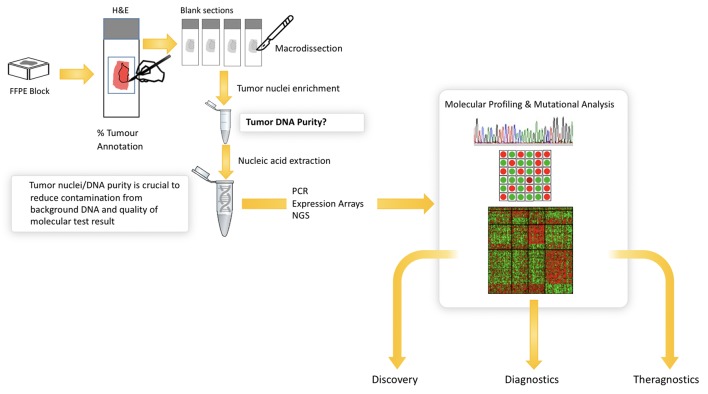
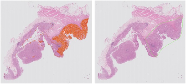
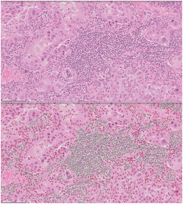
Verification
Claims the paper makes
The claims and statistics of this paper haven’t been checked yet.
Run the check — free, no account needed. It reads the open-access text and checks the sources it cites.
Claims other papers make about this one
Each is checked against this paper’s own text: does it say what the citing paper says it does?
In the citing paper
“On the other hand, the recent advancement in AI-based deep learning technologies focusing on cellular images can transform education in diagnostic pathology [24].”
From Artificial Intelligence Revolutionizing the Field of Medical Education (2023) · cites this paper as [24]
Self-check
Percentages, sums and reported differences re-computed from the paper’s own numbers.
The self-check needs the paper’s full text.
Citations
Cites
PD-1 and PD-L1 Immune Checkpoint Blockade to Treat Breast Cancer
Hartkopf, A. D., Taran, F., Wallwiener, M., Walter, C. B., Krämer, B., Grischke, E., & Brucker, S. Y. (2016) · Breast Care · doi:10.1159/000453569 · Cited by 30,989
Unpaired image-to-image translation using cycle-consistent adversarial networks
Zhu, J.-Y., Park, T., Isola, P., & Efros, A. A. (2017) · 2017 IEEE International Conference on Computer Vision (ICCV) · doi:10.1109/iccv.2017.244 · Cited by 23,602
Nishimura, R., Osako, T., Nishiyama, Y., Tashima, R., Nakano, M., Fujisue, M., Toyozumi, Y., & Arima, N. (2013) · Oncology · doi:10.1159/000353099 · Cited by 13,239
Comprehensive molecular portraits of human breast tumours
The Cancer Genome Atlas Network (2012) · Nature · doi:10.1038/nature11412 · Cited by 12,129
The Ki-67 protein: From the known and the unknown
Scholzen, T. E., & Gerdes, J. (2000) · Journal of Cellular Physiology · doi:10.1002/(sici)1097-4652(200003)182:3 · Cited by 4,707
Generative Adversarial Networks
Goodfellow, I. J., Pouget-Abadie, J., Mirza, M., Xu, B., Warde-Farley, D., Ozair, S., Courville, A., & Bengio, Y. (2014) · arXiv · doi:10.48550/arxiv.1406.2661 · Cited by 4,477
Deep Residual Learning for Image Recognition
He, K., Zhang, X., Ren, S., & Sun, J. (2015) · arXiv · doi:10.48550/arxiv.1512.03385 · Cited by 4,196
Ehteshami Bejnordi, B., Veta, M., Johannes van Diest, P., van Ginneken, B., Karssemeijer, N., Litjens, G., van der Laak, J. A. W. M., and the CAMELYON16 Consortium, Hermsen, M., Manson, Q. F., et al. (2017) · JAMA · doi:10.1001/jama.2017.14585 · Cited by 3,293
Epstein, J. I., Egevad, L., Amin, M. B., Delahunt, B., Srigley, J. R., & Humphrey, P. A. (2015) · The American Journal of Surgical Pathology · doi:10.1097/pas.0000000000000530 · Cited by 3,246
Coudray, N., Ocampo, P. S., Sakellaropoulos, T., Narula, N., Snuderl, M., Fenyö, D., Moreira, A. L., Razavian, N., & Tsirigos, A. (2018) · Nature Medicine · doi:10.1038/s41591-018-0177-5 · Cited by 2,930
Research works printed but not linked: 6
- Press Announcements - FDA Allows Marketing of First Whole Slide Imaging System for Digital Pathology2017
- Detection of mitosis and evaluation of nuclear atypia score in breast cancer histological images RouxL 22nd International Conference on Pattern Recognition 2014. MITOS-ATYPIA Contest.2014
- Mitosis detection in breast cancer histology images via deep cascaded networks11606 ChenH DouQ WangX QinJ HengP-A AAAI PressProceedings of the Thirtieth AAAI Conference on Artificial Intelligence2016
- Automated segmentation of epithelial tissue in prostatectomy slides using deep learning BultenW LitjensGJS Hulsbergen-van de KaaCA van der LaakJ Proceedings Medical Imaging 2018: Digital Pathology.2018
- Towards grading gleason score using generically trained deep convolutional neural networks11637 KallenH MolinJ HeydenA LundstromC AstromK In: 2016 IEEE 13th Int Symp on Biomed Im (ISBI).2016
- Automatic lymphocyte detection on gastric cancer ihc images using deep learning2004 GarciaE HermozaR CastanonCB CanoL CastilloM CastannedaC IEEE 30th International Symposium on Computer Based Medical Systems (CBMS)2017
Cited by
Sorted by citations
Items 201–271 of 271
Development of Enhance-Net Deep Learning Approach for Performance Boosting on Medical Images
Manoharan, G., Solanke, D. R., Acharjee, P. B., Nayak, C. K., Sharma, M. K., & Sahu, D. N. (2024) · Lecture notes in electrical engineering · doi:10.1007/978-981-97-8031-0_44 · Cited by 2
AnoGAN for Tabular Data: A Novel Approach to Anomaly Detection
Reddy, P., & Singh, A. (2024) · Communications in computer and information science · doi:10.1007/978-3-031-78561-0_32 · Cited by 4
Agrawal, H., Gupta, N., Tanwar, H., & Panesar, N. (2025) · Artificial Intelligence in Gastroenterology · doi:10.35712/aig.v6.i1.108198 · Cited by 2
Belciug, S. (2020) · Elsevier eBooks · doi:10.1016/b978-0-12-820201-2.00003-9 · Cited by 2
Assessment of parathyroid gland cellularity by digital slide analysis
Sagiv, R., Delgado, B., Lavon, O., Osipov, V., Sade, R., Shashar, S., Yegodayev, K. M., Elkabets, M., & Joshua, B. (2022) · Annals of Diagnostic Pathology · doi:10.1016/j.anndiagpath.2022.151907 · Cited by 2
Assessing Artificial Intelligence Models to Diagnose and Differentiate Common Liver CarcinomasPreprint
Thomas, M., Mastorides, S., Borkowski, S., Reed, J., Deland, L., Thomas, L., & Borkowski, A. (2022) · medRxiv · doi:10.1101/2022.08.30.22279347 · Cited by 2
Wong, B., & Yi, M. Y. (2025) · SSRN Electronic Journal · doi:10.2139/ssrn.5362908 · Cited by 2
Clinical and pathological diagnosis using AI and data-driven science
Hiraoka, S.-I. (2020) · Journal of Japanese Society of Oral Oncology · doi:10.5843/jsot.32.159 · Cited by 1
A Hybrid Approach to Osteosarcoma Detection Using Densenet201-SVM Model
Salih, D. K., Qadir, A. M., & Ghareb, M. I. (2023) · International Symposium on Digital Forensics and Security (ISDFS) · doi:10.1109/isdfs58141.2023.10131717 · Cited by 1
A Precise Survey on Multi-agent in Medical Domains
Alshehri, A., Alshahrani, F., & Shah, H. (2023) · International Journal of Advanced Computer Science and Applications · doi:10.14569/ijacsa.2023.01406107 · Cited by 2
Computational modeling of tumor invasion from limited and diverse data in Glioblastoma
Jonnalagedda, P., Weinberg, B. D., Min, T. L., Bhanu, S., & Bhanu, B. (2024) · Computerized Medical Imaging and Graphics · doi:10.1016/j.compmedimag.2024.102436 · Cited by 1
Mitosis detection and classification for breast cancer diagnosis: What we know and what is next
Khalil, R. U., Sajjad, M., Dhahbi, S., Bourouis, S., Hijji, M., & Muhammad, K. (2025) · Computers in Biology and Medicine · doi:10.1016/j.compbiomed.2025.110057 · Cited by 1
Evaluation of Post-Hoc Explainability Methods for Glaucoma Classification Using Fundus Images
Ming, H. K., Vong, W.-T., Loh, B. C. S., & Then, P. (2025) · Lecture notes in networks and systems · doi:10.1007/978-3-031-92605-1_40 · Cited by 1
Navigating AI-Driven Translation in Saudi Arabia's Media: Challenges and Opportunities
Alharbi, M. A. (2025) · Arabic Journal for Translation Studies · doi:10.63939/ajts.91zp6t15 · Cited by 1
Liu, J., Lu, S., Liu, J., Tian, H., Rahman, M. T., Bennis, I., Sayasone, S., Polessa, D., Mohteshamuddin, K., Li, Q., et al. (2025) · Science in One Health · doi:10.1016/j.soh.2025.100127 · Cited by 3
PathViT Model for Automated Disease Classification from Skeletal Muscle Histopathology
Akan, T., Alp, S., Aishwarya, R., Xing, D. G., Dicharry, D., Bhuiyan, M. S., & Bhuiyan, M. A. N. (2025) · American Journal Of Pathology · doi:10.1016/j.ajpath.2025.10.009 · Cited by 1
A systematic review and surgical anatomy of the greater omentum
Joseph, T. S., Hogan, E. A., Cesmebasi, A., Carrasco, M., Ang, C., Davis, N., Montalbano, M. J., & Loukas, M. (2025) · Morphologie · doi:10.1016/j.morpho.2025.101072 · Cited by 1
Beyond Broad Applications: Can Pathology Foundation Models Adapt to Hematopathology?
Rabah, C. B., & Serag, A. (2025) · Lecture notes in computer science · doi:10.1007/978-3-032-07845-2_13 · Cited by 1
Enhancing urine cytopathology with artificial intelligence: a systematic review
Nabiyouni, F., & Chiou, P. Z. (2025) · American Journal of Clinical Pathology · doi:10.1093/ajcp/aqaf135 · Cited by 1
El‐Hag, N. A., El‐Shafai, W., El-Hameed, H. A. A., Soliman, N. F., Algarni, A. D., & El‐Samie, F. E. A. (2026) · Scientific Reports · doi:10.1038/s41598-025-04606-z · Cited by 1
Matching Patients With Cell Surface–Targeted Clinical Trials Using Large Language Models
Callahan, S. C., Chrostek, M. R., Rydzewski, N. R., Bootsma, M. L., Helzer, K. T., Bakhtiar, H., Morris, Z. S., Kosoff, D., Lang, J. M., Ma, V. T.-L., et al. (2026) · JCO Precision Oncology · doi:10.1200/po-25-00757 · Cited by 1
AI‐Assisted Fungal Detection in Dermatopathology: A Study on Accuracy, Efficiency, and Usability
Schmidle, P., Lang, T., Springenberg, S., Calvopina, D. A., Köhler, T., Getova, V., Gebing, A.-M., Hetzer, S., Pierrone, J., Aebischer, V., et al. (2026) · JDDG Journal der Deutschen Dermatologischen Gesellschaft · doi:10.1111/ddg.70242 · Cited by 1
Dong, C., Liu, Y., Yin, J., Yan, W., Sun, J., Zhang, X., Huo, S., Yu, F., & Zhou, Y. (2026) · Digital Health · doi:10.1177/20552076261474637 · Cited by 3
The Challenges of Anatomic Pathology Medical Education in Indonesia
Ramkita, N., & Murti, K. (2021) · Jurnal RSMH Palembang · doi:10.37275/jrp.v2i1.16 · Cited by 1
Toward Automatic Mammography Auditing via Universal Language Model Fine Tuning
Issa, T. B., Uminsky, D., Shaw, A., Makipour, R., & Filice, R. W. (2021) · IEEE International Conference on Information Reuse and Integration (IRI) · doi:10.1109/iri51335.2021.00035 · Cited by 1
Classification of Diabetic Cardiomyopathy-Related Cells Using Machine Learning
Dalaman, U., Ayan, S. Ş., & Yaraş, N. (2022) · Moscow University Physics Bulletin · doi:10.3103/s0027134922060042 · Cited by 1
Prediction of Osteosarcoma Using Machine Learning Techniques
Srivastava, D. K., Batta, A., Gupta, T., & Shukla, A. (2023) · Lecture notes in networks and systems · doi:10.1007/978-981-19-6088-8_41 · Cited by 1
Sports Medicine: What Artificial Intelligence Can Do for Preventing Athlete Injuries
Sileo, M. (2024) · Mechanisms and machine science · doi:10.1007/978-3-031-63755-1_49 · Cited by 1
Digitalization is Fueling a Revolution in Precision Oncology
Cuendet, M. A., & Michielin, O. A. (2020) · Elsevier eBooks · doi:10.1016/b978-0-12-801238-3.11704-0 · Cited by 1
Abdukodirova, M., Abdullah, S. H., Alsadoon, A., & Prasad, P. W. C. (2020) · International Conference on Innovative Technologies in Intelligent Systems and Industrial Applications (CITISIA) · doi:10.1109/citisia50690.2020.9371844 · Cited by 1
Impact of Artificial Intelligence on Professional Autonomy of Pathologists
Feroze, S. (2022) · doi:10.32920/19008740 · Cited by 1
Signal, Image Processing and Embedded Systems Trends
(2022) · doi:10.5121/csit.2022.1220 · Cited by 1
Sammad, A., & Ding, Z. (2025) · Academic Radiology · doi:10.1016/j.acra.2025.02.015 · Cited by 1
Bilattice CADIAG-II: Theory and Experimental Results
Baldi, P., Ciabattoni, A., & Adlassnig, K.-P. (2021) · Studies in computational intelligence · doi:10.1007/978-3-030-76620-7_1
Sedick, Q., & Elyamany, G. (2022) · GLOBAL JOURNAL FOR RESEARCH ANALYSIS · doi:10.36106/gjra/4306231
Comparison of Ultra-Magnifying Endocytoscopic and Hematoxylin-Eosin-Stained Images of Lung Specimens
Kobayashi, M., Kurimoto, N., Tanino, R., Shiratsuki, Y., Okuno, T., Nakao, M., Hotta, T., Tsubata, Y., Nagasaki, M., Nishisaka, T., et al. (2023) · Diagnostics · doi:10.3390/diagnostics13051003
ЦИФРОВА ПАТОЛОГІЯ ПРИ МЕЛАНОМІ: ДОСЯГНЕННЯ, БАР’ЄРИ ТА ПЕРСПЕКТИВИ
Dudin, O. (2023) · Medical Informatics and Engineering · doi:10.11603/mie.1996-1960.2022.4.13411
Advanced Deep Learning for Skin Histoglyphics at Cellular Level
Kreher, R., Chitti, N. R., Hille, G., Hürtgen, J., Mengonie, M., Braun, A. D., Tüting, T., Preim, B., & Saalfeld, S. (2024) · Informatik aktuell · doi:10.1007/978-3-658-44037-4_20
Artificial Intelligence in Healthcare
Singh, K., Srivastava, V. K., & Shukla, A. (2024) · Advances in bioinformatics and biomedical engineering book series · doi:10.4018/979-8-3693-1922-2.ch001
Application of machine learning in identification of pathogenic microbes
Kutikuppala, L. V. S., Adhit, K. K., & Silva, R. G. D. (2023) · Digital Medicine · doi:10.1097/dm-2023-00016
-, N. R. S., -, V. V. N. S. G., & -, B. I. D. (2024) · International Journal For Multidisciplinary Research · doi:10.36948/ijfmr.2024.v06i03.20160
Utilization of Artificial Intelligence in Breast Pathology
Baclig, M. O. (2024) · Philippine Journal of Pathology · doi:10.21141/pjp2024.04
Expanding role and scope of artificial intelligence in the field of gastrointestinal pathology
Mubarak, M., Rashid, R., Sapna, F., & Shakeel, S. (2024) · Artificial Intelligence in Gastroenterology · doi:10.35712/aig.v5.i2.91550
Hossain, M. S. B., Piazza, Y., Braun, J., Bilic, A., Hsieh, M., Fouissi, S., Borowsky, A. D., Kaseb, H. O., Renduchintala, C., Fraser, A., et al. (2024) · medRxiv · doi:10.1101/2024.11.05.24316736
Sree, N. R., Lydia, E. L., & Kumari, P. A. (2024) · Lecture notes in networks and systems · doi:10.1007/978-981-97-6581-2_27
Poonacha, S. K., Sushma, Madhyastha, R., Nalilu, S. K., Bhandary, S. K., & Madhyastha, H. (2025) · Springer eBooks · doi:10.1007/978-981-97-5742-8_2
Shimokawa, I., Terasaki, M., Tanaka, S., Toda, E., Takakuma, S., Kajimoto, Y., Kunugi, S., Shimizu, A., & Terasaki, Y. (2025) · medRxiv · doi:10.1101/2025.05.21.25327411
Using AI to Address Demographic-Specific Challenges in Manual Handling Safety
da Silva, T. M. H. R. (2025) · Advances in computational intelligence and robotics book series · doi:10.4018/979-8-3693-9301-7.ch016
Hybrid Computational Intelligence Techniques for Automated Lung Cancer Detection
Shwetha, S., & Ramana, N. (2025) · International Conference on Research Methodologies in Knowledge Management, Artificial Intelligence and Telecommunication Engineering (RMKMATE) · doi:10.1109/rmkmate64874.2025.11042357
Gaur, Y., Chaudhary, A., & Abraham, A. (2025) · Elsevier eBooks · doi:10.1016/b978-0-443-27444-2.00013-7
Künstliche Intelligenz im Gesundheitswesen
Holle, A., Hering, V., Scheidt, M. V., & Arnold, U. (2025) · Springer eBooks · doi:10.1007/978-3-658-47498-0_5
PriorPath: Coarse-To-Fine Approach for Controlled De-Novo Pathology Semantic Masks Generation
Daniel, N., Nathan, M., Azeroual, E., Fisher, Y., & Savir, Y. (2025) · Annual International Conference of the IEEE Engineering in Medicine and Biology Society (EMBC) · doi:10.1109/embc58623.2025.11252679
Ameen, Y. A., Badary, D. M., Abonnoor, A. E. I., Hussain, K. F., & Sewisy, A. A. E.-M. (2023) · Figshare · doi:10.6084/m9.figshare.22622723
1Chapter 1 Artificial intelligence in cancer treatment and management
Malviya, R., Rajput, S., Roy, M., Sridhar, S. B., & Thomas, S. (2025) · De Gruyter eBooks · doi:10.1515/9783111670744-001
PAVR: High-Resolution Cellular Imaging via a Physics-Aware Volumetric Reconstruction FrameworkPreprint
Hua, X., Han, K., Ling, Z., Reid, O., Gao, Z., Zhang, H., Botchwey, E. A., Forghani, P., Liu, W., Sawant, M., et al. (2026) · bioRxiv (Cold Spring Harbor Laboratory) · doi:10.64898/2026.03.04.709609
Cordeiro‐Rodrigues, L. (2026) · Medical Humanities · doi:10.1136/medhum-2025-013526
The Future of IHC Diagnostics: Transformer-Driven Quantification of Tumor Microenvironments
Sghaier, A., Agrebi, M., Gueddas, A., Slama, A. B., Ksiaa, M., & Charfeddine, B. (2025) · IEEE International Conference on Advances in Data-Driven Analytics and Intelligent Systems (ADACIS) · doi:10.1109/adacis65663.2025.11436671
Weakly Supervised Segmentation and Context-Aware Classification in Computational Pathology
Lerousseau, M. (2022) · theses.fr (ABES) · doi:10.70675/8d10f89dz0f91z4e9dzb3a8zb5228b1172c1
Pye, H., Brettle, D. S., Kaye, D., Dunn, C. M., Humphries, M. P., & Treanor, D. E. (2026) · Journal of Histotechnology · doi:10.1080/01478885.2026.2643973
Akinrinwa, S. P. (2026) · Contributions to environmental sciences & innovative business technology · doi:10.1007/978-981-95-8473-4_6
AI Regulations in Medical Devices
Khinvasara, T. (2026) · Springer handbooks · doi:10.1007/978-3-032-01636-2_40
Helmy, H., Rabah, C. B., & Serag, A. (2026) · AI · doi:10.3390/ai7070261
The road ahead: innovations and future trends in artificial intelligence medical robotics
Manoharan, G., & Nivedha, M. (2026) · Elsevier eBooks · doi:10.1016/b978-0-443-27783-2.00002-8
Wins, J. A., Saranraj, P., Lokeshwari, B., Al‐Tawaha, A. R., Karnwal, A., & Sivasakthivelan, P. (2026) · Advances in computational intelligence and robotics book series · doi:10.4018/979-8-2600-1358-8.ch007
Albashrawi, M. A. (2026) · Information Discovery and Delivery · doi:10.1108/idd-09-2025-0235
Tham, J. A., Xie, F., Chen, B., Yu, P., Wu, J., Feng, T., Tse, G. M. K., To, K. F., & Kang, W. (2026) · Intelligent Systems with Applications · doi:10.1016/j.iswa.2026.200736
Stainless Steel vs Calcium Fluoride for biomedical Raman SpectroscopyPreprint
Ng, C., Blake, N., & Thomas, G. M. H. (2026) · bioRxiv (Cold Spring Harbor Laboratory) · doi:10.64898/2026.10.09.753418
Metadata
- Authors
- Serag, A., Ion-Margineanu, A., Qureshi, H., McMillan, R., Saint Martin, M.-J., Diamond, J., O'Reilly, P., & Hamilton, P.
- Affiliations
- Philips (United Kingdom) (GB)
- Corresponding author
- Hamilton, Peter
- Venue
- Frontiers in Medicine
- Publisher
- Frontiers Media SA
- Published
- 2019-10-01
- Volume
- 6
- Pages
- 185-185
- ISSN
- 2296-858X (ISSN-L)
- PMC
- PMC6779702
- OpenAlex
- W2965481926
- Cited by
- 274
- References
- 94
- Citations by year
- 2026: 22 · 2025: 43 · 2024: 35 · 2023: 46 · 2022: 43 · 2021: 51 · 2020: 33
- FWCI
- 2.06
- Type
- journal article
- Language
- en
- Open access
- gold
- doi.org
- Licence
- Locations
- Frontiers in Medicine (publishedVersion, cc-by)
- PubMed Central (publishedVersion, cc-by)
- PubMed (publishedVersion)
- DOAJ (DOAJ: Directory of Open Access Journals) (submittedVersion, cc-by-sa)
- Topics
- Physical Sciences › Computer Science › Artificial Intelligence › AI in cancer detection
- Health Sciences › Medicine › Health Informatics › Artificial Intelligence in Healthcare and Education
- Health Sciences › Medicine › Radiology, Nuclear Medicine and Imaging › Radiomics and Machine Learning in Medical Imaging
- Keywords
- artificial intelligence in pathology, deep learning, digital pathology, computational pathology, cell imaging, diagnostic consistency
About these checks
Registry, retraction, Cites and statistics results are computed, not judged, with one exception: a printed reference that no index lists is read once by a model before it counts as a citation error. Claims are extracted by a model and kept only when their quote appears verbatim in the text; each cited source is then checked, with a model’s help, for whether it says what the claim relies on. Those verdicts can be wrong — read the claim’s quote and the cited source before relying on one.
Statistics and arithmetic are recomputed, not judged.
Confidence bands are shown only for fields calibrated on our annotated test set. None is calibrated yet, so no band is shown.
What counts as a claim ›How we measure confidence ›How we check this ›
Are you an author, or do you see a problem with this page? Request a review
