

The DOI matches its Crossref record; 108 of the 108 references it cites were found in a registry or an index.
Translational AI and Deep Learning in Diagnostic Pathology
Open accessAbstract
There has been an exponential growth in the application of AI in health and in pathology. This is resulting in the innovation of deep learning technologies that are specifically aimed at cellular imaging and practical applications that could transform diagnostic pathology. This paper reviews the different approaches to deep learning in pathology, the public grand challenges that have driven this innovation and a range of emerging applications in pathology. The translation of AI into clinical practice will require applications to be embedded seamlessly within digital pathology workflows, driving an integrated approach to diagnostics and providing pathologists with new tools that accelerate workflow and improve diagnostic consistency and reduce errors. The clearance of digital pathology for primary diagnosis in the US by some manufacturers provides the platform on which to deliver practical AI. AI and computational pathology will continue to mature as researchers, clinicians, industry, regulatory organizations and patient advocacy groups work together to innovate and deliver new technologies to health care providers: technologies which are better, faster, cheaper, more precise, and safe.
Overview
The claims and statistics of this paper haven’t been checked yet.
Run the check — free, no account needed. It reads the open-access text and checks the sources it cites.
Integrity signals
Registry
Cites
Statistics
Figures
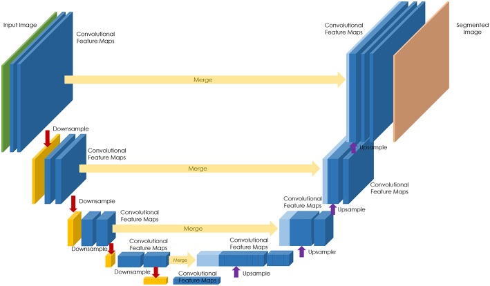
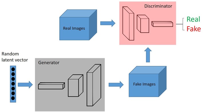
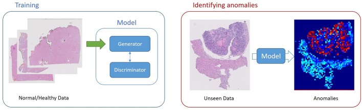
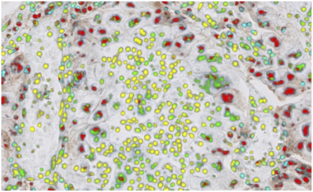
Verification
Claims the paper makes
The claims and statistics of this paper haven’t been checked yet.
Run the check — free, no account needed. It reads the open-access text and checks the sources it cites.
Claims other papers make about this one
Each is checked against this paper’s own text: does it say what the citing paper says it does?
In the citing paper
“On the other hand, the recent advancement in AI-based deep learning technologies focusing on cellular images can transform education in diagnostic pathology [24].”
From Artificial Intelligence Revolutionizing the Field of Medical Education (2023) · cites this paper as [24]
Self-check
Percentages, sums and reported differences re-computed from the paper’s own numbers.
The self-check needs the paper’s full text.
Citations
Cites
PD-1 and PD-L1 Immune Checkpoint Blockade to Treat Breast Cancer
Hartkopf, A. D., Taran, F., Wallwiener, M., Walter, C. B., Krämer, B., Grischke, E., & Brucker, S. Y. (2016) · Breast Care · doi:10.1159/000453569 · Cited by 30,989
Unpaired image-to-image translation using cycle-consistent adversarial networks
Zhu, J.-Y., Park, T., Isola, P., & Efros, A. A. (2017) · 2017 IEEE International Conference on Computer Vision (ICCV) · doi:10.1109/iccv.2017.244 · Cited by 23,602
Nishimura, R., Osako, T., Nishiyama, Y., Tashima, R., Nakano, M., Fujisue, M., Toyozumi, Y., & Arima, N. (2013) · Oncology · doi:10.1159/000353099 · Cited by 13,239
Comprehensive molecular portraits of human breast tumours
The Cancer Genome Atlas Network (2012) · Nature · doi:10.1038/nature11412 · Cited by 12,129
The Ki-67 protein: From the known and the unknown
Scholzen, T. E., & Gerdes, J. (2000) · Journal of Cellular Physiology · doi:10.1002/(sici)1097-4652(200003)182:3 · Cited by 4,707
Generative Adversarial Networks
Goodfellow, I. J., Pouget-Abadie, J., Mirza, M., Xu, B., Warde-Farley, D., Ozair, S., Courville, A., & Bengio, Y. (2014) · arXiv · doi:10.48550/arxiv.1406.2661 · Cited by 4,477
Deep Residual Learning for Image Recognition
He, K., Zhang, X., Ren, S., & Sun, J. (2015) · arXiv · doi:10.48550/arxiv.1512.03385 · Cited by 4,196
Ehteshami Bejnordi, B., Veta, M., Johannes van Diest, P., van Ginneken, B., Karssemeijer, N., Litjens, G., van der Laak, J. A. W. M., and the CAMELYON16 Consortium, Hermsen, M., Manson, Q. F., et al. (2017) · JAMA · doi:10.1001/jama.2017.14585 · Cited by 3,293
Epstein, J. I., Egevad, L., Amin, M. B., Delahunt, B., Srigley, J. R., & Humphrey, P. A. (2015) · The American Journal of Surgical Pathology · doi:10.1097/pas.0000000000000530 · Cited by 3,246
Coudray, N., Ocampo, P. S., Sakellaropoulos, T., Narula, N., Snuderl, M., Fenyö, D., Moreira, A. L., Razavian, N., & Tsirigos, A. (2018) · Nature Medicine · doi:10.1038/s41591-018-0177-5 · Cited by 2,930
Epstein, J. I., Allsbrook, W. C., Amin, M. B., & Egevad, L. L. (2005) · The American Journal of Surgical Pathology · doi:10.1097/01.pas.0000173646.99337.b1 · Cited by 2,763
Unsupervised Anomaly Detection with Generative Adversarial Networks to Guide Marker Discovery
Schlegl, T., Seeböck, P., Waldstein, S. M., Schmidt-Erfurth, U., & Langs, G. (2017) · Lecture Notes in Computer Science · doi:10.1007/978-3-319-59050-9_12 · Cited by 2,627
Unpaired Image-to-Image Translation using Cycle-Consistent Adversarial NetworksPreprint
Zhu, J.-Y., Park, T., Isola, P., et al. (2017) · arXiv (Cornell University) · doi:10.48550/arxiv.1703.10593 · Cited by 2,278
U-Net: deep learning for cell counting, detection, and morphometry
Falk, T., Mai, D., Bensch, R., Çiçek, Ö., Abdulkadir, A., Marrakchi, Y., Böhm, A., Deubner, J., Jäckel, Z., Seiwald, K., et al. (2018) · Nature Methods · doi:10.1038/s41592-018-0261-2 · Cited by 1,920
The Inevitable Application of Big Data to Health Care
Murdoch, T. B., & Detsky, A. S. (2013) · JAMA · doi:10.1001/jama.2013.393 · Cited by 1,676
f-AnoGAN: Fast unsupervised anomaly detection with generative adversarial networks
Schlegl, T., Seeböck, P., Waldstein, S. M., Langs, G., & Schmidt-Erfurth, U. M. (2019) · Medical Image Analysis · doi:10.1016/j.media.2019.01.010 · Cited by 1,645
Deep Learning Applications in Medical Image Analysis
Ker, J., Wang, L., Rao, J. P., & Lim, C. C. T. (2017) · IEEE Access · doi:10.1109/access.2017.2788044 · Cited by 1,574
Exome sequencing identifies recurrent SPOP, FOXA1 and MED12 mutations in prostate cancer
Barbieri, C. E., Baca, S. C., Lawrence, M. S., Demichelis, F., Blattner, M., Theurillat, J.-P. P., White, T. A., Stojanov, P., Allen, E. M. V., Stransky, N., et al. (2012) · Nature Genetics · doi:10.1038/ng.2279 · Cited by 1,550
Mitosis Detection in Breast Cancer Histology Images with Deep Neural Networks
Cireşan, D. C., Giusti, A., Gambardella, L. M., & Schmidhuber, J. (2013) · Lecture Notes in Computer Science · doi:10.1007/978-3-642-40763-5_51 · Cited by 1,536
Going Deeper with Convolutions
Szegedy, C., Liu, W., Jia, Y., Sermanet, P., Reed, S., Anguelov, D., Erhan, D., Vanhoucke, V., & Rabinovich, A. (2014) · arXiv · doi:10.48550/arxiv.1409.4842 · Cited by 1,461
A Dataset and a Technique for Generalized Nuclear Segmentation for Computational Pathology
Kumar, N., Verma, R., Sharma, S., Bhargava, S., Vahadane, A., & Sethi, A. (2017) · IEEE Transactions on Medical Imaging · doi:10.1109/tmi.2017.2677499 · Cited by 1,160
Deep Learning in Medical Image Analysis and Multimodal Learning for Clinical Decision Support
(2017) · Lecture notes in computer science · doi:10.1007/978-3-319-67558-9 · Cited by 1,149
AutoAugment: Learning Augmentation Policies from Data
Cubuk, E. D., Zoph, B., Mane, D., Vasudevan, V., & Le, Q. V. (2018) · arXiv · doi:10.48550/arxiv.1805.09501 · Cited by 1,106
U-Net: Convolutional Networks for Biomedical Image SegmentationPreprint
Ronneberger, O., Fischer, P., & Brox, T. (2015) · arXiv (Cornell University) · doi:10.48550/arxiv.1505.04597 · Cited by 1,079
Deep learning as a tool for increased accuracy and efficiency of histopathological diagnosis
Litjens, G., Sánchez, C. I., Timofeeva, N., Hermsen, M., Nagtegaal, I., Kovacs, I., Hulsbergen - van de Kaa, C., Bult, P., van Ginneken, B., & van der Laak, J. (2016) · Scientific Reports · doi:10.1038/srep26286 · Cited by 1,069
Nuclei Segmentation with Recurrent Residual Convolutional Neural Networks based U-Net (R2U-Net)
Alom, M. Z., Yakopcic, C., Taha, T. M., & Asari, V. K. (2018) · IEEE National Aerospace and Electronics Conference (NAECON) · doi:10.1109/naecon.2018.8556686 · Cited by 1,069
Classification of breast cancer histology images using Convolutional Neural Networks
Araújo, T., Aresta, G., Castro, E., Rouco, J., Aguiar, P., Eloy, C., Polónia, A., & Campilho, A. (2017) · PLoS ONE · doi:10.1371/journal.pone.0177544 · Cited by 1,044
Deep Learning for Identifying Metastatic Breast CancerPreprint
Wang, D., Khosla, A., Gargeya, R., Irshad, H., & Beck, A. H. (2016) · arXiv.org · doi:10.48550/arxiv.1606.05718 · Cited by 1,028
Gland segmentation in colon histology images: The glas challenge contest
Sirinukunwattana, K., Pluim, J. P. W., Chen, H., Qi, X., Heng, P., Guo, Y. B., Wang, L. Y., Matuszewski, B. J., Bruni, E., Sanchez, U., et al. (2016) · Medical Image Analysis · doi:10.1016/j.media.2016.08.008 · Cited by 1,014
BACH: Grand challenge on breast cancer histology images
Aresta, G., Araújo, T., Kwok, S., Chennamsetty, S. S., Safwan, M., Alex, V., Marami, B., Prastawa, M. W., Chan, M. S. M., Donovan, M. J., et al. (2019) · Medical Image Analysis · doi:10.1016/j.media.2019.05.010 · Cited by 755
An International Ki67 Reproducibility Study
Polley, M. C., Leung, S. C. Y., McShane, L. M., Gao, D., Hugh, J. C., Mastropasqua, M. G., Viale, G., Zabaglo, L. A., Penault-Llorca, F. M., Bartlett, J. M. S., et al. (2013) · JNCI Journal of the National Cancer Institute · doi:10.1093/jnci/djt306 · Cited by 593
Bandi, P., Geessink, O., Manson, Q., Van Dijk, M., Balkenhol, M., Hermsen, M., Ehteshami Bejnordi, B., Lee, B., Paeng, K., Zhong, A., et al. (2019) · IEEE Transactions on Medical Imaging · doi:10.1109/tmi.2018.2867350 · Cited by 569
DCAN: Deep contour-aware networks for object instance segmentation from histology images
Chen, H., Qi, X., Yu, L., Dou, Q., Qin, J., & Heng, P. (2016) · Medical Image Analysis · doi:10.1016/j.media.2016.11.004 · Cited by 514
Detecting Cancer Metastases on Gigapixel Pathology Images
Liu, Y., Gadepalli, K., Norouzi, M., Dahl, G. E., Kohlberger, T., Boyko, A., Venugopalan, S., Timofeev, A., Nelson, P. Q., Corrado, G. S., et al. (2017) · arXiv · doi:10.48550/arxiv.1703.02442 · Cited by 506
Nagpal, K., Foote, D., Liu, Y., Chen, P.-H. C., Wulczyn, E., Tan, F., Olson, N., Smith, J. L., Mohtashamian, A., Wren, J. H., et al. (2019) · npj Digital Medicine · doi:10.1038/s41746-019-0112-2 · Cited by 495
Artificial Intelligence in Drug Discovery and Development
Agrawal, P. (2018) · Journal of Pharmacovigilance · doi:10.4172/2329-6887.1000e173 · Cited by 493
Steiner, D. F., MacDonald, R., Liu, Y., Truszkowski, P., Hipp, J. D., Gammage, C., Thng, F., Peng, L., & Stumpe, M. C. (2018) · American Journal of Surgical Pathology · doi:10.1097/pas.0000000000001151 · Cited by 489
Assessment of algorithms for mitosis detection in breast cancer histopathology images
Veta, M., van Diest, P. J., Willems, S. M., Wang, H., Madabhushi, A., Cruz-Roa, Á., Gonzalez, F. A., Larsen, A., Vestergaard, J. S., Dahl, A. B., et al. (2014) · Medical Image Analysis · doi:10.1016/j.media.2014.11.010 · Cited by 480
Artificial intelligence in medicine: current trends and future possibilities
Buch, V., Ahmed, I., & Maruthappu, M. (2018) · British Journal of General Practice · doi:10.3399/bjgp18x695213 · Cited by 472
Automated Gleason grading of prostate cancer tissue microarrays via deep learning
Arvaniti, E., Fricker, K. S., Moret, M., Rupp, N. J., Hermanns, T., Fankhauser, C. D., Wey, N., Wild, P. J., Rüschoff, J. H., & Claassen, M. (2018) · Scientific Reports · doi:10.1038/s41598-018-30535-1 · Cited by 461
1399 H&E-stained sentinel lymph node sections of breast cancer patients: the CAMELYON dataset
Litjens, G. J. S., Bándi, P., Bejnordi, B. E., Geessink, O. G. F., Balkenhol, M. C. A., Bult, P., Halilović, A., Hermsen, M., van de Loo, R., Vogels, R. J., et al. (2018) · GigaScience · doi:10.1093/gigascience/giy065 · Cited by 441
Mukhopadhyay, S., Feldman, M. D., Abels, E., Ashfaq, R., Beltaifa, S., Cacciabeve, N. G., Cathro, H. P., Cheng, L., Cooper, K., Dickey, G. E., et al. (2018) · American Journal of Surgical Pathology · doi:10.1097/pas.0000000000000948 · Cited by 399
Staingan: Stain Style Transfer for Digital Histological Images
Shaban, M., Baur, C., Navab, N., & Albarqouni, S. (2019) · IEEE International Symposium on Biomedical Imaging (ISBI) · doi:10.1109/isbi.2019.8759152 · Cited by 371
Sharma, H., Zerbe, N., Klempert, I., Hellwich, O., & Hufnagl, P. (2017) · Computerized Medical Imaging and Graphics · doi:10.1016/j.compmedimag.2017.06.001 · Cited by 319
Deep Learning Algorithms for Detection of Lymph Node Metastases From Breast Cancer
Golden, J. A. (2017) · JAMA · doi:10.1001/jama.2017.14580 · Cited by 313
Predicting breast tumor proliferation from whole-slide images: The TUPAC16 challenge
Veta, M., Heng, Y. J., Stathonikos, N., Bejnordi, B. E., de Beça, F. F., Wollmann, T., Rohr, K., Shah, M., Wang, D., Rousson, M., et al. (2019) · Medical Image Analysis · doi:10.1016/j.media.2019.02.012 · Cited by 309
Mitosis detection in breast cancer histological images An ICPR 2012 contest
Roux, L., Racoceanu, D., Loménie, N., Kulikova, M. S., Irshad, H., Jacques, K., Capron, F., Genestie, C., Naour, G. L., & Gurcan Metin, N. (2013) · Journal of Pathology Informatics · doi:10.4103/2153-3539.112693 · Cited by 303
An application of machine learning to haematological diagnosis
Gunčar, G., Kukar, M., Notar, M., Brvar, M., Černelč, P., Notar, M., & Notar, M. (2018) · Scientific Reports · doi:10.1038/s41598-017-18564-8 · Cited by 237
Relevance of deep learning to facilitate the diagnosis of HER2 status in breast cancer
Vandenberghe, M. E., Scott, M. L. J., Scorer, P., Söderberg, M. P., Balcerzak, D., & Barker, C. (2017) · Scientific Reports · doi:10.1038/srep45938 · Cited by 215
An international study to increase concordance in Ki67 scoring
Polley, M. C., Leung, S. C. Y., Gao, D., Mastropasqua, M. G., Zabaglo, L. A., Bartlett, J. M. S., McShane, L. M., Enos, R. A., Badve, S., Bane, A. L., et al. (2015) · Modern Pathology · doi:10.1038/modpathol.2015.38 · Cited by 214
The estimation of tumor cell percentage for molecular testing by pathologists is not accurate
Smits, A. J. J., Kummer, J. A., de Bruin, P. C., Bol, M., van den Tweel, J. G., Seldenrijk, K. A., Willems, S. M., Offerhaus, G. J. A., de Weger, R. A., van Diest, P. J., et al. (2013) · Modern Pathology · doi:10.1038/modpathol.2013.134 · Cited by 213
Learning to Compose Domain-Specific Transformations for Data AugmentationPreprint
Ratner, A., Ehrenberg, H. R., Hussain, Z., Dunnmon, J., & Ré, C. (2017) · arXiv (Cornell University) · doi:10.48550/arxiv.1709.01643 · Cited by 212
Udall, M., Rizzo, M., Kenny, J., Doherty, J. P., Dahm, S., Robbins, P. B., & Faulkner, E. C. (2018) · Diagnostic Pathology · doi:10.1186/s13000-018-0689-9 · Cited by 209
Xie, Y., Xing, F., Kong, X., Su, H., & Yang, L. (2015) · Lecture notes in computer science · doi:10.1007/978-3-319-24574-4_43 · Cited by 203
Qaiser, T., Mukherjee, A., Chaitanya Reddy, P. B., Munugoti, S. D., Vamsi, T., Pitkäaho, T., Lehtimäki, T., Naughton, T. J., Berseth, M., Pedraza, A., et al. (2017) · Histopathology · doi:10.1111/his.13333 · Cited by 162
Leung, S. C. Y., Nielsen, T. O., Zabaglo, L. A., Arun, I., Badve, S., Bane, A. L., Bartlett, J. M. S., Borgquist, S., Chang, M. C., Dodson, A. R., et al. (2016) · npj Breast Cancer · doi:10.1038/npjbcancer.2016.14 · Cited by 153
Automated Image Analysis for High-Content Screening and Analysis
Shariff, A., Kangas, J. D., Coelho, L. P., Quinn, S. P., & Murphy, R. F. (2010) · SLAS DISCOVERY · doi:10.1177/1087057110370894 · Cited by 140
for The Trans-Atlantic Prostate Group, Berney, D. M., Gopalan, A., Kudahetti, S. C., Fisher, G., Ambroisine, L., Foster, C. S., Reuter, V., Eastham, J. A., Møller, H., et al. (2009) · British Journal of Cancer · doi:10.1038/sj.bjc.6604951 · Cited by 135
Evaluating the benefits of digital pathology implementation: time savings in laboratory logistics
Baidoshvili, A., Bucur, A., van Leeuwen, J., van der Laak, J. A. W. M., Kluin, P. M., & van Diest, P. J. (2018) · Histopathology · doi:10.1111/his.13691 · Cited by 129
Deep Learning Based Automatic Immune Cell Detection for Immunohistochemistry Images
Chen, T., & Chefd’hotel, C. (2014) · Lecture notes in computer science · doi:10.1007/978-3-319-10581-9_3 · Cited by 123
Kapil, A., Meier, A., Zuraw, A., Steele, K. E., Rebelatto, M. C., Schmidt, G., & Brieu, N. (2018) · Scientific Reports · doi:10.1038/s41598-018-35501-5 · Cited by 115
The molecular basis of breast cancer pathological phenotypes
Heng, Y. J., Lester, S. C., Tse, G. M.-K., Factor, R. E., Allison, K. H., Collins, L. C., Chen, Y., Jensen, K. C., Johnson, N. B., Jeong, J. C., et al. (2016) · The Journal of Pathology · doi:10.1002/path.4847 · Cited by 112
Viray, H., Li, K., Long, T. A., Vasalos, P., Bridge, J. A., Jennings, L. J., Halling, K. C., Hameed, M., & Rimm, D. L. (2013) · Archives of Pathology & Laboratory Medicine · doi:10.5858/arpa.2012-0561-cp · Cited by 107
McKenney, J. K., Simko, J. P., Bonham, M. J., True, L. D., Troyer, D. A., Hawley, S. J., Newcomb, L. F., Fazli, L., Kunju, L. P., Nicolas, M. M., et al. (2011) · The Journal of Urology · doi:10.1016/j.juro.2011.03.115 · Cited by 107
Klauschen, F., Wienert, S., Schmitt, W., Loibl, S., Gerber, B., Blohmer, J., Huober, J., Rüdiger, T., Erbstößer, E., Mehta, K. M., et al. (2014) · Clinical Cancer Research · doi:10.1158/1078-0432.ccr-14-1283 · Cited by 103
Prognostic role of Ki-67 score in localized prostate cancer: A systematic review and meta-analysis
Berlín, A., Castro-Mesta, J. F., Rodríguez‐Romo, L., Barajas, D. H., González-Guerrero, J. F., Rodríguez-Fernández, I. A., Conchas, G. A. G., Verdines-Perez, A. M., & Vera-Badillo, F. (2017) · Urologic Oncology Seminars and Original Investigations · doi:10.1016/j.urolonc.2017.05.004 · Cited by 98
Automatic Ki-67 Counting Using Robust Cell Detection and Online Dictionary Learning
Xing, F., Su, H., Neltner, J. H., & Yang, L. (2014) · IEEE Transactions on Biomedical Engineering · doi:10.1109/tbme.2013.2291703 · Cited by 94
BESNet: Boundary-Enhanced Segmentation of Cells in Histopathological Images
Oda, H., Roth, H. R., Chiba, K., et al. (2018) · Lecture notes in computer science · doi:10.1007/978-3-030-00934-2_26 · Cited by 90
Cell Counting by Regression Using Convolutional Neural Network
Xue, Y., Ray, N., Hugh, J. C., & Bigras, G. (2016) · Lecture notes in computer science · doi:10.1007/978-3-319-46604-0_20 · Cited by 89
A Fast and Refined Cancer Regions Segmentation Framework in Whole-slide Breast Pathological Images
Guo, Z., Liu, H., Ni, H., Wang, X., Su, M., Guo, W., Wang, K., Jiang, T., & Qian, Y. (2019) · Scientific Reports · doi:10.1038/s41598-018-37492-9 · Cited by 88
Deep Voting: A Robust Approach Toward Nucleus Localization in Microscopy Images
Xie, Y., Kong, X., Xing, F., Liu, F., Su, H., & Yang, L. (2015) · Lecture notes in computer science · doi:10.1007/978-3-319-24574-4_45 · Cited by 80
H&E-stained Whole Slide Image Deep Learning Predicts SPOP Mutation State in Prostate CancerPreprint
Schaumberg, A. J., Rubin, M. A., & Fuchs, T. J. (2016) · bioRxiv (Cold Spring Harbor Laboratory) · doi:10.1101/064279 · Cited by 79
Learning to Compose Domain-Specific Transformations for Data Augmentation.
Ratner, A., Ehrenberg, H. R., Hussain, Z., Dunnmon, J., & Ré, C. (2017) · PubMed · Cited by 79
Abubakar, M., Orr, N., Daley, F. M., Coulson, P., Ali, H. R., Blows, F. M., Benitez, J. J., Milne, R. L., Brenner, H., Stegmaier, C., et al. (2016) · Breast Cancer Research · doi:10.1186/s13058-016-0765-6 · Cited by 78
Lejeune, M., Jaén, J., Pons, L. E., López, C., Salvadó-Usach, T., Bosch, R., García, M., Escrivá, P., Baucells, J., Cugat, X., et al. (2008) · Journal of Anatomy · doi:10.1111/j.1469-7580.2008.00910.x · Cited by 76
A Unified Framework for Tumor Proliferation Score Prediction in Breast Histopathology
Paeng, K., Hwang, S., Park, S., & Kim, M. (2017) · Lecture notes in computer science · doi:10.1007/978-3-319-67558-9_27 · Cited by 73
Multiclass Classification of Breast Cancer in Whole-Slide Images
Kwok, S. (2018) · Lecture notes in computer science · doi:10.1007/978-3-319-93000-8_106 · Cited by 72
del Toro, Ó. J., Atzori, M., Otálora, S., Andersson, M., Eurén, K., Hedlund, M., Rönnquist, P., & Müller, H. (2017) · Proceedings of SPIE, the International Society for Optical Engineering/Proceedings of SPIE · doi:10.1117/12.2255710 · Cited by 69
Humphries, M. P., Hynes, S. O., Bingham, V., Cougot, D., James, J. A., Patell-Socha, F., Parkes, E. E., Blayney, J. K., O’Rorke, M. A., Irwin, G. W., et al. (2018) · Journal of Oncology · doi:10.1155/2018/2937012 · Cited by 63
Robboy, S. J., Gupta, S., Crawford, J. M., Cohen, M. B., Karcher, D. S., Leonard, D. G. B., Magnani, B., Novis, D. A., Prystowsky, M. B., Powell, S. Z.-E., et al. (2015) · Archives of Pathology & Laboratory Medicine · doi:10.5858/arpa.2014-0559-oa · Cited by 62
Automated tumor analysis for molecular profiling in lung cancer
Hamilton, P. W., Wang, Y., Boyd, C., James, J. A., Loughrey, M. B., Hougton, J. P., Boyle, D. P., Kelly, P., Maxwell, P., McCleary, D. F., et al. (2015) · Oncotarget · doi:10.18632/oncotarget.4391 · Cited by 58
From Machine Learning to Artificial Intelligence Applications in Cardiac Care
Tsay, D., & Patterson, C. (2018) · Circulation · doi:10.1161/circulationaha.118.031734 · Cited by 57
Humphries, M. P., McQuaid, S., Craig, S. G., Bingham, V., Maxwell, P., Maurya, M., McLean, F., Sampson, J., Higgins, P., Greene, C., et al. (2018) · Journal of Thoracic Oncology · doi:10.1016/j.jtho.2018.09.025 · Cited by 50
A Deep Learning Approach for Rapid Mutational Screening in MelanomaPreprint
Kim, R. H., Nomikou, S., Coudray, N., Jour, G., Dawood, Z., Hong, R., Esteva, E., Sakellaropoulos, T., Donnelly, D. M., Moran, U., et al. (2019) · bioRxiv (Cold Spring Harbor Laboratory) · doi:10.1101/610311 · Cited by 48
Vestjens, J. H. M. J., Pepels, M. J. A. E., de Boer, M., Borm, G. F., van Deurzen, C. H. M., van Diest, P. J., van Dijck, J. A. A. M., Adang, E., Nortier, J. W. R., Rutgers, E. J. T., et al. (2012) · Annals of Oncology · doi:10.1093/annonc/mds072 · Cited by 47
Kumar, A., Rao, A., Bhavani, S., Newberg, J. Y., & Murphy, R. F. (2014) · Proceedings of the National Academy of Sciences · doi:10.1073/pnas.1415120112 · Cited by 45
Reinforced Auto-Zoom Net: Towards Accurate and Fast Breast Cancer Segmentation in Whole-Slide Images
Dong, N., Kampffmeyer, M., Liang, X., Wang, Z., Dai, W., & Xing, E. P. (2018) · Lecture notes in computer science · doi:10.1007/978-3-030-00889-5_36 · Cited by 45
Cell Detection in Microscopy Images with Deep Convolutional Neural Network and Compressed SensingPreprint
Xue, Y., & Ray, N. (2017) · arXiv (Cornell University) · doi:10.48550/arxiv.1708.03307 · Cited by 42
Singh, R., Agashe, S., Gosavi, A., & Sulhyan, K. (2011) · Indian Journal of Cancer · doi:10.4103/0019-509x.92277 · Cited by 33
Patch-level Tumor Classification in Digital Histopathology Images with Domain Adapted Deep Learning
Xia, T., Kumar, A., Feng, D. D., & Kim, J. (2018) · Annual International Conference of the IEEE Engineering in Medicine and Biology Society (EMBC) · doi:10.1109/embc.2018.8512353 · Cited by 30
Zehntner, S. P., Chakravarty, M. M., Bolovan, R. J., Chan, C., & Bedell, B. J. (2008) · Journal of Histochemistry & Cytochemistry · doi:10.1369/jhc.2008.950345 · Cited by 26
Virtualization of Tissue Staining in Digital Pathology Using an Unsupervised Deep Learning Approach
Lahiani, A., Gildenblat, J., Klaman, I., Albarqouni, S., Navab, N., & Klaiman, E. (2019) · Lecture notes in computer science · doi:10.1007/978-3-030-23937-4_6 · Cited by 26
Zhou, N., Fedorov, A., Fennessy, F. M., Kikinis, R., & Gao, Y. D. (2017) · arXiv (Cornell University) · doi:10.48550/arxiv.1705.02678 · Cited by 23
Glory, E., Newberg, J. Y., & Murphy, R. F. (2008) · IEEE International Symposium on Biomedical Imaging: From Nano to Macro (ISBI) · doi:10.1109/isbi.2008.4540993 · Cited by 22
Pattern Recognition in Histopathological Images: An ICPR 2010 Contest
Gürcan, M. N., Madabhushi, A., & Rajpoot, N. (2010) · Lecture notes in computer science · doi:10.1007/978-3-642-17711-8_23 · Cited by 21
Automated Objective Determination of Percentage of Malignant Nuclei for Mutation Testing
Viray, H., Coulter, M., Li, K., Lane, K., Madan, A., Mitchell, K. A., Schalper, K. A., Hoyt, C. C., & Rimm, D. L. (2013) · Applied immunohistochemistry & molecular morphology · doi:10.1097/pai.0b013e318299a1f6 · Cited by 19
Is Ki67 Prognostic for Aggressive Prostate Cancer? A Multicenter Real-World Study
Fantony, J. J., Howard, L. E., Csizmadi, I., Armstrong, A. J., Lark, A. L., Galet, C., Aronson, W. J., & Freedland, S. J. (2018) · Biomarkers in Medicine · doi:10.2217/bmm-2017-0322 · Cited by 19
Wang, C., Lee, Y., Calista, E., Zhou, F., Hongtu Zhu, B., Suzuki, R. H., Komura, D., Ishikawa, S., & Cheng, S. (2017) · Bioinformatics · doi:10.1093/bioinformatics/btx838 · Cited by 18
Automating proliferation rate estimation from Ki-67 histology images
Al-Lahham, H. Z., Alomari, R. S., Hiary, H., & Chaudhary, V. (2012) · Proceedings of SPIE, the International Society for Optical Engineering/Proceedings of SPIE · doi:10.1117/12.911009 · Cited by 18
Jonnalagedda, P., Schmolze, D. B., & Bhanu, B. (2018) · IEEE International Conference on Bioinformatics and Bioengineering (BIBE) · doi:10.1109/bibe.2018.00044 · Cited by 15
Lahiani, A., Gildenblat, J., Klaman, I., Navab, N., & Klaiman, E. (2018) · arXiv (Cornell University) · doi:10.48550/arxiv.1805.06958 · Cited by 10
Digital and Computational Pathology for Biomarker Discovery
Hamilton, P., O’Reilly, P. G., Bankhead, P., Abels, E., & Salto‐Tellez, M. (2018) · Springer eBooks · doi:10.1007/978-3-319-95228-4_7 · Cited by 10
Reinforced Auto-Zoom Net: Towards Accurate and Fast Breast Cancer Segmentation in Whole-slide ImagesPreprint
Dong, N., Kampffmeyer, M., Liang, X., et al. (2018) · arXiv (Cornell University) · doi:10.48550/arxiv.1807.11113 · Cited by 9
DeepSDCS: Dissecting cancer proliferation heterogeneity in Ki67 digital whole slide imagesPreprint
Narayanan, P., Raza, S. E. A., Dodson, A. R., Gusterson, B. A., Dowsett, M., & Yuan, Y. (2018) · arXiv (Cornell University) · doi:10.48550/arxiv.1806.10850 · Cited by 8
A deep multiple instance model to predict prostate cancer metastasis from nuclear morphology
Ing, N., Tomczak, J. M., Miller, E., Garraway, I. P., Welling, M., Knudsen, B. S., & Gertych, A. (2018) · Cited by 7
A Unified Framework for Tumor Proliferation Score Prediction in Breast HistopathologyPreprint
Paeng, K., Hwang, S., Park, S., et al. (2016) · arXiv (Cornell University) · doi:10.48550/arxiv.1612.07180 · Cited by 5
Han, J. G., Shin, D., Arthur, G. L., & Shyu, C. (2010) · IEEE International Conference on Bioinformatics and Biomedicine Workshops (BIBMW) · doi:10.1109/bibmw.2010.5703949 · Cited by 4
Veta, M. (2019) · Medical Image Analysis · doi:10.1016/j.media.2019.05.008 · Cited by 3
Research works printed but not linked: 6
- Press Announcements - FDA Allows Marketing of First Whole Slide Imaging System for Digital Pathology2017
- Detection of mitosis and evaluation of nuclear atypia score in breast cancer histological images RouxL 22nd International Conference on Pattern Recognition 2014. MITOS-ATYPIA Contest.2014
- Mitosis detection in breast cancer histology images via deep cascaded networks11606 ChenH DouQ WangX QinJ HengP-A AAAI PressProceedings of the Thirtieth AAAI Conference on Artificial Intelligence2016
- Automated segmentation of epithelial tissue in prostatectomy slides using deep learning BultenW LitjensGJS Hulsbergen-van de KaaCA van der LaakJ Proceedings Medical Imaging 2018: Digital Pathology.2018
- Towards grading gleason score using generically trained deep convolutional neural networks11637 KallenH MolinJ HeydenA LundstromC AstromK In: 2016 IEEE 13th Int Symp on Biomed Im (ISBI).2016
- Automatic lymphocyte detection on gastric cancer ihc images using deep learning2004 GarciaE HermozaR CastanonCB CanoL CastilloM CastannedaC IEEE 30th International Symposium on Computer Based Medical Systems (CBMS)2017
Cited by
Sorted by citations
Digital pathology and artificial intelligence in translational medicine and clinical practice
Baxi, V., Edwards, R., Montalto, M., & Saha, S. (2022) · Modern Pathology · doi:10.1038/s41379-021-00919-2 · Cited by 592
Artificial intelligence and computational pathology
Cui, M., & Zhang, D. Y. (2021) · Laboratory Investigation · doi:10.1038/s41374-020-00514-0 · Cited by 544
Multimodal data fusion for cancer biomarker discovery with deep learning
Steyaert, S., Pizurica, M., Nagaraj, D., Khandelwal, P., Hernandez-Boussard, T., Gentles, A. J., & Gevaert, O. (2023) · Nature Machine Intelligence · doi:10.1038/s42256-023-00633-5 · Cited by 402
Artificial intelligence (AI) and big data in cancer and precision oncology
Dlamini, Z., Francies, F. Z., Hull, R., & Marima, R. M. (2020) · Computational and Structural Biotechnology Journal · doi:10.1016/j.csbj.2020.08.019 · Cited by 419
Medical image analysis based on deep learning approach
Puttagunta, M. K., & Ravi, S. (2021) · Multimedia Tools and Applications · doi:10.1007/s11042-021-10707-4 · Cited by 349
Metadata
- Authors
- Serag, A., Ion-Margineanu, A., Qureshi, H., McMillan, R., Saint Martin, M.-J., Diamond, J., O'Reilly, P., & Hamilton, P.
- Affiliations
- Philips (United Kingdom) (GB)
- Corresponding author
- Hamilton, Peter
- Venue
- Frontiers in Medicine
- Publisher
- Frontiers Media SA
- Published
- 2019-10-01
- Volume
- 6
- Pages
- 185-185
- ISSN
- 2296-858X (ISSN-L)
- PMC
- PMC6779702
- OpenAlex
- W2965481926
- Cited by
- 274
- References
- 94
- Citations by year
- 2026: 22 · 2025: 43 · 2024: 35 · 2023: 46 · 2022: 43 · 2021: 51 · 2020: 33
- FWCI
- 2.06
- Type
- journal article
- Language
- en
- Open access
- gold
- doi.org
- Licence
- Locations
- Frontiers in Medicine (publishedVersion, cc-by)
- PubMed Central (publishedVersion, cc-by)
- PubMed (publishedVersion)
- DOAJ (DOAJ: Directory of Open Access Journals) (submittedVersion, cc-by-sa)
- Topics
- Physical Sciences › Computer Science › Artificial Intelligence › AI in cancer detection
- Health Sciences › Medicine › Health Informatics › Artificial Intelligence in Healthcare and Education
- Health Sciences › Medicine › Radiology, Nuclear Medicine and Imaging › Radiomics and Machine Learning in Medical Imaging
- Keywords
- artificial intelligence in pathology, deep learning, digital pathology, computational pathology, cell imaging, diagnostic consistency
About these checks
Registry, retraction, Cites and statistics results are computed, not judged, with one exception: a printed reference that no index lists is read once by a model before it counts as a citation error. Claims are extracted by a model and kept only when their quote appears verbatim in the text; each cited source is then checked, with a model’s help, for whether it says what the claim relies on. Those verdicts can be wrong — read the claim’s quote and the cited source before relying on one.
Statistics and arithmetic are recomputed, not judged.
Confidence bands are shown only for fields calibrated on our annotated test set. None is calibrated yet, so no band is shown.
What counts as a claim ›How we measure confidence ›How we check this ›
Are you an author, or do you see a problem with this page? Request a review
