

The DOI matches its Crossref record; 3 of the 3 references it cites were found in a registry or an index.
Isolation and characterization of novel Fusobacterium nucleatum bacteriophages
Abstract
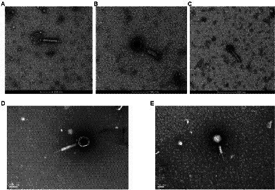
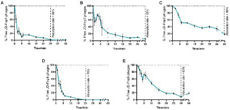
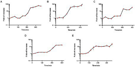
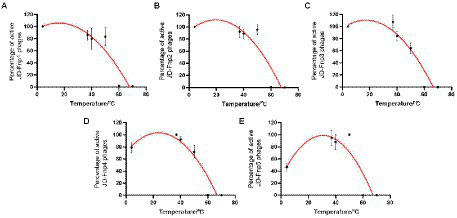
Fusobacterium nucleatum is a strictly anaerobic, Gram-negative bacterial species that is a member of the commensal flora in the oral cavity and gut. Recent studies suggested that the increase of abundance is associated with the development of various diseases, among which colorectal cancer is of the biggest concerns. Phage therapy is regarded as a potential approach to control the number of F. nucleatum, which may contribute to the prevention and treatment of related diseases. In this study, we isolated five isolates of bacteriophage targeting F. nucleatum. The morphological, biological, genomic and functional characteristics of five bacteriophages were investigated. Transmission electron microscopy revealed that JD-Fnp1 ~ JD-Fnp5 are all myoviruses. The size of the JD-Fnp1 ~ JD-Fnp5 genomes was 180,066 bp (JD-Fnp1), 41,329 bp (JD-Fnp2), 38,962 bp (JD-Fnp3), 180,231 bp (JD-Fnp4), and 41,353 bp (JD-Fnp5) respectively. The biological features including pH and heat stability, host range, growth characteristics of JD-Fnp1 ~ JD-Fnp5 displayed different patterns. Among them, JD-Fnp4 is considered to have the greatest clinical application value. The identification and characterization of JD-Fnp1 ~ JD-Fnp5 provides a basis for subsequent therapeutic strategy exploration of F. nucleatum-related diseases.
Overview
The claims and statistics of this paper haven’t been checked yet.
Run the check — free, no account needed. It reads the open-access text and checks the sources it cites.
Integrity signals
Registry
Cites
Statistics
Figures
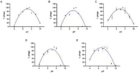
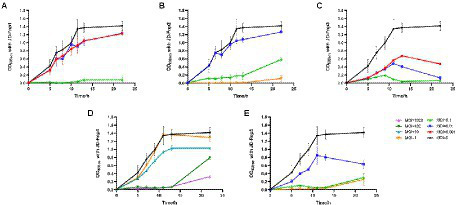
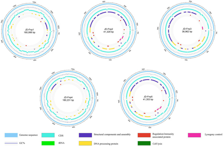
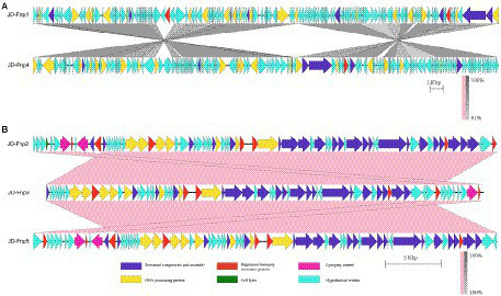
Verification
Claims the paper makes
The claims and statistics of this paper haven’t been checked yet.
Run the check — free, no account needed. It reads the open-access text and checks the sources it cites.
Claims other papers make about this one
Each is checked against this paper’s own text: does it say what the citing paper says it does?
In the citing paper
“Preclinical studies have demonstrated the efficacy of phages targeting Fusobacterium nucleatum [64,65]”
From Phage therapy in oncology: opportunities for cancer prevention and treatment (2026) · cites this paper as [65]
Self-check
Percentages, sums and reported differences re-computed from the paper’s own numbers.
The self-check needs the paper’s full text.
Citations
Cites
Fusobacterium nucleatum Promotes Chemoresistance to Colorectal Cancer by Modulating Autophagy
Yu, T., Guo, F., Yu, Y., Sun, T., Ma, D., Han, J., Qian, Y., Kryczek, I., Sun, D., Nagarsheth, N., et al. (2017) · Cell · doi:10.1016/j.cell.2017.07.008 · Cited by 2,198
Self-citationAnalysis of Fusobacterium persistence and antibiotic response in colorectal cancer
Bullman, S., Pedamallu, C. S., Sicinska, E., Clancy, T. E., Zhang, X., Cai, D., Neuberg, D., Huang, K., Guevara, F., Nelson, T., et al. (2017) · Science · doi:10.1126/science.aal5240 · Cited by 1,574
Zheng, D.-W., Dong, X., Pan, P., Chen, K.-W., Fan, J.-X., Cheng, S.-X., & Zhang, X.-Z. (2019) · Nature Biomedical Engineering · doi:10.1038/s41551-019-0423-2 · Cited by 495
Research works printed but not linked: 62
- Abed, 2016, Fap2 mediates fusobacterium nucleatum colorectal adenocarcinoma enrichment by binding to tumor-expressed gal-GalNAc, Cell Host Microbe, 20, 215, 10.1016/j.chom.2016.07.006 · DOI · Paper page
- Aghaee, 2021, Improving the inhibitory effect of phages against Pseudomonas aeruginosa isolated from a burn patient using a combination of phages and antibiotics, Viruses, 13, 1, 10.3390/v13020334 · DOI
- Aiewsakun, 2018, Evaluation of the genomic diversity of viruses infecting bacteria, archaea and eukaryotes using a common bioinformatic platform: steps towards a unified taxonomy, J. Gen. Virol., 99, 1331, 10.1099/jgv.0.001110 · DOI
- Aziz, 2008, The RAST server: rapid annotations using subsystems technology, BMC Genomics, 9, 75, 10.1186/1471-2164-9-75 · DOI
- Barrow, 1997, Bacteriophage therapy and prophylaxis: rediscovery and renewed assessment of potential, Trends Microbiol., 5, 268, 10.1016/s0966-842x(97)01054-8 · DOI
- Bin Jang, 2019, Taxonomic assignment of uncultivated prokaryotic virus genomes is enabled by gene-sharing networks, Nat. Biotechnol., 37, 632, 10.1038/s41587-019-0100-8 · DOI
- Bolstad, 1996, Taxonomy, biology, and periodontal aspects of fusobacterium nucleatum, Clin. Microbiol. Rev., 9, 55, 10.1128/cmr.9.1.55 · DOI
- Brennan, 2019, Fusobacterium nucleatum - symbiont, opportunist and oncobacterium, Nat. Rev. Microbiol., 17, 156, 10.1038/s41579-018-0129-6 · DOI · Paper page
- Castellarin, 2012, Fusobacterium nucleatum infection is prevalent in human colorectal carcinoma, Genome Res., 22, 299, 10.1101/gr.126516.111 · DOI · Paper page
- Chan, 2013, Phage cocktails and the future of phage therapy, Future Microbiol., 8, 769, 10.2217/fmb.13.47 · DOI
- Chen, Fusobacterium nucleatum promotes metastasis in colorectal cancer by activating autophagy signaling via the upregulation of CARD3 expression, Theranostics, 10, 323, 10.7150/thno.38870 · DOI
- Chen, Antibacterial activity of viable and heat-killed probiotic strains against oral pathogens, Lett. Appl. Microbiol., 70, 310, 10.1111/lam.13275 · DOI
- Chen, 2018, Fusobacterium nucleatum promotes M2 polarization of macrophages in the microenvironment of colorectal tumours via a TLR4-dependent mechanism, Cancer Immunol. Immunother., 67, 1635, 10.1007/s00262-018-2233-x · DOI
- Chen, Fusobacterium nucleatum promotes colorectal cancer metastasis by modulating KRT7-AS/KRT7, Gut Microbes, 11, 511, 10.1080/19490976.2019.1695494 · DOI
- Chen, 2005, VFDB: a reference database for bacterial virulence factors, Nucleic Acids Res., 33, D325, 10.1093/nar/gki008 · DOI
- Cochrane, 2016, Complete genome sequences and analysis of the fusobacterium nucleatum subspecies animalis 7-1 bacteriophage ɸFunu1 and ɸFunu2, Anaerobe, 38, 125, 10.1016/j.anaerobe.2015.10.013 · DOI
- Cui, 2017, Characterization and complete genome of the virulent Myoviridae phage JD007 active against a variety of Staphylococcus aureus isolates from different hospitals in Shanghai, China. Virol. J., 14, 26, 10.1186/s12985-017-0701-0 · DOI
- Dedrick, 2019, Engineered bacteriophages for treatment of a patient with a disseminated drug-resistant mycobacterium abscessus, Nat. Med., 25, 730, 10.1038/s41591-019-0437-z · DOI
- Dong, 2020, Bioinorganic hybrid bacteriophage for modulation of intestinal microbiota to remodel tumor-immune microenvironment against colorectal cancer, Sci. Adv., 6, eaba1590, 10.1126/sciadv.aba1590 · DOI
- Duan, 2019, Bacteriophage targeting of gut bacterium attenuates alcoholic liver disease, Nature, 575, 505, 10.1038/s41586-019-1742-x · DOI
- Feuille, 1996, Mixed infection with Porphyromonas gingivalis and fusobacterium nucleatum in a murine lesion model: potential synergistic effects on virulence, Infect. Immun., 64, 2094, 10.1128/iai.64.6.2094-2100.1996 · DOI
- Geier, 1973, Fate of bacteriophage lambda in non-immune germ-free mice, Nature, 246, 221, 10.1038/246221a0 · DOI
- Gur, Immunity, 42, 344, 10.1016/j.immuni.2015.01.010 · DOI · Paper page
- Han, 2015, Fusobacterium nucleatum: a commensal-turned pathogen, Curr. Opin. Microbiol., 23, 141, 10.1016/j.mib.2014.11.013 · DOI
- Hernández, 2020, Phages in anaerobic systems, Viruses, 12, 1, 10.3390/v12101091 · DOI
- Hesse, 2021, Bacteriophage treatment rescues mice infected with multidrug-resistant Klebsiella pneumoniae ST258, MBio, 12, 1, 10.1128/mBio.00034-21 · DOI
- Hsu, 2019, Dynamic modulation of the gut microbiota and metabolome by bacteriophages in a mouse model, Cell Host Microbe, 25, 803, 10.1016/j.chom.2019.05.001 · DOI · Paper page
- Jang, 2016, Comparative study on the characteristics of Weissella cibaria CMU and probiotic strains for Oral care, Molecules, 21, 1, 10.3390/molecules21121752 · DOI
- Jault, 2019, Efficacy and tolerability of a cocktail of bacteriophages to treat burn wounds infected by Pseudomonas aeruginosa (PhagoBurn): a randomised, controlled, double-blind phase 1/2 trial, Lancet Infect. Dis., 19, 35, 10.1016/s1473-3099(18)30482-1 · DOI
- Kabwe, 2019, Genomic, morphological and functional characterisation of novel bacteriophage FNU1 capable of disrupting fusobacterium nucleatum biofilms, Sci. Rep., 9, 9107, 10.1038/s41598-019-45549-6 · DOI
- Kifelew, 2020, Efficacy of phage cocktail AB-SA01 therapy in diabetic mouse wound infections caused by multidrug-resistant Staphylococcus aureus, BMC Microbiol., 20, 204, 10.1186/s12866-020-01891-8 · DOI
- Koskella, 2014, Bacteria-phage coevolution as a driver of ecological and evolutionary processes in microbial communities, FEMS Microbiol. Rev., 38, 916, 10.1111/1574-6976.12072 · DOI
- Kostic, 2013, Fusobacterium nucleatum potentiates intestinal tumorigenesis and modulates the tumor-immune microenvironment, Cell Host Microbe, 14, 207, 10.1016/j.chom.2013.07.007 · DOI · Paper page
- Kostic, 2012, Genomic analysis identifies association of fusobacterium with colorectal carcinoma, Genome Res., 22, 292, 10.1101/gr.126573.111 · DOI · Paper page
- Leshkasheli, 2019, Efficacy of newly isolated and highly potent bacteriophages in a mouse model of extensively drug-resistant Acinetobacter baumannii bacteraemia, J. Glob. Antimicrob. Resist., 19, 255, 10.1016/j.jgar.2019.05.005 · DOI
- Letunic, 2021, Interactive tree of life (iTOL) v5: an online tool for phylogenetic tree display and annotation, Nucleic Acids Res., 49, W293, 10.1093/nar/gkab301 · DOI
- Liu, 2020, Fusobacterium nucleatum exacerbates colitis by damaging epithelial barriers and inducing aberrant inflammation, J. Dig. Dis., 21, 385, 10.1111/1751-2980.12909 · DOI
- Liu, 2009, ARDB--antibiotic resistance genes database, Nucleic Acids Res., 37, D443, 10.1093/nar/gkn656 · DOI
- Loh, 2020, Encapsulation and delivery of therapeutic phages, Appl. Environ. Microbiol., 87, 1, 10.1128/aem.01979-20 · DOI
- Lu, 2020, CDD/SPARCLE: the conserved domain database in 2020, Nucleic Acids Res., 48, D265, 10.1093/nar/gkz991 · DOI
- Machuca, 2010, Isolation of a novel bacteriophage specific for the periodontal pathogen fusobacterium nucleatum, Appl. Environ. Microbiol., 76, 7243, 10.1128/aem.01135-10 · DOI
- Mima, 2015, Fusobacterium nucleatum and T cells in colorectal carcinoma, JAMA Oncol., 1, 653, 10.1001/jamaoncol.2015.1377 · DOI · Paper page
- Nishimura, 2017, ViPTree: the viral proteomic tree server, Bioinformatics, 33, 2379, 10.1093/bioinformatics/btx157 · DOI
- Page, 2015, Roary: rapid large-scale prokaryote pan genome analysis, Bioinformatics, 31, 3691, 10.1093/bioinformatics/btv421 · DOI · Paper page
- Parhi, 2020, Breast cancer colonization by fusobacterium nucleatum accelerates tumor growth and metastatic progression, Nat. Commun., 11, 3259, 10.1038/s41467-020-16967-2 · DOI · Paper page
- Rhoads, 2009, Bacteriophage therapy of venous leg ulcers in humans: results of a phase I safety trial, J. Wound Care, 18, 237, 10.12968/jowc.2009.18.6.42801 · DOI
- Roux, 2021, IMG/VR v3: an integrated ecological and evolutionary framework for interrogating genomes of uncultivated viruses, Nucleic Acids Res., 49, D764, 10.1093/nar/gkaa946 · DOI
- Rubinstein, Fusobacterium nucleatum promotes colorectal carcinogenesis by modulating E-cadherin/beta-catenin signaling via its FadA adhesin, Cell Host Microbe, 14, 195, 10.1016/j.chom.2013.07.012 · DOI · Paper page
- Rubinstein, Fusobacterium nucleatum promotes colorectal carcinogenesis by modulating E-cadherin/β-catenin signaling via its FadA adhesin, Cell Host Microbe, 14, 195, 10.1016/j.chom.2013.07.012 · DOI · Paper page
- Shen, 2021, Phage genome annotation: where to begin and end, Phage, 2, 183, 10.1089/phage.2021.0015 · DOI
- Signat, 2011, Fusobacterium nucleatum in periodontal health and disease, Curr. Issues Mol. Biol., 13, 25, 10.21775/cimb.013.025 · DOI
- Strauss, 2011, Invasive potential of gut mucosa-derived fusobacterium nucleatum positively correlates with IBD status of the host, Inflamm. Bowel Dis., 17, 1971, 10.1002/ibd.21606 · DOI
- Szabo, 2013, Tumor-derived hydrogen sulfide, produced by cystathionine-β-synthase, stimulates bioenergetics, cell proliferation, and angiogenesis in colon cancer, Proc. Natl. Acad. Sci. U. S. A., 110, 12474, 10.1073/pnas.1306241110 · DOI
- Turner, Phage annotation guide: guidelines for assembly and high-quality annotation, Phage (New Rochelle), 2, 170, 10.1089/phage.2021.0013 · DOI
- Turner, A roadmap for genome-based phage taxonomy, Viruses, 13, 1, 10.3390/v13030506 · DOI
- Wong, 2019, Gut microbiota in colorectal cancer: mechanisms of action and clinical applications, Nat. Rev. Gastroenterol. Hepatol., 16, 690, 10.1038/s41575-019-0209-8 · DOI
- Wright, 2009, A controlled clinical trial of a therapeutic bacteriophage preparation in chronic otitis due to antibiotic-resistant Pseudomonas aeruginosa; a preliminary report of efficacy, Clin. Otolaryngol., 34, 349, 10.1111/j.1749-4486.2009.01973.x · DOI
- Wu, 2018, Fusobacterium nucleatum potentiates intestinal tumorigenesis in mice via a toll-like receptor 4/p21-activated kinase 1 Cascade, Dig. Dis. Sci., 63, 1210, 10.1007/s10620-018-4999-2 · DOI
- Xue, 2018, Indoleamine 2,3-dioxygenase expression regulates the survival and proliferation of fusobacterium nucleatum in THP-1-derived macrophages, Cell Death Dis., 9, 355, 10.1038/s41419-018-0389-0 · DOI
- Yang, 2017, Fusobacterium nucleatum increases proliferation of colorectal cancer cells and tumor development in mice by activating toll-like receptor 4 signaling to nuclear factor-κB, and up-regulating expression of MicroRNA-21, Gastroenterology, 152, 851, 10.1053/j.gastro.2016.11.018 · DOI
- Yu, 2015, Berberine may rescue fusobacterium nucleatum-induced colorectal tumorigenesis by modulating the tumor microenvironment, Oncotarget, 6, 32013, 10.18632/oncotarget.5166 · DOI
- Zhu, 2010, Competition between yogurt probiotics and periodontal pathogens in vitro, Acta Odontol. Scand., 68, 261, 10.3109/00016357.2010.492235 · DOI
Cited by
Sorted by citations
Phage therapy in oncology: opportunities for cancer prevention and treatment
Mutalik, V. K., Inman, J. L., Chang, H., Arkin, A., & Mao, J.-H. (2026) · Trends in Molecular Medicine · doi:10.1016/j.molmed.2026.02.001 · Cited by 7
Metadata
- Authors
- Wang, Y., Liu, Z., Chen, Q., Yi, L., Xu, Z., Cai, M., Qin, J., Zhang, Y., Du, G., Hong, J., Guo, X., & Liu, C.
- Venue
- Frontiers in Microbiology
- Publisher
- Frontiers Media SA
- Published
- 2022-11-03
- Volume
- 13
- ISSN
- 1664-302X
- PMC
- PMC9670143
- Cited by
- 23
- References
- 3
About these checks
Registry, retraction, Cites and statistics results are computed, not judged, with one exception: a printed reference that no index lists is read once by a model before it counts as a citation error. Claims are extracted by a model and kept only when their quote appears verbatim in the text; each cited source is then checked, with a model’s help, for whether it says what the claim relies on. Those verdicts can be wrong — read the claim’s quote and the cited source before relying on one.
Statistics and arithmetic are recomputed, not judged.
Confidence bands are shown only for fields calibrated on our annotated test set. None is calibrated yet, so no band is shown.
What counts as a claim ›How we measure confidence ›How we check this ›
Are you an author, or do you see a problem with this page? Request a review
