

The DOI matches its Crossref record; 61 of the 61 references it cites were found in a registry or an index.
Role of mitochondria ROS generation in ethanol-induced NLRP3 inflammasome activation and cell death in astroglial cells
Open accessAbstract
Toll-like receptors (TLRs) and NOD-like receptors (NLRs) are innate immunity sensors that provide an early/effective response to pathogenic or injury conditions. We have reported that ethanol-induced TLR4 activation triggers signaling inflammatory responses in glial cells, causing neuroinflammation and brain damage. However, it is uncertain if ethanol is able to activate NLRs/inflammasome in astroglial cells, which is the mechanism of activation, and whether there is crosstalk between both immune sensors in glial cells. Here we show that chronic ethanol treatment increases the co-localization of caspase-1 with GFAP(+) cells, and up-regulates IL-1β and IL-18 in the frontal medial cortex in WT, but not in TLR4 knockout mice. We further show that cultured cortical astrocytes expressed several inflammasomes (NLRP3, AIM2, NLRP1, and IPAF), although NLRP3 mRNA is the predominant form. Ethanol, as ATP and LPS treatments, up-regulates NLRP3 expression, and causes caspase-1 cleavage and the release of IL-1β and IL-18 in astrocytes supernatant. Ethanol-induced NLRP3/caspase-1 activation is mediated by mitochondrial (m) reactive oxygen species (ROS) generation because when using a specific mitochondria ROS scavenger, the mito-TEMPO (500 μM) or NLRP3 blocking peptide (4 μg/ml) or a specific caspase-1 inhibitor, Z-YVAD-FMK (10 μM), abrogates mROS release and reduces the up-regulation of IL-1β and IL-18 induced by ethanol or LPS or ATP. Confocal microscopy studies further confirm that ethanol, ATP or LPS promotes NLRP3/caspase-1 complex recruitment within the mitochondria to promote cell death by caspase-1-mediated pyroptosis, which accounts for ≈73% of total cell death (≈22%) and the remaining (≈25%) die by caspase-3-dependent apoptosis. Suppression of the TLR4 function abrogates most ethanol effects on NLRP3 activation and reduces cell death. These findings suggest that NLRP3 participates, in ethanol-induced neuroinflammation and highlight the NLRP3/TLR4 crosstalk in ethanol-induced brain injury.
Overview
The claims and statistics of this paper haven’t been checked yet.
Run the check — free, no account needed. It reads the open-access text and checks the sources it cites.
Integrity signals
Registry
Cites
Statistics
Figures
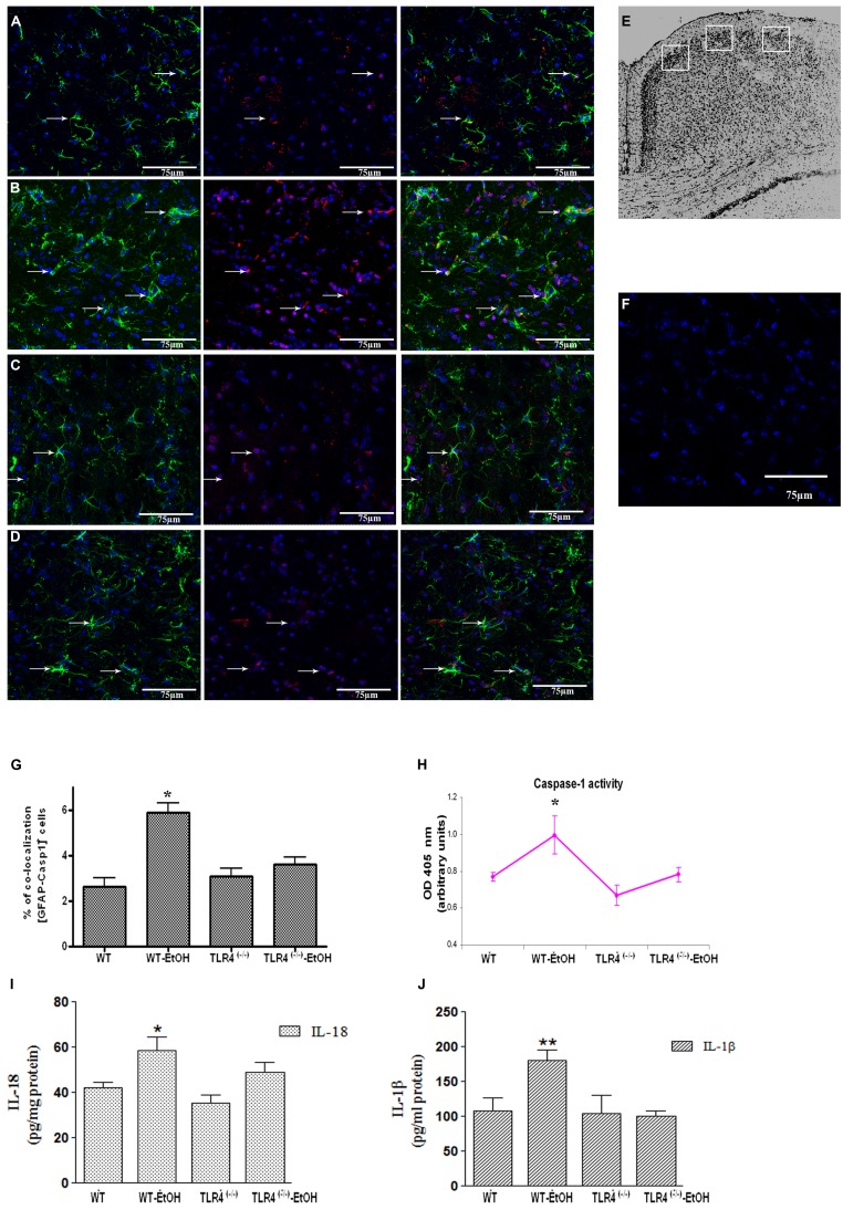
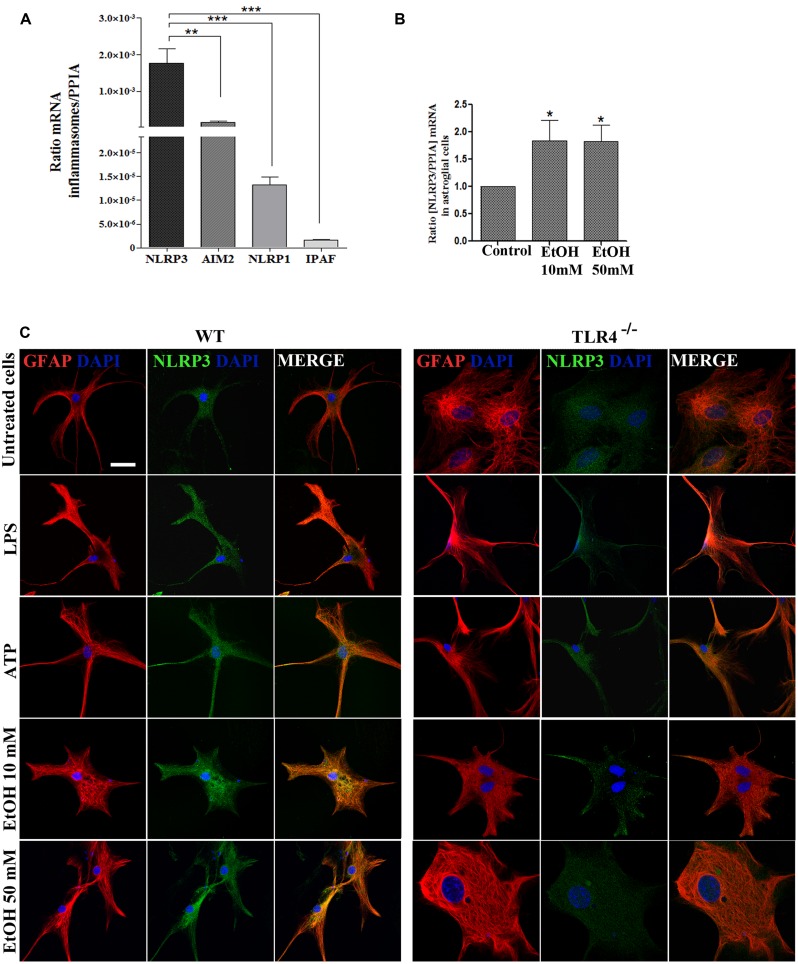
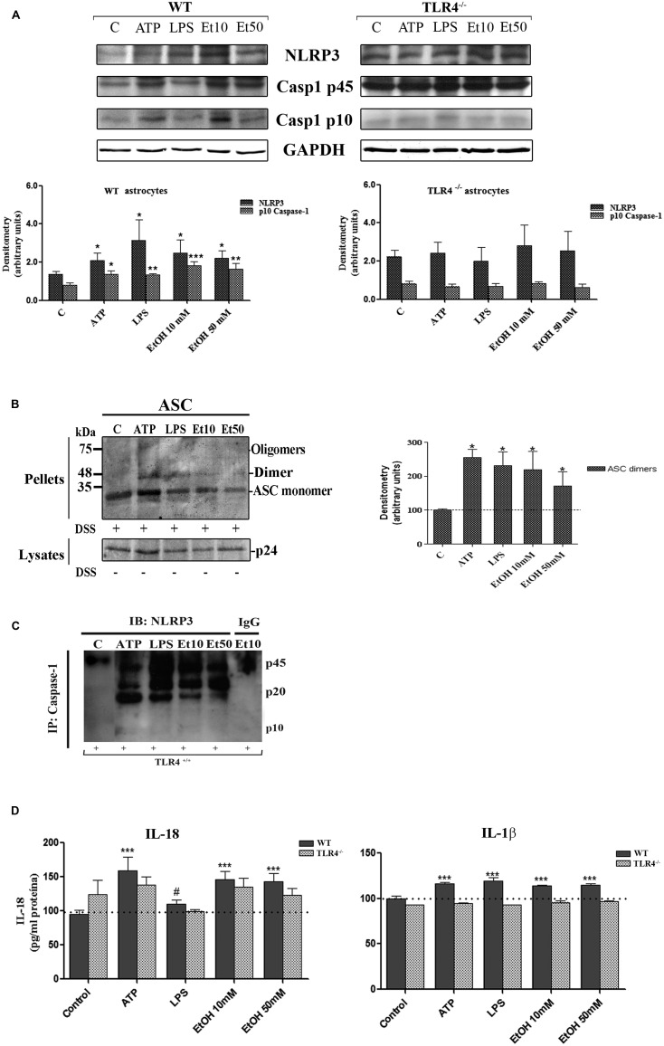
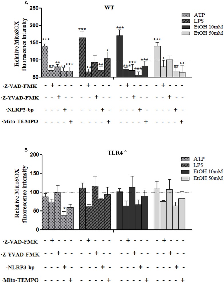
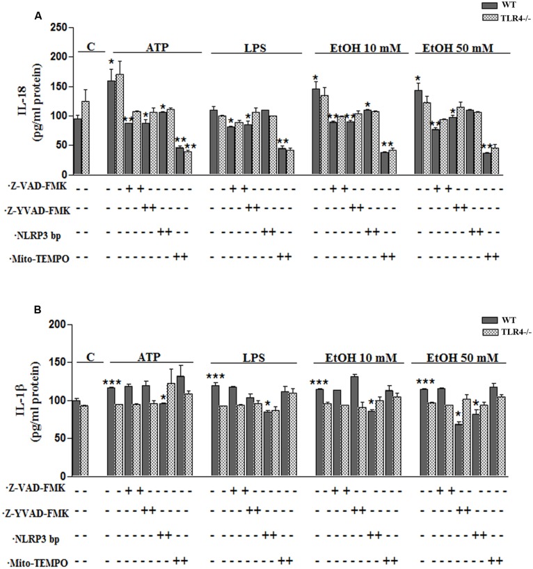
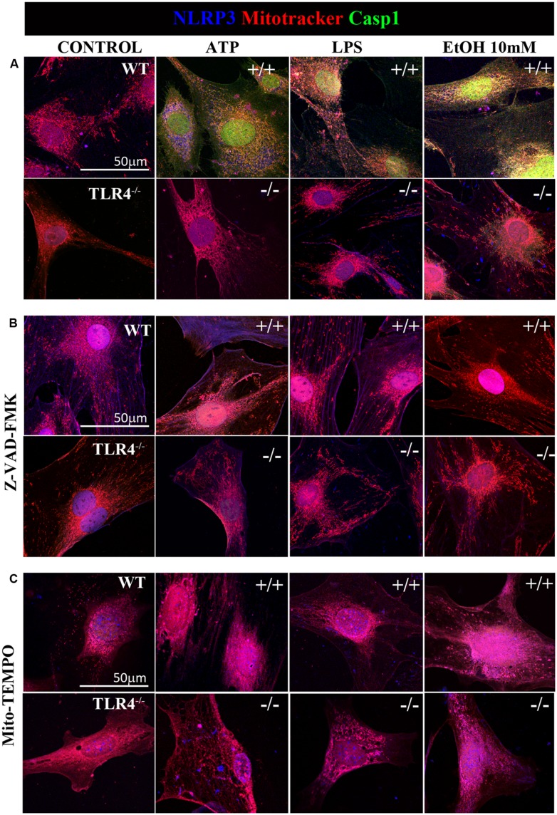
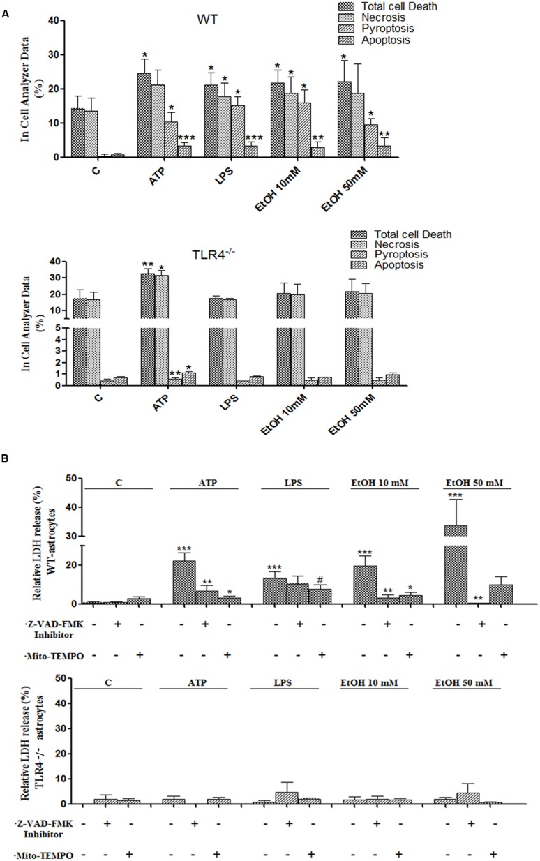
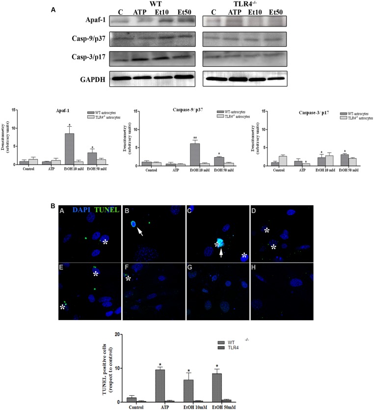
Verification
Claims the paper makes
The claims and statistics of this paper haven’t been checked yet.
Run the check — free, no account needed. It reads the open-access text and checks the sources it cites.
Claims other papers make about this one
Each is checked against this paper’s own text: does it say what the citing paper says it does?
In the citing paper
“Following decades of research, the most well‐characterized mechanism underlying activation of the NLRP3 inflammasome is reactive oxygen species (ROS), particularly mitochondrial ROS (mtROS), generation.”
From Trimethylamine‐N‐Oxide Induces Vascular Inflammation by Activating the NLRP3 Inflammasome Through the SIRT3‐SOD2‐mtROS Signaling Pathway (2017) · cites this paper as [15]
In the citing paper
“The majority of NLRP3 activators increase mtROS levels, and chemical scavengers of mtROS effectively inhibit NLRP3 activation.14, 15”
From Trimethylamine‐N‐Oxide Induces Vascular Inflammation by Activating the NLRP3 Inflammasome Through the SIRT3‐SOD2‐mtROS Signaling Pathway (2017) · cites this paper as [15]
Self-check
Percentages, sums and reported differences re-computed from the paper’s own numbers.
The self-check needs the paper’s full text.
Citations
Cites
A new mathematical model for relative quantification in real-time RT-PCR
Pfaffl, M. W. (2001) · Nucleic Acids Research · doi:10.1093/nar/29.9.e45 · Cited by 34,610
The Mouse Brain in Stereotaxic Coordinates
Paxinos, G. (2001) · Cited by 12,859
A role for mitochondria in nlrp3 inflammasome activation
Zhou, R., Yazdi, A. S., Menu, P., & Tschopp, J. (2011) · Nature · doi:10.1038/nature09663 · Cited by 5,602
Schroder, K., & Tschopp, J. (2010) · Cell · doi:10.1016/j.cell.2010.01.040 · Cited by 5,475
mouse brain in stereotaxic coordinates
Franklin, K. B. J., & Paxinos, G. (1997) · Cited by 3,258
Nakahira, K., Haspel, J. A., Rathinam, V. A. K., Lee, S.-J., Dolinay, T., Lam, H. C., Englert, J. A., Rabinovitch, M., Cernadas, M., Kim, H. P., et al. (2011) · Nature Immunology · doi:10.1038/ni.1980 · Cited by 3,049
NLRP3 is activated in Alzheimer’s disease and contributes to pathology in APP/PS1 mice
Heneka, M. T., Kummer, M. P., Stutz, A., Delekate, A., Schwartz, S., Vieira-Saecker, A., Griep, A., Axt, D., Remus, A., Tzeng, T.-C., et al. (2013) · Nature · doi:10.1038/nature11729 · Cited by 2,898
Activation and regulation of the inflammasomes
Latz, E., Xiao, T. S., & Stutz, A. (2013) · Nature Reviews Immunology · doi:10.1038/nri3452 · Cited by 2,884
Oxidized mitochondrial DNA activates the NLRP3 inflammasome during apoptosis
Shimada, K., Crother, T. R., Karlin, J., Dagvadorj, J., Chiba, N., Chen, S., Ramanujan, V. K., Wolf, A. J., Vergnes, L., Ojcius, D. M., et al. (2012) · Immunity · doi:10.1016/j.immuni.2012.01.009 · Cited by 2,282
NLRP3 inflammasome activation: the convergence of multiple signalling pathways on ROS production?
Tschopp, J., & Schroder, K. (2010) · Nature reviews. Immunology · doi:10.1038/nri2725 · Cited by 1,859
Cited by
Sorted by citations
Dopamine controls systemic inflammation through inhibition of NLRP3 inflammasome
Yan, Y., Jiang, W., Liu, L., Wang, X., Ding, C., Tian, Z., & Zhou, R. (2015) · Cell · doi:10.1016/j.cell.2014.11.047 · Cited by 1,014
Chen, M., Zhu, X., Ran, L., Lang, H., Yi, L., & Mi, M. (2017) · Journal of the American Heart Association · doi:10.1161/jaha.117.006347 · Cited by 600
The role of mitochondria in NLRP3 inflammasome activation
Liu, Q., Zhang, D., Hu, D., Zhou, X., & Zhou, Y. (2018) · Molecular Immunology · doi:10.1016/j.molimm.2018.09.010 · Cited by 475
NLRP3 inflammasome in neurological diseases, from functions to therapies
Song, L., Pei, L., Yao, S., Wu, Y., & Shang, Y. (2017) · Frontiers in Cellular Neuroscience · doi:10.3389/fncel.2017.00063 · Cited by 467
Mitochondrial ROS promote macrophage pyroptosis by inducing GSDMD oxidation
Wang, Y., Shi, P., Chen, Q., Huang, Z., Zou, D., Zhang, J., Gao, X., & Lin, Z. (2019) · Journal of Molecular Cell Biology · doi:10.1093/jmcb/mjz020 · Cited by 365
Metadata
- Authors
- Alfonso-Loeches, S., Ureña-Peralta, J. R., Morillo-Bargues, M. J., Oliver-De La Cruz, J., & Guerri, C.
- Affiliations
- Centro de Investigacion Principe Felipe (ES)
- Corresponding author
- Guerri, Consuelo
- ORCID
- Guerri, Consuelo
- Venue
- Frontiers in Cellular Neuroscience
- Publisher
- Frontiers Media SA
- Published
- 2014-08-01
- Volume
- 8
- Pages
- 216-216
- ISSN
- 1662-5102 (ISSN-L)
- PMC
- PMC4118026
- OpenAlex
- W2081653529
- Cited by
- 265
- References
- 61
- Citations by year
- 2026: 11 · 2025: 19 · 2024: 21 · 2023: 22 · 2022: 33 · 2021: 33 · 2020: 27 · 2019: 24 · 2018: 22 · 2017: 22 · 2016: 21 · 2015: 8 · 2014: 2
- FWCI
- 7.25
- Type
- journal article
- Language
- en
- Open access
- gold
- doi.org
- Licence
- cc-by
- Locations
- Frontiers in Cellular Neuroscience (publishedVersion, cc-by)
- Europe PMC (PubMed Central) (publishedVersion, cc-by)
- PubMed Central (publishedVersion, cc-by)
- PubMed (publishedVersion)
- DOAJ (DOAJ: Directory of Open Access Journals) (submittedVersion, cc-by-sa)
- Funders
- Ministerio de Economía y Competitividad
- Ministerio de Ciencia e Innovación
- Topics
- Life Sciences › Biochemistry, Genetics and Molecular Biology › Molecular Biology › Inflammasome and immune disorders
- Life Sciences › Neuroscience › Neurology › Neuroinflammation and Neurodegeneration Mechanisms
- Life Sciences › Immunology and Microbiology › Immunology › Immune Response and Inflammation
- Keywords
- ethanol, NLRP3 inflammasome, reactive oxygen species, mitochondria, astrocytes, caspase-1, pyroptosis, TLR4, neuroinflammation, IL-1β, IL-18, innate immunity, brain injury, apoptosis, LPS, ATP
About these checks
Registry, retraction, Cites and statistics results are computed, not judged, with one exception: a printed reference that no index lists is read once by a model before it counts as a citation error. Claims are extracted by a model and kept only when their quote appears verbatim in the text; each cited source is then checked, with a model’s help, for whether it says what the claim relies on. Those verdicts can be wrong — read the claim’s quote and the cited source before relying on one.
Statistics and arithmetic are recomputed, not judged.
Confidence bands are shown only for fields calibrated on our annotated test set. None is calibrated yet, so no band is shown.
What counts as a claim ›How we measure confidence ›How we check this ›
Are you an author, or do you see a problem with this page? Request a review
