

The DOI matches its Crossref record; 231 of the 235 references it cites were found in a registry or an index, and 1 are not checked yet.
Potential Mechanisms Underlying Centralized Pain and Emerging Therapeutic Interventions
Open accessAbstract
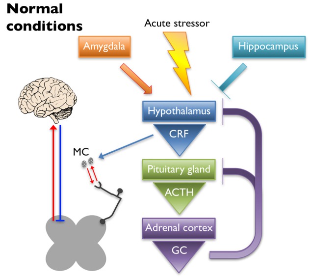
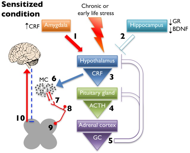
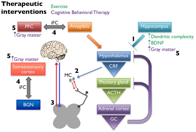
Centralized pain syndromes are associated with changes within the central nervous system that amplify peripheral input and/or generate the perception of pain in the absence of a noxious stimulus. Examples of idiopathic functional disorders that are often categorized as centralized pain syndromes include fibromyalgia, chronic pelvic pain syndromes, migraine, and temporomandibular disorder. Patients often suffer from widespread pain, associated with more than one specific syndrome, and report fatigue, mood and sleep disturbances, and poor quality of life. The high degree of symptom comorbidity and a lack of definitive underlying etiology make these syndromes notoriously difficult to treat. The main purpose of this review article is to discuss potential mechanisms of centrally-driven pain amplification and how they may contribute to increased comorbidity, poorer pain outcomes, and decreased quality of life in patients diagnosed with centralized pain syndromes, as well as discuss emerging non-pharmacological therapies that improve symptomology associated with these syndromes. Abnormal regulation and output of the hypothalamic-pituitary-adrenal (HPA) axis is commonly associated with centralized pain disorders. The HPA axis is the primary stress response system and its activation results in downstream production of cortisol and a dampening of the immune response. Patients with centralized pain syndromes often present with hyper- or hypocortisolism and evidence of altered downstream signaling from the HPA axis including increased Mast cell (MC) infiltration and activation, which can lead to sensitization of nearby nociceptive afferents. Increased peripheral input via nociceptor activation can lead to "hyperalgesic priming" and/or "wind-up" and eventually to central sensitization through long term potentiation in the central nervous system. Other evidence of central modifications has been observed through brain imaging studies of functional connectivity and magnetic resonance spectroscopy and are shown to contribute to the widespreadness of pain and poor mood in patients with fibromyalgia and chronic urological pain. Non-pharmacological therapeutics, including exercise and cognitive behavioral therapy (CBT), have shown great promise in treating symptoms of centralized pain.
Overview
The claims and statistics of this paper haven’t been checked yet.
Run the check — free, no account needed. It reads the open-access text and checks the sources it cites.
Integrity signals
Registry
Cites
Statistics
Figures
Verification
Claims the paper makes
The claims and statistics of this paper haven’t been checked yet.
Run the check — free, no account needed. It reads the open-access text and checks the sources it cites.
Claims other papers make about this one
Each is checked against this paper’s own text: does it say what the citing paper says it does?
The source backs part of the claim, not all of it — or backs it for a different population, condition or comparison.
In the citing paper
“Local infiltration and migration of mast cells, leading to sensitization of nearby nociceptive afferents, are considered vital contributors to NPP pathogenesis (69).”
From Nociplastic pain: the prominent role of non-neuronal cells in central and peripheral sensitization (2026) · cites this paper as [69]
Experimental — accuracy not yet measuredThe cited source says what the claim attributes to it.
In the citing paper
“NPP is widely reported to be correlated with mast cell activation.”
From Nociplastic pain: the prominent role of non-neuronal cells in central and peripheral sensitization (2026) · cites this paper as [69]
Self-check
Percentages, sums and reported differences re-computed from the paper’s own numbers.
The self-check needs the paper’s full text.
Citations
Cites
(1985) · Europe PMC (PubMed Central) · Cited by 9,143
(1968) · American Association of Industrial Nurses Journal · doi:10.1177/216507996801601208 · Cited by 8,811
Heart Disease and Stroke Statistics—2016 Update: A Report From the American Heart Association
Mozaffarian, D., Benjamin, E. J., Go, A. S., Arnett, D. K., Blaha, M. J., Cushman, M., Das, S. R., de Ferranti, S., Després, J.-P., Fullerton, H. J., et al. (2016) · Circulation · doi:10.1161/cir.0000000000000350 · Cited by 6,596
A new and sensitive method for measuring thermal nociception in cutaneous hyperalgesia
Hargreaves, K., Dubner, R. A., Brown, F. J., Flores, C., & Joris, J. L. (1988) · Pain · doi:10.1016/0304-3959(88)90026-7 · Cited by 5,155
Central sensitization: implications for the diagnosis and treatment of pain
Woolf, C. J. (2011) · Pain · doi:10.1016/j.pain.2010.09.030 · Cited by 4,771
Neural regulation of endocrine and autonomic stress responses
Ulrich-Lai, Y. M., & Herman, J. P. (2009) · Nature Reviews Neuroscience · doi:10.1038/nrn2647 · Cited by 3,318
Classification of chronic pain
Merskey, H. (1994) · Medical Entomology and Zoology · Cited by 2,952
Running enhances neurogenesis, learning, and long-term potentiation in mice
van Praag, H., Christie, B. R., Sejnowski, T. J., & Gage, F. H. (1999) · Proceedings of the National Academy of Sciences · doi:10.1073/pnas.96.23.13427 · Cited by 2,904
The Concepts of Stress and Stress System Disorders: Overview of Physical and Behavioral Homeostasis
Chrousos, G. P. (1992) · JAMA · doi:10.1001/jama.1992.03480090092034 · Cited by 2,689
Molecular mechanisms of nociception
Julius, D., & Basbaum, A. I. (2001) · Nature · doi:10.1038/35093019 · Cited by 2,685 · cited 1×
“…enhances pain-related afferent input to the central nervous system; however, they also generate and maintain peripheral neurogenic inflammation by releasing neuropeptides, including SP and CGRP, which perpetuates inflammatory mediator release in the proximate milieu (Julius and Basbaum, 2001).”
Background
Research works printed but not linked: 1
- 2016, Statistics About Diabetes.
Cited by
Sorted by citations
The anatomy of pain and suffering in the brain and its clinical implications
Ridder, D. D., Adhia, D. B., & Vanneste, S. (2021) · Neuroscience & Biobehavioral Reviews · doi:10.1016/j.neubiorev.2021.08.013 · Cited by 298
Reframing chronic pain as a disease, not a symptom: rationale and implications for pain management
Clauw, D. J., Essex, M. N., Pitman, V. W., & Jones, K. D. (2019) · Postgraduate Medicine · doi:10.1080/00325481.2019.1574403 · Cited by 300
Altered microbiome composition in individuals with fibromyalgia
Minerbi, A., Gonzalez, E., Brereton, N. J. B., Anjarkouchian, A., Dewar, K., Fitzcharles, M.-A., Chevalier, S., & Shir, Y. (2019) · Pain · doi:10.1097/j.pain.0000000000001640 · Cited by 220
Myofascial trigger points in migraine and tension-type headache
Do, T. P., Heldarskard, G. F., Kolding, L. T., Hvedstrup, J., & Schytz, H. (2018) · The Journal of Headache and Pain · doi:10.1186/s10194-018-0913-8 · Cited by 188
Central sensitization, chronic pain, and other symptoms: Better understanding, better management
Volcheck, M. M., Graham, S. M., Fleming, K. C., Mohabbat, A. B., & Luedtke, C. A. (2023) · Cleveland Clinic Journal of Medicine · doi:10.3949/ccjm.90a.22019 · Cited by 204
Metadata
- Authors
- Eller-Smith, O. C., Nicol, A. L., & Christianson, J. A.
- Affiliations
- University of Kansas Medical Center (US)
- Corresponding author
- Christianson, Julie A.
- Venue
- Frontiers in Cellular Neuroscience
- Publisher
- Frontiers Media SA
- Published
- 2018-02-13
- Volume
- 12
- Pages
- 35-35
- ISSN
- 1662-5102 (ISSN-L)
- PMC
- PMC5816755
- OpenAlex
- W2793570991
- Cited by
- 227
- References
- 235
- Citations by year
- 2026: 31 · 2025: 24 · 2024: 20 · 2023: 31 · 2022: 31 · 2021: 31 · 2020: 26 · 2019: 23 · 2018: 10
- FWCI
- 2.95
- Type
- journal article
- Language
- en
- Open access
- gold
- doi.org
- Locations
- Frontiers in Cellular Neuroscience (publishedVersion, cc-by)
- Europe PMC (PubMed Central) (publishedVersion, cc-by)
- PubMed Central (publishedVersion, cc-by)
- PubMed (publishedVersion)
- DOAJ (DOAJ: Directory of Open Access Journals) (submittedVersion, cc-by-sa)
- Funders
- National Institutes of Health (DK099611, DK103872, GM123320, HD057850)
- Topics
- Health Sciences › Medicine › Physiology (medical) › Pain Mechanisms and Treatments
- Health Sciences › Medicine › Psychiatry and Mental health › Fibromyalgia and Chronic Fatigue Syndrome Research
- Health Sciences › Medicine › Anesthesiology and Pain Medicine › Pain Management and Treatment
- Keywords
- fibromyalgia, chronic pelvic pain, migraine, temporomandibular disorders, central sensitization, HPA axis dysregulation, mast cells, hyperalgesic priming, wind-up, long-term potentiation, functional connectivity, non-pharmacological treatment, physical activity, cognitive behavioral therapy, comorbidities, quality of life
About these checks
Registry, retraction, Cites and statistics results are computed, not judged, with one exception: a printed reference that no index lists is read once by a model before it counts as a citation error. Claims are extracted by a model and kept only when their quote appears verbatim in the text; each cited source is then checked, with a model’s help, for whether it says what the claim relies on. Those verdicts can be wrong — read the claim’s quote and the cited source before relying on one.
Statistics and arithmetic are recomputed, not judged.
Confidence bands are shown only for fields calibrated on our annotated test set. None is calibrated yet, so no band is shown.
What counts as a claim ›How we measure confidence ›How we check this ›
Are you an author, or do you see a problem with this page? Request a review
