

The DOI matches its Crossref record; 234 of the 235 references it cites were found in a registry or an index, and 1 are not checked yet.
Microglia-orchestrated neuroinflammation and synaptic remodeling: roles of pro-inflammatory cytokines and receptors in neurodegeneration
Open accessAbstract
Microglia, the innate immune cells of the central nervous system (CNS), play essential roles in maintaining neural homeostasis through dynamic interactions with neurons and other brain structures. While their protective functions are well-established, recent studies have illuminated the detrimental consequences of sustained microglial activation in the context of neurodegeneration. In particular, overactivated microglia contribute to neuroinflammation and induce synaptic alterations through the release of pro-inflammatory cytokines and engagement of specific receptors. These interactions disrupt synaptic structure and function, compromising connectivity, plasticity, and cognitive processes. Notably, neuronal synapses are primary targets of such inflammation-driven dysfunction, where prolonged exposure to cytokines such as interleukin-1β (IL-1β), interleukin-6 (IL-6), and tumor necrosis factor- α (TNF-α), and signaling via receptor systems including cluster of differentiation-200 (CD200)/CD200 receptor (CD200R), C-X3-C motif chemokine ligand 1 (CX3CL1)/CX3C receptor 1 (CX3CR1), colony-stimulating factor 1 (CSF1)/CSF1 receptor (CSF1R), and interferon- γ (IFN-γ)/IFN-γ receptor (IFN-γR), lead to impaired learning, excitotoxicity, and neurodegenerative progression. This review synthesizes emerging evidence on the mechanisms by which microglia-mediated immune responses regulate synaptic remodeling, emphasizing the roles of pro-inflammatory cytokines and their receptors in neurodegenerative disorders.
Overview
The claims and statistics of this paper haven’t been checked yet.
Run the check — free, no account needed. It reads the open-access text and checks the sources it cites.
Integrity signals
Registry
Cites
Statistics
Figures
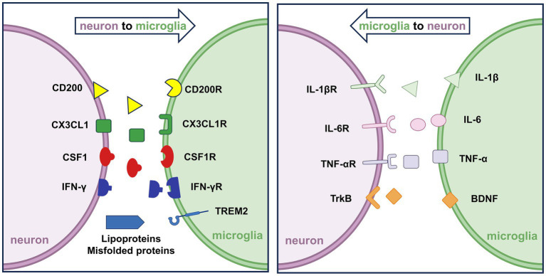
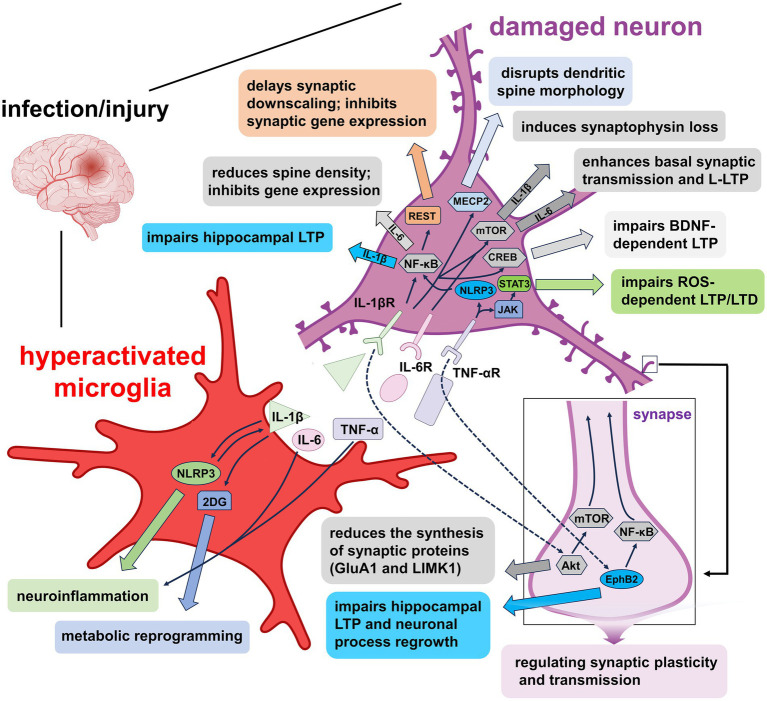
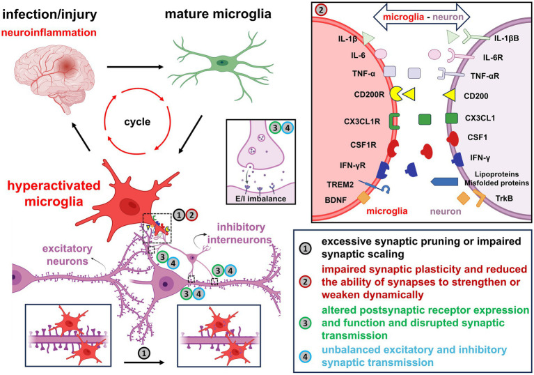
Verification
Claims the paper makes
The claims and statistics of this paper haven’t been checked yet.
Run the check — free, no account needed. It reads the open-access text and checks the sources it cites.
Claims other papers make about this one
Each is checked against this paper’s own text: does it say what the citing paper says it does?
The source backs part of the claim, not all of it — or backs it for a different population, condition or comparison.
In the citing paper
“This provides a plausible link between EDC-induced neuroinflammation and large-scale disturbances in connectivity and plasticity (Lara de Deus et al., 2025; Yang et al., 2025).”
From Environmental endocrine disruptors at the synapse: mechanisms linking chemical exposure to cognitive and behavioral dysfunction (2026) · cites this paper as Yang et al., 2025
Experimental — accuracy not yet measuredThe cited source says what the claim attributes to it.
In the citing paper
“Microglia are essential for synaptic pruning and circuit refinement during development, but chronic inflammatory activation can shift them toward maladaptive synapse elimination and aberrant remodeling.”
From Environmental endocrine disruptors at the synapse: mechanisms linking chemical exposure to cognitive and behavioral dysfunction (2026) · cites this paper as Yang et al., 2025
Self-check
Percentages, sums and reported differences re-computed from the paper’s own numbers.
The self-check needs the paper’s full text.
Citations
Cites
Fate Mapping Analysis Reveals That Adult Microglia Derive from Primitive Macrophages
Ginhoux, F., Greter, M., Leboeuf, M., Nandi, S., See, P., Gokhan, S., Mehler, M. F., Conway, S. J., Ng, L. G., Stanley, E. R., et al. (2010) · Science · doi:10.1126/science.1194637 · Cited by 5,175
Krasemann, S., Madore, C., Cialic, R., Baufeld, C., Calcagno, N., El Fatimy, R., Beckers, L., O’Loughlin, E., Xu, Y., Fanek, Z., et al. (2017) · Immunity · doi:10.1016/j.immuni.2017.08.008 · Cited by 2,979
Self-citationMicroglia states and nomenclature: A field at its crossroads
Paolicelli, R. C., Sierra, A., Stevens, B., Tremblay, M.-E., Aguzzi, A., Ajami, B., Amit, I., Audinat, E., Bechmann, I., Bennett, M., et al. (2022) · Neuron · doi:10.1016/j.neuron.2022.10.020 · Cited by 1,973
NLRP3 inflammasome activation drives tau pathology
Ising, C., Venegas, C., Zhang, S., Scheiblich, H., Schmidt, S. V., Vieira-Saecker, A., Schwartz, S., Albasset, S., McManus, R. M., Tejera, D., et al. (2019) · Nature · doi:10.1038/s41586-019-1769-z · Cited by 1,487
Immune modulation of learning, memory, neural plasticity and neurogenesis
Yirmiya, R., & Goshen, I. (2011) · Brain, Behavior, and Immunity · doi:10.1016/j.bbi.2010.10.015 · Cited by 1,483
Interleukin-6, a Major Cytokine in the Central Nervous System
Erta, M., Quintana, A., & Hidalgo, J. (2012) · International Journal of Biological Sciences · doi:10.7150/ijbs.4679 · Cited by 1,093
Unexpected role of interferon-γ in regulating neuronal connectivity and social behaviour
Filiano, A. J., Xu, Y., Tustison, N. J., Marsh, R. L., Baker, W., Smirnov, I., Overall, C. C., Gadani, S. P., Turner, S. D., Weng, Z., et al. (2016) · Nature · doi:10.1038/nature18626 · Cited by 697
Self-citationFilipello, F., Morini, R., Corradini, I., Zerbi, V., Canzi, A., Michalski, B., Erreni, M., Markicevic, M., Starvaggi-Cucuzza, C., Otero, K., et al. (2018) · Immunity · doi:10.1016/j.immuni.2018.04.016 · Cited by 695
Injured sensory neuron–derived CSF1 induces microglial proliferation and DAP12-dependent pain
Guan, Z., Kuhn, J., Wang, X., Colquitt, B. M., Solórzano, C., Vaman, S., Guan, A. K., Evans-Reinsch, Z., Braz, J. M., Devor, M. G., et al. (2015) · Nature Neuroscience · doi:10.1038/nn.4189 · Cited by 554
Self-citationOlmos-Alonso, A., Schetters, S. T. T., Sri, S., Askew, K., Mancuso, R., Vargas-Caballero, M., Holscher, C., Perry, V. H., & Gomez-Nicola, D. (2016) · Brain · doi:10.1093/brain/awv379 · Cited by 532
Research works printed but not linked: 2
- Han, 2016, Seizure induced synaptic plasticity alteration in hippocampus is mediated by IL-1β receptor through PI3K/Akt pathway, Am. J. Transl. Res., 15, 4499
- Ngwa, 2019, CD200-CD200R signaling and diseases: a potential therapeutic target, Int J Physiol Pathophysiol Pharmacol, 11, 297
Cited by
Sorted by citations
Rethinking early Alzheimer’s disease: An impaired internal model updating disorder?
Antonioni, A. (2026) · Neuroscience & Biobehavioral Reviews · doi:10.1016/j.neubiorev.2026.106790 · Cited by 8
Yoo, Y., Baek, S. Y., Lee, H., Lee, J., Lee, H., Lee, H., Ma, H., Kim, Y., Choi, H., Lee, J. T., et al. (2026) · Journal of Medicinal Chemistry · doi:10.1021/acs.jmedchem.5c02981 · Cited by 6
Ferreira, A. F. F., Santos-Silva, A. C., Muratori, B. G., & Britto, L. R. G. (2026) · Frontiers in Aging Neuroscience · doi:10.3389/fnagi.2026.1733682 · Cited by 5
Yen, C., & Chiang, M. (2026) · International Journal of Molecular Sciences · doi:10.3390/ijms27021080 · Cited by 4
Messina, A., Bella, F., Maccarone, G., Avincola, G., & Signorelli, M. S. (2026) · Journal of Psychiatric Research · doi:10.1016/j.jpsychires.2026.01.007 · Cited by 3
Kim, H., Vasquez, F. J. A., Bello, A. B., & Lee, S.-H. (2026) · Tissue and Cell · doi:10.1016/j.tice.2026.103401 · Cited by 3
Dual immune armies in glaucoma: microglia and monocyte-derived macrophages
Tan, K., Zhang, Y., Jiang, P., Yu, Y., Wu, Z. G., Peng, J. Q., Liu, P., & Peng, Q. (2026) · Frontiers in Immunology · doi:10.3389/fimmu.2026.1801497 · Cited by 3
The Microbiota–Gut–Brain Axis in Insomnia: Mechanisms and Intervention Strategies
Yang, M., Chen, Q., Meng, Z., Gu, X., & Bai, C. (2026) · Life · doi:10.3390/life16040583 · Cited by 3
Lipid metabolic regulation of neuroinflammation in Alzheimer’s disease
Li, T., Guo, K., Ma, Y., Zhao, J., Cao, Y., Zhang, R., Li, X., Wei, J., Ma, Y., Zhu, Z., et al. (2026) · Frontiers in Immunology · doi:10.3389/fimmu.2026.1815719 · Cited by 3
Zhu, T., Ye, M., Huang, C., Jiang, Y., Gu, Y., Cao, Z., Xu, F., Su, J., & Yang, R. (2026) · Neuropharmacology · doi:10.1016/j.neuropharm.2026.110893 · Cited by 2
Lee, B. M., Yoo, J.-R., & Jung, Y. (2026) · Cells · doi:10.3390/cells15080692 · Cited by 2
Targeting Pain and Depression in Alzheimer’s Disease: Translational Insights and Emerging Treatments
Costăchescu, I., Stanciu, G. D., Gogu, M.-R., & Tamba, B.-I. (2026) · Pharmaceuticals · doi:10.3390/ph19040626 · Cited by 2
Cui, D., Wang, X., & Shang, X. (2026) · Journal of Inflammation Research · doi:10.2147/jir.s606362 · Cited by 2
Self-citationde Souza, M. R. M., Lindner, M. R., Mladinich, M. C., Gorbunova, E. E., Kirillov, V., Byrne, A. G., Laird, A. Y., Finnerty, C. E., Ikeda, P., Vorkas, C. K., et al. (2026) · mBio · doi:10.1128/mbio.01294-26 · Cited by 2
Davtyan, H., Naguib, S. A., Voskobiynyk, Y., Chadarevian, J. P., Capocchi, J. K., Giacchino, J. L., Eskandari‐Sedighi, G., DeNittis, V. R., Ford, J. B., Agababian, A., et al. (2026) · Molecular Neurodegeneration · doi:10.1186/s13024-026-00978-6 · Cited by 2
Azimzadeh, M., & Azimzadeh, M. (2026) · Behavioural Brain Research · doi:10.1016/j.bbr.2026.116404 · Cited by 2
Jocsak, G., Kiss, D. S., Alymbaeva, D., Diebald, A., Zsarnovszky, A., Heinzlmann, A., & Bartha, T. (2026) · Frontiers in Behavioral Neuroscience · doi:10.3389/fnbeh.2026.1821381 · Cited by 1
Wang, Z., Yao, Y., Liu, N., Wei, T., Huang, X., Zou, L., Hu, Y., Fang, Q., Guan, S., Niu, Y., et al. (2025) · Small · doi:10.1002/smll.202510266 · Cited by 1
Self-citationHuang, C., Zhang, X., Yu, J., Xuan, C., Xu, C., Li, X., Cui, H., & Xu, D. (2026) · Neuropharmacology · doi:10.1016/j.neuropharm.2026.110992 · Cited by 1
Xu, L., Zhou, Y., Yang, H. K., Chen, X., & Ouyang, Q. (2025) · Experimental Neurology · doi:10.1016/j.expneurol.2025.115596 · Cited by 2
Low-Dose Naltrexone in Chronic Pain Management: Mechanisms, Evidence, and Clinical Implications
McKenzie, A., Bittar, T. G., Dombrower, R., Raman, D., Hussain, H., Theeraphapphong, N., McKenzie, S. M., & Abd-Elsayed, A. A. (2026) · Journal of Personalized Medicine · doi:10.3390/jpm16030151 · Cited by 1
Chen, W., Wang, X., Zhang, Y., Sun, P., Jiang, Y., Shen, J. S., Xu, T., Shen, J., Ke, K., & Gu, X. (2026) · Neural Regeneration Research · doi:10.4103/nrr.nrr-d-25-00608 · Cited by 2
Self-citationZhang, H., Yu, J., Sheng, R., Zhang, S., Chen, L., & Wang, H. Y. (2026) · Frontiers in Marine Science · doi:10.3389/fmars.2026.1832655 · Cited by 1
Fraer, A., Saleh, S., Tasabehji, D., Sharma, D. S., & Mokadem, M. (2026) · Frontiers in Human Neuroscience · doi:10.3389/fnhum.2026.1857087 · Cited by 1
Fawzy, M. N. (2026) · Inflammopharmacology · doi:10.1007/s10787-026-02363-7 · Cited by 1
Histone Deacetylase Inhibitors Improve Memory Function and Decrease Neuropathology in APP/PS1 MicePreprint
McClarty, B. M., Monteiro, R., & Dong, H. (2026) · Research Square · doi:10.21203/rs.3.rs-10816126/v1 · Cited by 1
Berdasco, C., Cho, W.-H., Lee, J., Annu, A., Ledo, J. H., Jeong, Y. Y., Kim, Y.-S., An, S. S., Tanaka, Y., Ahn, J.-H., et al. (2026) · bioRxiv (Cold Spring Harbor Laboratory) · doi:10.64898/2026.01.14.699391
Infectious vs. Sterile Neuroinflammation: Differential Consequences on Neuronal CircuitryPreprint
Talero-López, D. (2026) · Qeios · doi:10.32388/1h63qt
PDCD4: A Double-Edged Sword in Neurological Diseases
Li, C., Zheng, K., Qiu, M. S., & Yang, J. (2026) · Molecular Neurobiology · doi:10.1007/s12035-026-05768-1
Bridging Regenerative and Restorative Stem Cell Therapies in Parkinson’s Disease
Onuigbo, C. C., Martínez-Lemus, J. D., Tharp, E., & Schiess, M. C. (2026) · Cells · doi:10.3390/cells15070578
Joo, J. D., Yang, W. S., & Koh, H. J. (2026) · International Journal of Molecular Sciences · doi:10.3390/ijms27073307
Mopuru, R., Elliott, B. L., Hoffman, L. J., Tani, N., Kring, A. M., Breen, E. C., Cohn, B. A., Cirillo, P. M., Krigbaum, N. Y., D’Esposito, M., et al. (2026) · bioRxiv (Cold Spring Harbor Laboratory) · doi:10.64898/2026.04.06.716489
Çağlar, Ö. (2026) · Molecular Biology Reports · doi:10.1007/s11033-026-11811-0
Greco, D., Das, D., Novotný, J., Olsen, M. J., & Telenský, P. (2026) · Scientific Reports · doi:10.1038/s41598-026-47881-0
Advancing Alzheimer’s Disease Research Using Brain Organoids and Organ-on-Chip Technologies
Singh, S. (2026) · American journal of student research. · doi:10.70251/hyjr2348.42413419
Liu, Q., Wang, Y., Wang, B. Q., Wang, J., Zhang, M., Liu, Y., & Min, X. (2026) · Mediators of Inflammation · doi:10.1155/mi/8871660
Bhamare, J. S., & Kaur, G. (2026) · Network Modeling Analysis in Health Informatics and Bioinformatics · doi:10.1007/s13721-026-00793-5
Neuroimmune Dysregulation and the Role of IL-10 in Depression: A Systematic Review
Cortes-Altamirano, J. L., Alfaro-Rodríguez, A., González-Maciel, A., Pérez-Guillé, B., Soriano‐Rosales, R. E., Jaime, H. B., Ávila-Luna, A., Bueno-Nava, A., Sánchez-Aparicio, P., & Dotor-Llerena, A. L. (2026) · Brain Sciences · doi:10.3390/brainsci16060548
Saifi, M. A., Javed, M., Jindal, G., Agarwal, N. B., & Raisuddin, S. (2026) · Naunyn-Schmiedeberg s Archives of Pharmacology · doi:10.1007/s00210-026-05445-z
Gnoupa, K.-G. K., Ngoupaye, G. T., Foutsop, A. F., Kom, B. T. D., Ngoufack, S. B. K., Noubouwo, J. C., & Diane, R. B. (2026) · IBRO Neuroscience Reports · doi:10.1016/j.ibneur.2026.05.008
Khene, M. A., Belabbassi, O., Chabane, K., Kechkoul, L., Ousmaal, M. E. F., & Benaziza, D. (2026) · International Journal of Computational and Experimental Science and Engineering · doi:10.22399/ijcesen.5294
Yao, M., Liu, J., Dong, Y., Li, M., Yuan, Y., Wang, B., Liu, W., Zhang, B., Dong, R., Sun, X., et al. (2026) · Scientific Reports · doi:10.1038/s41598-026-57741-6
Li, J., Ma, X., Liu, J., Xu, W., Zhang, X., Liang, F., & Dou, Y. (2026) · Psychoradiology · doi:10.1093/psyrad/kkag023
Gu, D., & Yuan, G. (2026) · Immuno · doi:10.3390/immuno6030042
Hoehne, C. L., Salinas, V. H., Shirani, A., Stüve, O., & Stopschinski, B. E. (2026) · medRxiv · doi:10.64898/2026.06.27.26355593
Hammad, M. Y., Fardoos, S., & Shakoor, K. (2026) · Exploration of Neuroprotective Therapy · doi:10.37349/ent.2026.1004162
Endogenous Neurotoxicity: A Pathophysiological Consequence of Homeostatic Dysfunction
Yanglem, S., Loushambam, B., Singh, S. M., & Vijayaraghavalu, S. (2026) · Neuroglia · doi:10.3390/neuroglia7030029
Colostrum Extracellular Vesicle Isolation, Characterization, and Function
Akter, S., Fayez, N., Kumar, M., Sil, S., & Gendelman, H. E. (2026) · Biomedicines · doi:10.3390/biomedicines14071549
Microglial synaptic pruning in early Alzheimer’s disease: emerging roles of the IL-1β-NLRP3 axis
Kaushik, A. S., & Singh, N. (2026) · Inflammopharmacology · doi:10.1007/s10787-026-02335-x
The drug discovery landscape of Alzheimer’s disease
Chinnathambi, S., & Prakash, L. E. (2026) · Advances in protein chemistry and structural biology · doi:10.1016/bs.apcsb.2026.07.005
Chemokine levels in the murine brain from early postnatal development to aging
Dantas, D. P., Campos, R. M. P., Galvani, R. G. A., Bonomo, A., & Pimentel‐Coelho, P. M. (2026) · Brain Behavior & Immunity - Health · doi:10.1016/j.bbih.2026.101308
Infectious vs. Sterile Neuroinflammation: Differential Consequences on Neuronal CircuitryPreprint
Talero-López, D. (2026) · Qeios · doi:10.32388/1h63qt.3
Chakraborty, W., Anand, A., & Roy, K. K. (2026) · Progress in Neuro-Psychopharmacology and Biological Psychiatry · doi:10.1016/j.pnpbp.2026.111846
Chen, J., & Chen, L.-H. (2026) · Current Opinion in Pharmacology · doi:10.1016/j.coph.2026.102650
Brito, A., Carneiro, B. D., & Pozza, D. H. (2026) · Pathophysiology · doi:10.3390/pathophysiology33030054
Hillal, J. P. D., Weber, T. A., Staub, H. L., & Bauer, M. E. (2026) · Autoimmunity Reviews · doi:10.1016/j.autrev.2026.104144
Xiao, X., Shen, S., Cui, T., Tan, H., Hu, S., Deng, X., Huang, Y., Cui, C., Yu, B., Qiu, W., et al. (2026) · Journal of Translational Medicine · doi:10.1186/s12967-026-08735-5
Self-citationBungarus multicinctus venom induces pyroptosis via the NLRP3/Caspase-1/GSDMD pathway in BV2 cells
Luo, M., Wang, Z.-R., Tan, X.-L., Yang, X., & Ning, Z. (2026) · Cell Biology and Toxicology · doi:10.1007/s10565-026-10240-w
Aditi, Blackwell, T. A., Fang, X., Sharma, S., Mendoza, M., LANDAY, A. L., Golovko, G., & Samir, P. (2026) · Journal of Neurology · doi:10.1007/s00415-026-14017-3
Targeting the Complement–Microglia Axis for Neuroprotection in Pediatric Epilepsy
Karayanni, M., Mitsoudis, N., Vanakliotou, M., Bakirtzis, C., Kesidou, E., Polyzoidou, E., & Liana, E. (2026) · Biomedicines · doi:10.3390/biomedicines14081788
Neural glial network instability in epilepsy
Naffaa, M. M. (2026) · Exploration of neuroscience · doi:10.37349/en.2026.1006144
Yang, E., Lin, H., Alvarez, J., Russo, S. J., Toro-Chacon, C., & Pasinetti, G. M. (2026) · Research Square · doi:10.21203/rs.3.rs-10425197/v1
Wang, Y., Li, P., Dong, Y., Zhang, X., & Shang, K. (2026) · Frontiers in Immunology · doi:10.3389/fimmu.2026.1934162
Liu, Y., Yu, H., Zhang, J., Li, X., Ma, Y., Guang, C., Mu, W., Chen, H., & Hou, H. (2026) · Frontiers in Immunology · doi:10.3389/fimmu.2026.1901868
Vallés, A. S., & Barrantes, F. J. (2026) · Frontiers in Neurology · doi:10.3389/fneur.2026.1860052
Mamedova, D. I., Nedogreeva, O. A., Aniol, V. A., Kvichansky, A. A., Tret’yakova, L. V., Riga, V. D., Лазарева, Н. А., Gulyaeva, N. V., & Stepanichev, M. Y. (2026) · Neurochemical Research · doi:10.1007/s11064-026-04889-2
Aslanlar, D. A., Atalık, K. E. N., Öz, M., Yıldız, İ., & Yılmaz, M. T. (2026) · Neurochemical Research · doi:10.1007/s11064-026-04883-8
Алексеевич, П. М., Skovorodko, P., Dorchenko, E., Milorava, G., Ivanova, E., Aigubova, Z., & Sarkisova, M. (2026) · Scripta Medica · doi:10.5937/scriptamed57-67911
Choi, G.-Y., Lee, S., Je, A. R., Choi, H., Moon, E., Cho, E., Kwon, S., Lee, K., & Kweon, H. (2026) · Phytomedicine · doi:10.1016/j.phymed.2026.158788
Li, S., Wang, S., Ren, Y., & Yan, J. (2026) · Journal of Multidisciplinary Healthcare · doi:10.2147/jmdh.s637471
Yu, Y., Chen, M.-H., Ding, S., Zhang, G., Kong, L., Liu, Y., Zang, J., Zheng, Y., Zhang, Y.-N., Li, X., et al. (2026) · Chemical Engineering Journal · doi:10.1016/j.cej.2026.182033
Li, J., Wang, Y., Cai, W., Chen, S., Zhang, L., & Wang, T. (2026) · Frontiers in Neuroscience · doi:10.3389/fnins.2026.1850015
Gao, J., Xie, R., Rao, C., Jiao, J., Zhang, W., Hu, Y., Yang, B., Zhao, H., Yuan, P., Xie, T., et al. (2026) · Journal of International Medical Research · doi:10.1177/03000605261483860
Si, J., Zhu, M., & Mingzhe, D. (2026) · Brain and Behavior · doi:10.1002/brb3.71777
Cystatin F as a neuroimmune effector in CNS inflammation and neurodegeneration
Murray, K., Fernandez, K., & Lane, T. E. (2026) · Frontiers in Cell and Developmental Biology · doi:10.3389/fcell.2026.1959727
Neuroplasticity under constraint: From sensory ecology to neuroinflammation
Diniz, D. G., Bento-Torres, N. V. O., Bento‐Torres, J., Jardim, N. Y. V., Sosthenes, M. C. K., Krejčová, L. V., dos Santos, Z. A., de Sousa, A. A., da Costa, E. R., Pereira, P. D. C., et al. (2026) · Brain Behavior and Immunity Integrative · doi:10.1016/j.bbii.2026.100199
Gabr, R. I. (2026) · Developmental Neurobiology · doi:10.1002/dneu.70063
Liu, X., Song, X., Zhang, J., Ma, S., Wang, X., Guo, Y., Gao, B., Wang, X., & Duan, J. (2026) · Journal of Ethnopharmacology · doi:10.1016/j.jep.2026.122426
Self-citationGlia-Informed Design of Smart Biomimetic Biomaterials for Brain Repair and Neural Tissue Engineering
Naffaa, M. M. (2026) · European Cells and Materials · doi:10.22203/ecm.v059a02
Targeting glial modulation: molecular mechanisms of Polygonatum in neurological disorders
Liu, Z., Luo, Y., Zheng, C., Long, X., Zhao, Y., Pan, Z., Xu, B., & Sun, J. (2026) · Frontiers in Pharmacology · doi:10.3389/fphar.2026.1969384
Self-citationAyyubova, G., Gasimov, E. K., Rzayev, F., Mehraliyeva, G., Sadigi, I., Guliyeva, N., Ismayilova, S. H., & Ayyubova, N. (2026) · Scientific Reports · doi:10.1038/s41598-026-73800-4
NF-ΚB-ASSOCIATED INFLAMMATION AND COGNITIVE IMPAIRMENT IN SCHIZOPHRENIA: A REVIEW
Mahardika, A., Tanra, A. J., & Limo, E. (2026) · Acta Innovations · doi:10.62441/actainnovations.v63i.794
Han, Z., Cheng, F., Wang, Y., Zhang, H., Wang, L., & Zhao, C. (2026) · Frontiers in Immunology · doi:10.3389/fimmu.2026.1920083
Chen, H., Yong, H., Hou, L., Liu, L., & Ma, C. (2026) · NeuroImage · doi:10.1016/j.neuroimage.2026.122267
Liu, F., Xu, T.-S., Zhao, J.-Z., Xu, P., Jiang, P.-P., Xu, Q., Liu, L., & Dai, Q. (2026) · Natural Product Research · doi:10.1080/14786419.2026.2738221
Sharoar, M. G., Zaman, Z., Singh, N., Fathi, E., Graham, S. F., Zhu, X., & Homayouni, R. (2026) · npj Aging · doi:10.1038/s41514-026-00529-9
Lekshmi, S. S. A., Palathingal, M., Manikkath, J., Sarath Chandran, C., & Nampoothiri, M. (2026) · Journal of Drug Delivery Science and Technology · doi:10.1016/j.jddst.2026.109009
Cognitive Impairment in Asthma: Emerging Evidence, Clinical Consequences, and Future Directions
Mengash, A. S., & Siraj, R. A. (2026) · Journal of Clinical Medicine · doi:10.3390/jcm15197761
Glial lipid reprogramming in brain aging: Lipid identity, organelle stress, and redox vulnerability
Rana, M., Ko, W., Nepali, K., Ko, C.-Y., Yen, T., Jan, J., Taliyan, R., Li, C.-H., & Yang, C. (2026) · Redox Biology · doi:10.1016/j.redox.2026.104453
Liu, Y., Guo, C., & Yu, X. (2026) · International Journal of Molecular Sciences · doi:10.3390/ijms27219431
Wang, D., Zhigang, Z., & Ding, Y. (2026) · Acta Pharmaceutica · doi:10.2478/acph-2026-0023
Role of macrophages in immunometabolism and neurodegeneration
Mishra, P., Khan, A., Doshi, Z., Tungare, K., & Nand, S. (2026) · Elsevier eBooks · doi:10.1016/b978-0-443-44253-7.00018-7
Samaila, S., Christopher, C. H., Williams, I. K., Michael, J. C., Adedayo, L. D., & Danborno, A. M. (2026) · Research Square · doi:10.21203/rs.3.rs-11224606/v1
Metadata
- Authors
- Yang, G., Xu, X., Gao, W., Wang, X., Zhao, Y., & Xu, Y.
- Affiliations
- Shanghai University of Traditional Chinese Medicine (CN)
- Corresponding author
- Zhao, Yan
- Xu, Ying
- Venue
- Frontiers in Cellular Neuroscience
- Publisher
- Frontiers Media SA
- Published
- 2025-11-10
- Volume
- 19
- Pages
- 1700692-1700692
- ISSN
- 1662-5102 (ISSN-L)
- PMC
- PMC12641016
- OpenAlex
- W4416096320
- Cited by
- 85
- References
- 234
- Citations by year
- 2026: 83 · 2025: 2
- FWCI
- 8.24
- Type
- journal article
- Language
- en
- Open access
- gold
- doi.org
- Licence
- Locations
- Frontiers in Cellular Neuroscience (publishedVersion, cc-by)
- Europe PMC (PubMed Central) (publishedVersion, cc-by)
- PubMed (publishedVersion)
- DOAJ (DOAJ: Directory of Open Access Journals) (submittedVersion, cc-by-sa)
- Funders
- National Natural Science Foundation of China
- China Postdoctoral Science Foundation
- Topics
- Life Sciences › Neuroscience › Neurology › Neuroinflammation and Neurodegeneration Mechanisms
- Life Sciences › Neuroscience › Cellular and Molecular Neuroscience › Neuroscience and Neuropharmacology Research
- Life Sciences › Neuroscience › Neurology › Neurological Disease Mechanisms and Treatments
- Keywords
- microglia, neuroinflammation, synaptic remodeling, inflammatory cytokines, neurodegeneration, IL-1β, interleukin-6, TNF-α, CX3CL1/CX3CR1, excitotoxicity, learning disabilities, neurodegenerative diseases
About these checks
Registry, retraction, Cites and statistics results are computed, not judged, with one exception: a printed reference that no index lists is read once by a model before it counts as a citation error. Claims are extracted by a model and kept only when their quote appears verbatim in the text; each cited source is then checked, with a model’s help, for whether it says what the claim relies on. Those verdicts can be wrong — read the claim’s quote and the cited source before relying on one.
Statistics and arithmetic are recomputed, not judged.
Confidence bands are shown only for fields calibrated on our annotated test set. None is calibrated yet, so no band is shown.
What counts as a claim ›How we measure confidence ›How we check this ›
Are you an author, or do you see a problem with this page? Request a review
