

The DOI matches its Crossref record; 80 of the 81 references it cites were found in a registry or an index; none of the 43 sources its claims cite is checked for support yet.
Brain mechanisms underlying the impact of attachment-related stress on social cognition
Open accessAbstract
Mentalizing, in particular the successful attribution of complex mental states to others, is crucial for navigating social interactions. This ability is highly influenced by external factors within one's daily life, such as stress. We investigated the impact of stress on the brain basis of mentalization in adults. Using a novel modification of the Reading the Mind in the Eyes Test (RMET-R) we compared the differential effects of two personalized stress induction procedures: a general stress induction (GSI) and an attachment-related stress induction (ASI). Participants performed the RMET-R at baseline and after each of the two inductions. Baseline results replicated and extended previous findings regarding the neural correlates of the RMET-R. Additionally, we identified brain regions associated with making complex age judgments from the same stimuli. Results after stress exposure showed that the ASI condition resulted in reduced mentalization-related activation in the left posterior superior temporal sulcus (STS), left inferior frontal gyrus and left temporoparietal junction (TPJ). Moreover, the left middle frontal gyrus and left anterior insula showed greater functional connectivity to the left posterior STS after the ASI. Our findings indicate that attachment-related stress has a unique effect on the neural correlates of mentalization.
Overview
AI snapshot
Written from the paper’s full text.
TL;DR
This fMRI study compared attachment-related stress induction (ASI) with general stress induction (GSI) on mentalization using a revised Reading the Mind in the Eyes Test. ASI reduced mental-state accuracy and decreased activation in left posterior STS, left IFG, and left TPJ, with altered left posterior STS functional connectivity.
Key findings
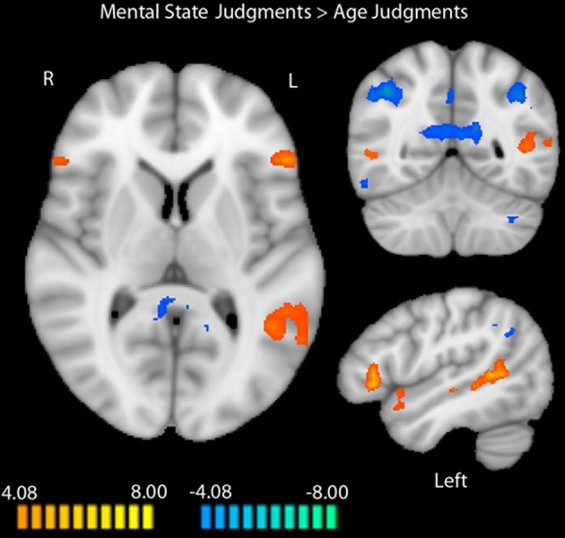
- At baseline, mental state > age judgments activated bilateral IFG (right t = 6.75; left t = 10.04), right posterior STS (t = 5.43), left posterior STS (t = 8.56), and dorsoMPFC (t = 6.51), among other regions; age > mental state judgments activated bilateral inferior parietal lobules, PCC/precuneus, right DLPFC, right inferior temporal gyrus, and left cerebellum.
- Mental-state correct responses differed across conditions (F(2,24) = 3.59, p = 0.04), with lowest accuracy post-ASI (67.03%) vs baseline (74.70%) and post-GSI (74.98%); baseline vs post-ASI t(12) = 2.30, p < 0.05, and post-GSI vs post-ASI t(12) = 2.20, p < 0.05. Mental-state reaction times also differed (F(2,24) = 10.13, p = 0.01), with longest at baseline and shortest post-ASI. Age judgments showed no significant differences.
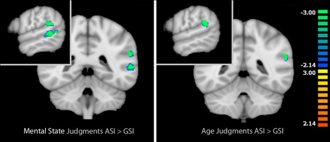
- Post-ASI vs post-GSI during mental-state judgments showed reduced activation in left IFG (t = −2.93, p = 0.011; did not survive cluster correction), left posterior STS (t = −5.02, p < 0.001), and left TPJ (t = −4.83, p < 0.001). During age judgments, only left TPJ differed (t = −5.08, p < 0.001).
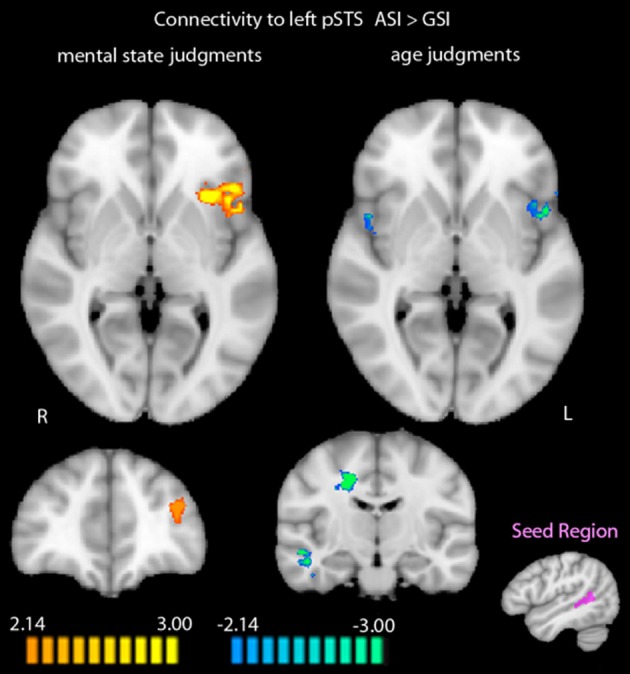
- After ASI vs GSI, mental-state judgments showed greater functional connectivity to left posterior STS in left MFG (t = 4.90, p < 0.001) and left anterior insula (t = 5.24, p < 0.001). During age judgments, cuneus showed greater post-ASI connectivity, while bilateral anterior insula, right MTG/PHG/FFG, right dorsal ACC, and left FFG showed greater post-GSI connectivity.
Methods
- Eighteen healthy right-handed adults were recruited; 15 remained for fMRI analysis (8 male; mean age 20.73 years, SD = 1.20, range 18–23), and behavioral analyses included 13 participants. Participants completed three fMRI runs: baseline, post-GSI, and post-ASI, with order balanced across participants. Personalized stress imagery scripts were created from recalled events rated 8 or above on a 10-point perceived stress scale.
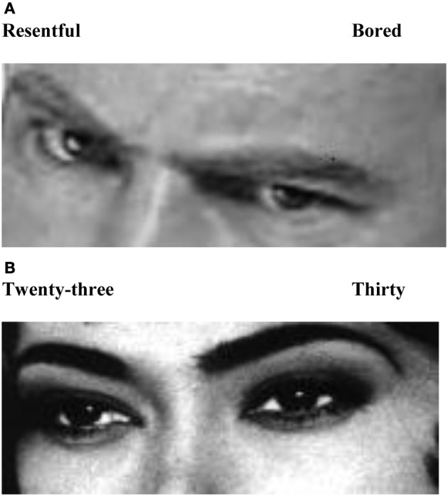
- The task was a revised RMET (RMET-R) with mental-state and age-judgment blocks; stimuli were shown for 3500 ms with 500 ms fixation, six trials per block, four blocks per task per condition. fMRI data were analyzed in Brain Voyager QX using random-effects GLMs; baseline mental state vs age contrast used FDR q < 0.05 with clusters ≥ 4 voxels. Stressor comparisons used a baseline-derived mask and paired t-tests (p < 0.05, cluster ≥ 4 voxels); PPI connectivity used a left posterior STS seed with cluster threshold 34 voxels.
Limitations
- The paper states: the sample was relatively small (15 fMRI, 13 behavioral, 12 overlapping), preventing direct behavior–fMRI correlations and gender-difference analyses; the age control task’s difficulty/cognitive load was not well quantified; the sample lacked ethnic and developmental diversity; no psychophysiological indices such as heart rate variability or cortisol were reported; and the design cannot rule out other induced emotions, general social-script effects, positive-script priming, or whether effects are due to social vs non-social stressor nature or individual attachment differences.
- The text also makes evident that the left IFG stress effect did not survive cluster correction, and behavioral data were lost for three participants due to software errors.
Model-written summary. Check it against the paper before relying on it.
Integrity signals
Registry
Cites
Statistics
Figures
Verification
Claims the paper makes
23 Own result · 22 Not checked yet · 17 Own method · 11 Own interpretation · 3 No basis given
A result the paper reports from its own data — it cites no other source for it, so there is none to check. The quote shows it is in the paper.
In the paper
“Scores for subjective stress experience (Wolpe, 1969) were obtained again at the beginning of the debriefing after scanning and showed no difference in mean scores when comparing both stress conditions in a paired samples t-test [t(14) = 1.07, p = 0.34].”
§ Brain mechanisms underlying the impact of attachment-related stress on social cognition > Methods > Experimental design
plain- Asserted: passes — The authors put it forward as true.
- Checkable: passes — It can be checked against evidence.
- Load-bearing: unclear — It is unclear whether the paper’s conclusions depend on it.
- Based: passes — The paper gives a reason why it holds.
A result the paper reports from its own data — it cites no other source for it, so there is none to check. The quote shows it is in the paper.
In the paper
“Results after stress exposure showed that the ASI condition resulted in reduced mentalization-related activation in the left posterior superior temporal sulcus (STS), left inferior frontal gyrus and left temporoparietal junction (TPJ).”
§ Brain mechanisms underlying the impact of attachment-related stress on social cognition > Abstract
plaindirect- Asserted: passes — The authors put it forward as true.
- Checkable: passes — It can be checked against evidence.
- Load-bearing: passes — The paper’s conclusions depend on it.
- Based: passes — The paper gives a reason why it holds.
A result the paper reports from its own data — it cites no other source for it, so there is none to check. The quote shows it is in the paper.
In the paper
“A random-effects whole-brain GLM analysis identified a left posterior STS region (136 functional voxels) with greater activation to mental state vs. age judgments during the baseline RMET-R.”
§ Brain mechanisms underlying the impact of attachment-related stress on social cognition > Methods > Data analysis
plaincorrelational- Asserted: passes — The authors put it forward as true.
- Checkable: passes — It can be checked against evidence.
- Load-bearing: passes — The paper’s conclusions depend on it.
- Based: passes — The paper gives a reason why it holds.
A result the paper reports from its own data — it cites no other source for it, so there is none to check. The quote shows it is in the paper.
In the paper
“We identified a network of brain regions that responds differentially to age and mental state judgments during the RMET-R in the absence of any pre-task stress induction (Figure 1).”
§ Brain mechanisms underlying the impact of attachment-related stress on social cognition > Results > fMRI
plaincorrelational- Asserted: passes — The authors put it forward as true.
- Checkable: passes — It can be checked against evidence.
- Load-bearing: unclear — It is unclear whether the paper’s conclusions depend on it.
- Based: passes — The paper gives a reason why it holds.
The paper states this without a source or data behind it here, so there is nothing to check it against. The quote shows it is in the paper.
In the paper
“Baseline results replicated and extended previous findings regarding the neural correlates of the RMET-R.”
§ Brain mechanisms underlying the impact of attachment-related stress on social cognition > Abstract
plaincorrelational- Asserted: passes — The authors put it forward as true.
- Checkable: passes — It can be checked against evidence.
- Load-bearing: unclear — It is unclear whether the paper’s conclusions depend on it.
- Based: passes — The paper gives a reason why it holds.
A result the paper reports from its own data — it cites no other source for it, so there is none to check. The quote shows it is in the paper.
In the paper
“Moreover, the left middle frontal gyrus and left anterior insula showed greater functional connectivity to the left posterior STS after the ASI.”
§ Brain mechanisms underlying the impact of attachment-related stress on social cognition > Abstract
plaincorrelational- Asserted: passes — The authors put it forward as true.
- Checkable: passes — It can be checked against evidence.
- Load-bearing: passes — The paper’s conclusions depend on it.
- Based: passes — The paper gives a reason why it holds.
A result the paper reports from its own data — it cites no other source for it, so there is none to check. The quote shows it is in the paper.
In the paper
“Regions that showed more activation to age judgments included bilateral inferior parietal lobules (right: t = −11.76, p < 0.0001; left: t = −7.73, p < 0.0001), right middle frontal gyrus (MFG; t = −5.92, p < 0.0001), PCC extending into precuneus (t = −10.14, p < 0.0001), right dorsolateral prefrontal cortex (t = −6.19, p < 0.0001), right inferior temporal gyrus (t = −9.72, p < 0.0001), and left c”
§ Brain mechanisms underlying the impact of attachment-related stress on social cognition > Results > fMRI
plaincorrelational- Asserted: passes — The authors put it forward as true.
- Checkable: passes — It can be checked against evidence.
- Load-bearing: unclear — It is unclear whether the paper’s conclusions depend on it.
- Based: passes — The paper gives a reason why it holds.
A result the paper reports from its own data — it cites no other source for it, so there is none to check. The quote shows it is in the paper.
In the paper
“In sum, using a novel modification of the RMET, we identified the left posterior STS and left IFG as brain regions that were modulated by attachment stressors during mental state judgments accompanying compromised behavioral performance, as well as the left TPJ, which showed an effect of stressor type during both mental state and age judgments.”
§ Brain mechanisms underlying the impact of attachment-related stress on social cognition > Discussion > Conclusion
plaincorrelational- Asserted: passes — The authors put it forward as true.
- Checkable: passes — It can be checked against evidence.
- Load-bearing: passes — The paper’s conclusions depend on it.
- Based: passes — The paper gives a reason why it holds.
A result the paper reports from its own data — it cites no other source for it, so there is none to check. The quote shows it is in the paper.
In the paper
“Again, no regions were more active during age judgments after the ASI compared with the GSI.”
§ Brain mechanisms underlying the impact of attachment-related stress on social cognition > Results > fMRI
plain- Asserted: passes — The authors put it forward as true.
- Checkable: passes — It can be checked against evidence.
- Load-bearing: unclear — It is unclear whether the paper’s conclusions depend on it.
- Based: passes — The paper gives a reason why it holds.
A result the paper reports from its own data — it cites no other source for it, so there is none to check. The quote shows it is in the paper.
In the paper
“When we compared functional connectivity to the functionally-defined left posterior STS during mental state judgments after the two stress inductions, regions that showed greater functional connectivity to the left posterior STS after the ASI included the left MFG (t = 4.90, p < 0.001) and left anterior insula (AI; t = 5.24, p < 0.001).”
§ Brain mechanisms underlying the impact of attachment-related stress on social cognition > Results > fMRI
plaincorrelational- Asserted: passes — The authors put it forward as true.
- Checkable: passes — It can be checked against evidence.
- Load-bearing: passes — The paper’s conclusions depend on it.
- Based: passes — The paper gives a reason why it holds.
A result the paper reports from its own data — it cites no other source for it, so there is none to check. The quote shows it is in the paper.
In the paper
“Bilateral AI (right: t = −3.94, p < 0.01; left: t = −5.66, p < 0.001), right middle temporal gyrus extending into parahippocampal gyrus and fusiform gyrus (t = −5.37, p < 0.001), right dorsal anterior cingulate cortex (t = −6.28, p < 0.001), and left fusiform gyrus (t = −4.58, p < 0.001) were more functionally connected to the left posterior STS during age judgments post-GSI vs. post-ASI.”
§ Brain mechanisms underlying the impact of attachment-related stress on social cognition > Results > fMRI
plaincorrelational- Asserted: passes — The authors put it forward as true.
- Checkable: passes — It can be checked against evidence.
- Load-bearing: passes — The paper’s conclusions depend on it.
- Based: passes — The paper gives a reason why it holds.
A result the paper reports from its own data — it cites no other source for it, so there is none to check. The quote shows it is in the paper.
In the paper
“When we compared activation during mental state judgments between the two types of stress inductions in the identified task-related areas, we localized regions that were more active during mental state judgments after the GSI compared with the ASI. These regions included the left IFG (t = −2.93, p < 0.05), left posterior STS (t = −5.02, p < 0.001), and left temporal parietal junction (t = −4.83, p”
- Also stated in: § fMRI · plain
“Thus, post-ASI, activation in these three regions decreased significantly in comparison to post-GSI.”
§ Brain mechanisms underlying the impact of attachment-related stress on social cognition > Results > fMRI
plain- Asserted: passes — The authors put it forward as true.
- Checkable: passes — It can be checked against evidence.
- Load-bearing: passes — The paper’s conclusions depend on it.
- Based: passes — The paper gives a reason why it holds.
- Also stated in: § fMRI · plain
A result the paper reports from its own data — it cites no other source for it, so there is none to check. The quote shows it is in the paper.
In the paper
“No regions were more functionally connected to the left posterior STS during mental state judgments post-GSI.”
§ Brain mechanisms underlying the impact of attachment-related stress on social cognition > Results > fMRI
plain- Asserted: passes — The authors put it forward as true.
- Checkable: passes — It can be checked against evidence.
- Load-bearing: passes — The paper’s conclusions depend on it.
- Based: passes — The paper gives a reason why it holds.
A result the paper reports from its own data — it cites no other source for it, so there is none to check. The quote shows it is in the paper.
In the paper
“There were no regions that were more active during mental state judgments after the ASI compared with the GSI.”
§ Brain mechanisms underlying the impact of attachment-related stress on social cognition > Results > fMRI
plain- Asserted: passes — The authors put it forward as true.
- Checkable: passes — It can be checked against evidence.
- Load-bearing: passes — The paper’s conclusions depend on it.
- Based: passes — The paper gives a reason why it holds.
A result the paper reports from its own data — it cites no other source for it, so there is none to check. The quote shows it is in the paper.
In the paper
“The cuneus showed increased functional connectivity to the left posterior STS following the ASI compared with the GSI (t = 3.78, p < 0.01).”
§ Brain mechanisms underlying the impact of attachment-related stress on social cognition > Results > fMRI
plaindirect- Asserted: passes — The authors put it forward as true.
- Checkable: passes — It can be checked against evidence.
- Load-bearing: passes — The paper’s conclusions depend on it.
- Based: passes — The paper gives a reason why it holds.
A result the paper reports from its own data — it cites no other source for it, so there is none to check. The quote shows it is in the paper.
In the paper
“The same analysis within age judgments identified only one region that was more active during age judgments after the GSI compared with the ASI: the left TPJ (t = −5.08, p < 0.001).”
§ Brain mechanisms underlying the impact of attachment-related stress on social cognition > Results > fMRI
plain- Asserted: passes — The authors put it forward as true.
- Checkable: passes — It can be checked against evidence.
- Load-bearing: passes — The paper’s conclusions depend on it.
- Based: passes — The paper gives a reason why it holds.
None of the sources cited for this claim have been checked yet.
In the paper
“Indeed, implicit mentalization, defined as reflexive, automatic processing of mental states that does not require conscious, verbal efforts, engages the fusiform face area, STS, inferior frontal gyrus (IFG), and premotor areas (Allison et al., 2000; Pelphrey et al., 2003; Vuilleumier and Pourtois, 2007; Iacoboni, 2009; Vander Wyk et al., 2009).”
§ Brain mechanisms underlying the impact of attachment-related stress on social cognition > Introduction
What the sources say
- Not checked yetcites Allison et al., 2000
- Not checked yetcites Pelphrey et al., 2003
- Not checked yetcites Vuilleumier and Pourtois, 2007
plaindirect- Asserted: passes — The authors put it forward as true.
- Checkable: passes — It can be checked against evidence.
- Load-bearing: unclear — It is unclear whether the paper’s conclusions depend on it.
- Based: passes — The paper gives a reason why it holds.
None of the sources cited for this claim have been checked yet.
In the paper
“The neural system that underpins mentalizing comprises the medial prefrontal cortex (MPFC), temporoparietal junction (TPJ), temporal poles, superior temporal sulcus (STS), and posterior cingulate cortex (PCC)/precuneus (Gallagher and Frith, 2003; Saxe and Kanwisher, 2003; Frith and Frith, 2006; Lieberman, 2007).”
§ Brain mechanisms underlying the impact of attachment-related stress on social cognition > Introduction
What the sources say
- Not checked yetcites Gallagher and Frith, 2003
- Not checked yetcites Saxe and Kanwisher, 2003
- Not checked yetcites Frith and Frith, 2006
plain- Asserted: passes — The authors put it forward as true.
- Checkable: passes — It can be checked against evidence.
- Load-bearing: passes — The paper’s conclusions depend on it.
- Based: passes — The paper gives a reason why it holds.
None of the sources cited for this claim have been checked yet.
In the paper
“Previous research supports the latter hypothesis, as associations have been reported between childhood parental maltreatment and impaired mentalization (Cicchetti et al., 2003; Pears and Fisher, 2005).”
§ Brain mechanisms underlying the impact of attachment-related stress on social cognition > Introduction
What the sources say
- Not checked yetcites Cicchetti et al., 2003
- Not checked yetcites Pears and Fisher, 2005
plaincorrelational- Asserted: passes — The authors put it forward as true.
- Checkable: passes — It can be checked against evidence.
- Load-bearing: passes — The paper’s conclusions depend on it.
- Based: passes — The paper gives a reason why it holds.
None of the sources cited for this claim have been checked yet.
In the paper
“As expected, the RMET reliably engages key nodes of the mentalizing system, including the MPFC, posterior STS, and IFG (Baron-Cohen et al., 1999; Russell et al., 2000; Adams et al., 2009; Moor et al., 2012).”
§ Brain mechanisms underlying the impact of attachment-related stress on social cognition > Introduction
What the sources say
- Not checked yetcites Baron-Cohen et al., 1999
- Not checked yetcites Russell et al., 2000
- Not checked yetcites Adams et al., 2009
plaindirect- Asserted: passes — The authors put it forward as true.
- Checkable: passes — It can be checked against evidence.
- Load-bearing: passes — The paper’s conclusions depend on it.
- Based: passes — The paper gives a reason why it holds.
In the paper
“Increases in salivary cortisol and subjective ratings of experienced stress post-induction also supported the validity of the procedure (Nolte et al., submitted).”
§ Brain mechanisms underlying the impact of attachment-related stress on social cognition > Introduction
What the sources say
- Not checked yetcites Nolte et al., submitted
plaindirect- Asserted: passes — The authors put it forward as true.
- Checkable: passes — It can be checked against evidence.
- Load-bearing: passes — The paper’s conclusions depend on it.
- Based: passes — The paper gives a reason why it holds.
In the paper
“Using this technique, we previously provided evidence that exposure to an attachment-related stressor affects participants' accuracy scores on the RMET-R, compared with performance under no stress.”
§ Brain mechanisms underlying the impact of attachment-related stress on social cognition > Introduction
What the sources say
- Not checked yetcites Nolte et al., submitted
plaindirect- Asserted: passes — The authors put it forward as true.
- Checkable: passes — It can be checked against evidence.
- Load-bearing: passes — The paper’s conclusions depend on it.
- Based: passes — The paper gives a reason why it holds.
What the paper did — it cites no source for this, so there is none to check. The quote shows it is in the paper.
In the paper
“This calculated cluster threshold only eliminates the significance of the result in left IFG.”
§ Brain mechanisms underlying the impact of attachment-related stress on social cognition > Methods > Data analysis
plain- Asserted: passes — The authors put it forward as true.
- Checkable: passes — It can be checked against evidence.
- Load-bearing: unclear — It is unclear whether the paper’s conclusions depend on it.
- Based: passes — The paper gives a reason why it holds.
In the paper
“As an example, when one experiences interpersonal stress that activates this attachment system, the capacity to understand someone else's mental state may be reduced, distorted, and less flexible, potentially corresponding to reduced attention to social cues (Luyten et al., under review).”
§ Brain mechanisms underlying the impact of attachment-related stress on social cognition > Introduction
What the sources say
- Not checked yetcites Luyten et al., under review
hedgedconditional- Asserted: passes — The authors put it forward as true.
- Checkable: passes — It can be checked against evidence.
- Load-bearing: passes — The paper’s conclusions depend on it.
- Based: passes — The paper gives a reason why it holds.
The paper states this without a source or data behind it here, so there is nothing to check it against. The quote shows it is in the paper.
In the paper
“However, The TSST does not operationalize stress in a truly individualized induction paradigm based upon individually significant interpersonal life events.”
§ Brain mechanisms underlying the impact of attachment-related stress on social cognition > Introduction
plain- Asserted: passes — The authors put it forward as true.
- Checkable: passes — It can be checked against evidence.
- Load-bearing: passes — The paper’s conclusions depend on it.
- Based: passes — The paper gives a reason why it holds.
In the paper
“Although behavioral studies employing the Trier Social Stress Test (TSST; Kirschbaum et al., 1993) have shown that stress affects subsequent performance on a variety of cognitive and physiological processes (Kudielka et al., 2004, 2007; Kuhlmann et al., 2005; Roelofs et al., 2005), the TSST was not shown to affect performance on the RMET (Smeets et al., 2009).”
§ Brain mechanisms underlying the impact of attachment-related stress on social cognition > Introduction
What the sources say
- Not checked yetcites Smeets et al., 2009
plain- Asserted: passes — The authors put it forward as true.
- Checkable: passes — It can be checked against evidence.
- Load-bearing: passes — The paper’s conclusions depend on it.
- Based: passes — The paper gives a reason why it holds.
None of the sources cited for this claim have been checked yet.
In the paper
“In line with this theory, there is growing evidence confirming that emotional arousal and psychosocial stress, especially when related to attachment relationships (with parents, romantic partners, close family members in situations entailing loss, abandonment, abuse etc.), behaviorally impair both explicit and implicit mentalizing (Repacholi and Slaughter, 2003; Moriguchi et al., 2006; Lieberman,”
§ Brain mechanisms underlying the impact of attachment-related stress on social cognition > Introduction
What the sources say
- Not checked yetcites Repacholi and Slaughter, 2003
- Not checked yetcites Moriguchi et al., 2006
- Not checked yetcites Lieberman, 2007
boosteddirect- Asserted: passes — The authors put it forward as true.
- Checkable: passes — It can be checked against evidence.
- Load-bearing: passes — The paper’s conclusions depend on it.
- Based: passes — The paper gives a reason why it holds.
A conclusion the paper draws from its own results — it cites no other source for it, so there is none to check. The quote shows it is in the paper.
In the paper
“Our findings indicate that attachment-related stress has a unique effect on the neural correlates of mentalization.”
§ Brain mechanisms underlying the impact of attachment-related stress on social cognition > Abstract
hedgeddirect- Asserted: passes — The authors put it forward as true.
- Checkable: passes — It can be checked against evidence.
- Load-bearing: passes — The paper’s conclusions depend on it.
- Based: passes — The paper gives a reason why it holds.
In the paper
“Perceived threats, fear and psychosocial stress have been shown to behaviorally activate an individual's attachment system, prompting a series of brain-modulated processes that ultimately aim to regulate the stress response by drawing on one's mental representations of attachment figures (i.e., parents, romantic partners) for comfort (Mikulincer and Shaver, 2007).”
§ Brain mechanisms underlying the impact of attachment-related stress on social cognition > Introduction
What the sources say
- Not checked yetcites Mikulincer and Shaver, 2007
boosteddirect- Asserted: passes — The authors put it forward as true.
- Checkable: passes — It can be checked against evidence.
- Load-bearing: passes — The paper’s conclusions depend on it.
- Based: passes — The paper gives a reason why it holds.
What the paper did — it cites no source for this, so there is none to check. The quote shows it is in the paper.
In the paper
“Although our results do not provide direct evidence regarding a link between neural and behavioral findings, previous work has established reliable relationships between the behavioral performance of the RMET and its neural correlates (Baron-Cohen et al., 1999; Russell et al., 2000; Adams et al., 2009; Moor et al., 2012).”
§ Brain mechanisms underlying the impact of attachment-related stress on social cognition > Discussion > Limitations
plain- Asserted: passes — The authors put it forward as true.
- Checkable: passes — It can be checked against evidence.
- Load-bearing: unclear — It is unclear whether the paper’s conclusions depend on it.
- Based: passes — The paper gives a reason why it holds.
A conclusion the paper draws from its own results — it cites no other source for it, so there is none to check. The quote shows it is in the paper.
In the paper
“Additionally, differential functional connectivity to the left posterior STS during both tasks post-ASI, compared with post-GSI, corroborated the hypothesis that attachment-related stress uniquely alters the engagement of brain networks involved in mentalizing.”
§ Brain mechanisms underlying the impact of attachment-related stress on social cognition > Discussion > Attachment-related stress and mentalizing
plaindirect- Asserted: passes — The authors put it forward as true.
- Checkable: passes — It can be checked against evidence.
- Load-bearing: passes — The paper’s conclusions depend on it.
- Based: passes — The paper gives a reason why it holds.
- Rests on an assumption: The attachment-related and general stress inductions induced comparable levels of general stress, so differences between them can be attributed to attachment-related content rather than stress intensity.
A conclusion the paper draws from its own results — it cites no other source for it, so there is none to check. The quote shows it is in the paper.
In the paper
“On a neural level, our findings are, furthermore, indicative of a substantial reduction in activation during mental state judgments, due to attachment-related stress. This reduction in activation occurred in several regions of the core neural system that integrates controlled, effortful (explicit) and automatic, reflexive (implicit) mentalization (the TPJ, IFG, and STS).”
§ Brain mechanisms underlying the impact of attachment-related stress on social cognition > Discussion > Attachment-related stress and mentalizing
hedgeddirect- Asserted: passes — The authors put it forward as true.
- Checkable: passes — It can be checked against evidence.
- Load-bearing: passes — The paper’s conclusions depend on it.
- Based: passes — The paper gives a reason why it holds.
- Rests on an assumption: The attachment-related and general stress inductions induced comparable levels of general stress, so differences between them can be attributed to attachment-related content rather than stress intensity.
None of the sources cited for this claim have been checked yet.
In the paper
“This provides support for Fonagy and Luyten's model (Fonagy and Luyten, 2009; Nolte et al., 2011; Luyten et al., under review), which proposes that stress-induced affective states compromise social cognition in the context of intimate relationships.”
§ Brain mechanisms underlying the impact of attachment-related stress on social cognition > Discussion > Attachment-related stress and mentalizing
What the sources say
- Not checked yetcites Fonagy and Luyten, 2009
- Not checked yetcites Nolte et al., 2011
- Not checked yetcites Luyten et al., under review
plaindirect- Asserted: passes — The authors put it forward as true.
- Checkable: passes — It can be checked against evidence.
- Load-bearing: passes — The paper’s conclusions depend on it.
- Based: passes — The paper gives a reason why it holds.
- Rests on an assumption: The attachment-related and general stress inductions induced comparable levels of general stress, so differences between them can be attributed to attachment-related content rather than stress intensity.
What the paper did — it cites no source for this, so there is none to check. The quote shows it is in the paper.
In the paper
“While we took efforts to match the inductions on the level of induced stress, we do not have data that speak to the simultaneous induction of other emotions and underpinning neural networks that may have occurred with more intensity during ASI (such as fear, anger, sadness, or a complex combination thereof).”
§ Brain mechanisms underlying the impact of attachment-related stress on social cognition > Discussion > Limitations
hedged- Asserted: passes — The authors put it forward as true.
- Checkable: passes — It can be checked against evidence.
- Load-bearing: unclear — It is unclear whether the paper’s conclusions depend on it.
- Based: passes — The paper gives a reason why it holds.
A conclusion the paper draws from its own results — it cites no other source for it, so there is none to check. The quote shows it is in the paper.
In the paper
“This finding follows behavioral work showing the specific effects of attachment-related stress on mentalizing, and thus suggests a neurobiological basis by which these effects likely occur.”
§ Brain mechanisms underlying the impact of attachment-related stress on social cognition > Discussion > Conclusion
hedgedconditional- Asserted: passes — The authors put it forward as true.
- Checkable: passes — It can be checked against evidence.
- Load-bearing: passes — The paper’s conclusions depend on it.
- Based: passes — The paper gives a reason why it holds.
- Rests on an assumption: The attachment-related and general stress inductions induced comparable levels of general stress, so differences between them can be attributed to attachment-related content rather than stress intensity.
A conclusion the paper draws from its own results — it cites no other source for it, so there is none to check. The quote shows it is in the paper.
In the paper
“While our aforementioned results showed that left posterior STS activation is compromised following the attachment-related stressor, increased temporal coupling with the AI and MFG post-ASI is in line with decreased performance accuracy after experiencing attachment-related stress.”
§ Brain mechanisms underlying the impact of attachment-related stress on social cognition > Discussion > Attachment-related stress and mentalizing
plaincorrelational- Asserted: passes — The authors put it forward as true.
- Checkable: passes — It can be checked against evidence.
- Load-bearing: passes — The paper’s conclusions depend on it.
- Based: passes — The paper gives a reason why it holds.
- Rests on an assumption: The attachment-related and general stress inductions induced comparable levels of general stress, so differences between them can be attributed to attachment-related content rather than stress intensity.
What the paper did — it cites no source for this, so there is none to check. The quote shows it is in the paper.
In the paper
“Left IFG did not survive cluster correction to α < 0.05.”
§ Brain mechanisms underlying the impact of attachment-related stress on social cognition > Results > fMRI
plain- Asserted: passes — The authors put it forward as true.
- Checkable: passes — It can be checked against evidence.
- Load-bearing: unclear — It is unclear whether the paper’s conclusions depend on it.
- Based: passes — The paper gives a reason why it holds.
In the paper
“Indeed, reduced accuracy and the reduction in activation of areas underpinning mentalization might be related to the setting in motion of the behavioral attachment system (Mikulincer and Shaver, 2007).”
§ Brain mechanisms underlying the impact of attachment-related stress on social cognition > Discussion > Attachment-related stress and mentalizing
What the sources say
- Not checked yetcites Mikulincer and Shaver, 2007
hedgedconditional- Asserted: passes — The authors put it forward as true.
- Checkable: passes — It can be checked against evidence.
- Load-bearing: passes — The paper’s conclusions depend on it.
- Based: passes — The paper gives a reason why it holds.
- Rests on an assumption: The attachment-related and general stress inductions induced comparable levels of general stress, so differences between them can be attributed to attachment-related content rather than stress intensity.
A conclusion the paper draws from its own results — it cites no other source for it, so there is none to check. The quote shows it is in the paper.
In the paper
“Overall, these results suggest that when performing our extended version of the revised RMET with a modified control task, the previously established mentalization neural network is engaged.”
§ Brain mechanisms underlying the impact of attachment-related stress on social cognition > Discussion > Neural correlates of the revised RMET
hedgeddirect- Asserted: passes — The authors put it forward as true.
- Checkable: passes — It can be checked against evidence.
- Load-bearing: passes — The paper’s conclusions depend on it.
- Based: passes — The paper gives a reason why it holds.
What the paper did — it cites no source for this, so there is none to check. The quote shows it is in the paper.
In the paper
“the small number of participants with fMRI and behavioral data (12) occluded the possibility of performing direct investigations of correlations between behavioral responses and fMRI data.”
§ Brain mechanisms underlying the impact of attachment-related stress on social cognition > Discussion > Limitations
plaindirect- Asserted: passes — The authors put it forward as true.
- Checkable: passes — It can be checked against evidence.
- Load-bearing: unclear — It is unclear whether the paper’s conclusions depend on it.
- Based: passes — The paper gives a reason why it holds.
A conclusion the paper draws from its own results — it cites no other source for it, so there is none to check. The quote shows it is in the paper.
In the paper
“In sum, using a novel modification of the RMET, we identified the left posterior STS and left IFG as brain regions that were modulated by attachment stressors during mental state judgments accompanying compromised behavioral performance, as well as the left TPJ, which showed an effect of stressor type during both mental state and age judgments.”
§ Brain mechanisms underlying the impact of attachment-related stress on social cognition > Discussion > Conclusion
plaincorrelational- Asserted: passes — The authors put it forward as true.
- Checkable: passes — It can be checked against evidence.
- Load-bearing: passes — The paper’s conclusions depend on it.
- Based: passes — The paper gives a reason why it holds.
- Rests on an assumption: The attachment-related and general stress inductions induced comparable levels of general stress, so differences between them can be attributed to attachment-related content rather than stress intensity.
None of the sources cited for this claim have been checked yet.
In the paper
“The results obtained from the RMET-R at baseline replicated findings from previous studies on the neural processes underpinning mental state decoding operationalized with the RMET (Baron-Cohen et al., 1999; Russell et al., 2000; Adams et al., 2009).”
§ Brain mechanisms underlying the impact of attachment-related stress on social cognition > Discussion > Neural correlates of the revised RMET
What the sources say
- Not checked yetcites Baron-Cohen et al., 1999
- Not checked yetcites Russell et al., 2000
- Not checked yetcites Adams et al., 2009
plaincorrelational- Asserted: passes — The authors put it forward as true.
- Checkable: passes — It can be checked against evidence.
- Load-bearing: unclear — It is unclear whether the paper’s conclusions depend on it.
- Based: passes — The paper gives a reason why it holds.
A conclusion the paper draws from its own results — it cites no other source for it, so there is none to check. The quote shows it is in the paper.
In the paper
“In contrast, decreased temporal coupling of the left posterior STS and AI during age judgments post-ASI supports the specificity of this hypothesis to mentalization processes.”
§ Brain mechanisms underlying the impact of attachment-related stress on social cognition > Discussion > Attachment-related stress and mentalizing
plaincorrelational- Asserted: passes — The authors put it forward as true.
- Checkable: passes — It can be checked against evidence.
- Load-bearing: passes — The paper’s conclusions depend on it.
- Based: passes — The paper gives a reason why it holds.
- Rests on an assumption: The age judgment task and the mental state judgment task are comparable in difficulty or cognitive load, so differential activation can be interpreted as task-specific.
What the paper did — it cites no source for this, so there is none to check. The quote shows it is in the paper.
In the paper
“Finally, even though we were specifically interested in the subjective experience of distress, we do not provide evidence for alterations of psychophysiological indices such as heart rate variability or cortisol levels associated with the different stress conditions.”
§ Brain mechanisms underlying the impact of attachment-related stress on social cognition > Discussion > Limitations
plain- Asserted: passes — The authors put it forward as true.
- Checkable: passes — It can be checked against evidence.
- Load-bearing: unclear — It is unclear whether the paper’s conclusions depend on it.
- Based: passes — The paper gives a reason why it holds.
A conclusion the paper draws from its own results — it cites no other source for it, so there is none to check. The quote shows it is in the paper.
In the paper
“In addition, these results provide a more detailed understanding of the effects of stress on social cognition, suggesting that factors including the nature of the stressor and the type of social reasoning interact in meaningful ways to shape neural correlates of social cognition.”
§ Brain mechanisms underlying the impact of attachment-related stress on social cognition > Discussion > Conclusion
hedgedcorrelational- Asserted: passes — The authors put it forward as true.
- Checkable: passes — It can be checked against evidence.
- Load-bearing: passes — The paper’s conclusions depend on it.
- Based: passes — The paper gives a reason why it holds.
- Rests on an assumption: The attachment-related and general stress inductions induced comparable levels of general stress, so differences between them can be attributed to attachment-related content rather than stress intensity.
- Probably not a substantial claim. 18
- It may state two separate claims as one. 18
- The quote only partly states this claim. 1
A result the paper reports from its own data — it cites no other source for it, so there is none to check. The quote shows it is in the paper.
In the paper
“Additionally, we identified brain regions associated with making complex age judgments from the same stimuli.”
§ Brain mechanisms underlying the impact of attachment-related stress on social cognition > Abstract
plaincorrelational- Asserted: passes — The authors put it forward as true.
- Checkable: passes — It can be checked against evidence.
- Load-bearing: fails — The paper’s conclusions may not depend on it.
- Based: passes — The paper gives a reason why it holds.
Probably not a substantial claim. The paper’s conclusions may not depend on it.
A result the paper reports from its own data — it cites no other source for it, so there is none to check. The quote shows it is in the paper.
In the paper
“Post-hoc paired samples comparisons for correct responses in mental state judgments indicated that there were significant differences in the percentage of correct responses between baseline and post-ASI [t(12) = 2.30, p < 0.05] and between post-GSI and post-ASI [t(12) = 2.20, p < 0.05] with highest accuracy at baseline and lowest post-ASI.”
§ Brain mechanisms underlying the impact of attachment-related stress on social cognition > Results > Behavior
hedged- Asserted: passes — The authors put it forward as true.
- Checkable: passes — It can be checked against evidence.
- Load-bearing: passes — The paper’s conclusions depend on it.
- Based: passes — The paper gives a reason why it holds.
It may state two separate claims as one.
A result the paper reports from its own data — it cites no other source for it, so there is none to check. The quote shows it is in the paper.
In the paper
“Mean percentages of correct responses [t(12) = 0.21, p = 0.84] and reaction times [t(12) = 1.89, p = 0.08] comparing mental state and age judgments at baseline did not differ significantly.”
§ Brain mechanisms underlying the impact of attachment-related stress on social cognition > Results > Behavior
plain- Asserted: passes — The authors put it forward as true.
- Checkable: passes — It can be checked against evidence.
- Load-bearing: unclear — It is unclear whether the paper’s conclusions depend on it.
- Based: passes — The paper gives a reason why it holds.
It may state two separate claims as one.
A result the paper reports from its own data — it cites no other source for it, so there is none to check. The quote shows it is in the paper.
In the paper
“For age judgments, this analysis did not yield significant differences for either correct responses or reaction times.”
§ Brain mechanisms underlying the impact of attachment-related stress on social cognition > Results > Behavior
plain- Asserted: passes — The authors put it forward as true.
- Checkable: passes — It can be checked against evidence.
- Load-bearing: unclear — It is unclear whether the paper’s conclusions depend on it.
- Based: passes — The paper gives a reason why it holds.
It may state two separate claims as one.
A result the paper reports from its own data — it cites no other source for it, so there is none to check. The quote shows it is in the paper.
In the paper
“Results of the ANOVA for mental state judgments indicated significant differences between task conditions for correct responses (F = 3.59, p = 0.04) and reaction times [F(2, 24) = 10.13, p = 0.01].”
§ Brain mechanisms underlying the impact of attachment-related stress on social cognition > Results > Behavior
hedged- Asserted: passes — The authors put it forward as true.
- Checkable: passes — It can be checked against evidence.
- Load-bearing: passes — The paper’s conclusions depend on it.
- Based: passes — The paper gives a reason why it holds.
It may state two separate claims as one.
A result the paper reports from its own data — it cites no other source for it, so there is none to check. The quote shows it is in the paper.
In the paper
“Post-hoc t-tests for reaction times during mental state judgments revealed significant differences between baseline and post-GSI [t(12) = 4.12, p < 0.01] and between baseline and post-ASI [t(12) = 3.65, p < 0.01] with longest reaction times at baseline and shortest reaction times post-ASI.”
§ Brain mechanisms underlying the impact of attachment-related stress on social cognition > Results > Behavior
plain- Asserted: passes — The authors put it forward as true.
- Checkable: passes — It can be checked against evidence.
- Load-bearing: passes — The paper’s conclusions depend on it.
- Based: passes — The paper gives a reason why it holds.
It may state two separate claims as one.
A result the paper reports from its own data — it cites no other source for it, so there is none to check. The quote shows it is in the paper.
In the paper
“Additional regions that were more active during mental state judgments included the left parahippocampal gyrus (t = 5.85, p < 0.0001) and dorsoMPFC (t = 6.51, p < 0.0001).”
§ Brain mechanisms underlying the impact of attachment-related stress on social cognition > Results > fMRI
plaincorrelational- Asserted: passes — The authors put it forward as true.
- Checkable: passes — It can be checked against evidence.
- Load-bearing: unclear — It is unclear whether the paper’s conclusions depend on it.
- Based: passes — The paper gives a reason why it holds.
It may state two separate claims as one. The quote only partly states this claim.
A result the paper reports from its own data — it cites no other source for it, so there is none to check. The quote shows it is in the paper.
In the paper
“Attachment-related stress also significantly modulated activation during age judgments, but only in one region of the mentalizing network we identified (TPJ), and without significant behavioral changes.”
§ Brain mechanisms underlying the impact of attachment-related stress on social cognition > Discussion > Attachment-related stress and mentalizing
plaindirect- Asserted: passes — The authors put it forward as true.
- Checkable: passes — It can be checked against evidence.
- Load-bearing: passes — The paper’s conclusions depend on it.
- Based: passes — The paper gives a reason why it holds.
It may state two separate claims as one.
- Rests on an assumption: The age judgment task and the mental state judgment task are comparable in difficulty or cognitive load, so differential activation can be interpreted as task-specific.
None of the sources cited for this claim have been checked yet.
In the paper
“In contrast, explicit mentalization, defined as controlled, conscious, verbal efforts to decode one's mental states, involves the MPFC and TPJ (Amodio and Frith, 2006; Saxe, 2006).”
§ Brain mechanisms underlying the impact of attachment-related stress on social cognition > Introduction
What the sources say
- Not checked yetcites Amodio and Frith, 2006
- Not checked yetcites Saxe, 2006
plaincorrelational- Asserted: passes — The authors put it forward as true.
- Checkable: passes — It can be checked against evidence.
- Load-bearing: fails — The paper’s conclusions may not depend on it.
- Based: passes — The paper gives a reason why it holds.
Probably not a substantial claim. The paper’s conclusions may not depend on it.
What the paper did — it cites no source for this, so there is none to check. The quote shows it is in the paper.
In the paper
“The presentation order of induction type was balanced between participants in order to control for carry-over effects so that half of them re-experienced a GSI first and the other half re-experienced an ASI first.”
§ Brain mechanisms underlying the impact of attachment-related stress on social cognition > Methods > Experimental design
plain- Asserted: passes — The authors put it forward as true.
- Checkable: passes — It can be checked against evidence.
- Load-bearing: passes — The paper’s conclusions depend on it.
- Based: passes — The paper gives a reason why it holds.
Probably not a substantial claim.
What the paper did — it cites no source for this, so there is none to check. The quote shows it is in the paper.
In the paper
“Two additional participants were excluded for failing to respond during the task for more than six consecutive trials (one full condition block).”
§ Brain mechanisms underlying the impact of attachment-related stress on social cognition > Methods > Participants
plainn = 2- Asserted: passes — The authors put it forward as true.
- Checkable: passes — It can be checked against evidence.
- Load-bearing: fails — The paper’s conclusions may not depend on it.
- Based: passes — The paper gives a reason why it holds.
Probably not a substantial claim. The paper’s conclusions may not depend on it.
In the paper
“After the stressor, accuracy increased in the gender-detection control task and decreased in the mentalization task.”
§ Brain mechanisms underlying the impact of attachment-related stress on social cognition > Introduction
What the sources say
- Not checked yetcites Nolte et al., submitted
plaindirect- Asserted: passes — The authors put it forward as true.
- Checkable: passes — It can be checked against evidence.
- Load-bearing: passes — The paper’s conclusions depend on it.
- Based: passes — The paper gives a reason why it holds.
It may state two separate claims as one.
None of the sources cited for this claim have been checked yet.
In the paper
“Based on this and related evidence, it has been hypothesized that strong triggering of the attachment system may be associated with a relative deactivation of the blood oxygenation level-dependent (BOLD) response in brain regions underpinning the mentalizing system (Bartels and Zeki, 2000, 2004; Mayes, 2006; Satpute and Lieberman, 2006; Lieberman, 2007).”
§ Brain mechanisms underlying the impact of attachment-related stress on social cognition > Introduction
What the sources say
- Not checked yetcites Bartels and Zeki, 2000
- Not checked yetcites Mayes, 2006
- Not checked yetcites Satpute and Lieberman, 2006
hedgedcorrelational- Asserted: fails — The authors may not put it forward as true.
- Checkable: passes — It can be checked against evidence.
- Load-bearing: passes — The paper’s conclusions depend on it.
- Based: passes — The paper gives a reason why it holds.
Probably not a substantial claim. The authors may not put it forward as true.
What the paper did — it cites no source for this, so there is none to check. The quote shows it is in the paper.
In the paper
“This two-volume discrepancy was due to a scanner operator inconsistency, and did not overlap with data acquisition during the presentation of task stimuli.”
§ Brain mechanisms underlying the impact of attachment-related stress on social cognition > Methods > MRI data acquisition
plaindirect- Asserted: passes — The authors put it forward as true.
- Checkable: passes — It can be checked against evidence.
- Load-bearing: fails — The paper’s conclusions may not depend on it.
- Based: passes — The paper gives a reason why it holds.
Probably not a substantial claim. The paper’s conclusions may not depend on it. It may state two separate claims as one.
What the paper did — it cites no source for this, so there is none to check. The quote shows it is in the paper.
In the paper
“After these exclusions, 15 participants remained for analysis (eight male) with a mean age of 20.73 years (SD = 1.20), range 18–23 years.”
§ Brain mechanisms underlying the impact of attachment-related stress on social cognition > Methods > Participants
plainn = 15- Asserted: passes — The authors put it forward as true.
- Checkable: passes — It can be checked against evidence.
- Load-bearing: fails — The paper’s conclusions may not depend on it.
- Based: passes — The paper gives a reason why it holds.
Probably not a substantial claim. The paper’s conclusions may not depend on it. It may state two separate claims as one.
The paper states this without a source or data behind it here, so there is nothing to check it against. The quote shows it is in the paper.
In the paper
“The RMET assesses aspects of both explicit and implicit mentalization, as it entails effortful and conscious decoding of intentional mental states as well as automatic processing of facial and emotionally salient stimulus components.”
§ Brain mechanisms underlying the impact of attachment-related stress on social cognition > Introduction
plain- Asserted: passes — The authors put it forward as true.
- Checkable: passes — It can be checked against evidence.
- Load-bearing: fails — The paper’s conclusions may not depend on it.
- Based: passes — The paper gives a reason why it holds.
Probably not a substantial claim. The paper’s conclusions may not depend on it.
What the paper did — it cites no source for this, so there is none to check. The quote shows it is in the paper.
In the paper
“One participant was excluded for having head motion drift greater than 3 mm or degrees from position at the first volume acquisition during the functional scan.”
§ Brain mechanisms underlying the impact of attachment-related stress on social cognition > Methods > Participants
plainSingle case- Asserted: passes — The authors put it forward as true.
- Checkable: passes — It can be checked against evidence.
- Load-bearing: fails — The paper’s conclusions may not depend on it.
- Based: passes — The paper gives a reason why it holds.
Probably not a substantial claim. The paper’s conclusions may not depend on it.
What the paper did — it cites no source for this, so there is none to check. The quote shows it is in the paper.
In the paper
“For one participant, the first block of the ASI condition was cut out due to an isolated period of motion.”
§ Brain mechanisms underlying the impact of attachment-related stress on social cognition > Methods > Participants
plaindirectSingle case- Asserted: passes — The authors put it forward as true.
- Checkable: passes — It can be checked against evidence.
- Load-bearing: fails — The paper’s conclusions may not depend on it.
- Based: passes — The paper gives a reason why it holds.
Probably not a substantial claim. The paper’s conclusions may not depend on it.
None of the sources cited for this claim have been checked yet.
In the paper
“The AI has been associated with subjective awareness (Craig, 2009) and emotion processing (Critchley et al., 2005; Hennenlotter et al., 2005; Jabbi et al., 2007), and has been suggested to hold a key role in empathy (for review, see Singer et al., 2009).”
§ Brain mechanisms underlying the impact of attachment-related stress on social cognition > Discussion > Attachment-related stress and mentalizing
What the sources say
- Not checked yetcites Craig, 2009
- Not checked yetcites Critchley et al., 2005
- Not checked yetcites Hennenlotter et al., 2005
hedgedcorrelational- Asserted: passes — The authors put it forward as true.
- Checkable: passes — It can be checked against evidence.
- Load-bearing: fails — The paper’s conclusions may not depend on it.
- Based: passes — The paper gives a reason why it holds.
Probably not a substantial claim. The paper’s conclusions may not depend on it. It may state two separate claims as one.
What the paper did — it cites no source for this, so there is none to check. The quote shows it is in the paper.
In the paper
“With the current design it is also undecided whether the behavioral effects (faster and less accurate) are specifically related to the negative social scripts or whether positive social scripts might have yielded similar priming responses.”
§ Brain mechanisms underlying the impact of attachment-related stress on social cognition > Discussion > Limitations
hedged- Asserted: passes — The authors put it forward as true.
- Checkable: passes — It can be checked against evidence.
- Load-bearing: fails — The paper’s conclusions may not depend on it.
- Based: passes — The paper gives a reason why it holds.
Probably not a substantial claim. The paper’s conclusions may not depend on it.
What the paper did — it cites no source for this, so there is none to check. The quote shows it is in the paper.
In the paper
“First, for a non-clinical sample, 15 participants may be considered a relatively small sample size. Further, behavioral analyses were limited to only 13 participants, 12 of which overlapped with the fMRI group.”
§ Brain mechanisms underlying the impact of attachment-related stress on social cognition > Discussion > Limitations
hedgedn = 15- Asserted: passes — The authors put it forward as true.
- Checkable: passes — It can be checked against evidence.
- Load-bearing: passes — The paper’s conclusions depend on it.
- Based: passes — The paper gives a reason why it holds.
It may state two separate claims as one.
In the paper
“Further, along with the MFG (which was also found in the present study to show increased functional connectivity with the left posterior STS during mental state judgments post-ASI), AI has been shown to be preferentially active in difficult vs. easy moral judgments (Greene et al., 2004).”
§ Brain mechanisms underlying the impact of attachment-related stress on social cognition > Discussion > Attachment-related stress and mentalizing
What the sources say
- Not checked yetcites Greene et al., 2004
plaincorrelational- Asserted: passes — The authors put it forward as true.
- Checkable: passes — It can be checked against evidence.
- Load-bearing: fails — The paper’s conclusions may not depend on it.
- Based: passes — The paper gives a reason why it holds.
Probably not a substantial claim. The paper’s conclusions may not depend on it.
What the paper did — it cites no source for this, so there is none to check. The quote shows it is in the paper.
In the paper
“Second, there are limitations due to the lack of diversity in the specific sample investigated. Future research should assess the neural processes at the focus of the current study in populations more ethnically and developmentally diverse to allow for generalizability.”
§ Brain mechanisms underlying the impact of attachment-related stress on social cognition > Discussion > Limitations
plain- Asserted: passes — The authors put it forward as true.
- Checkable: passes — It can be checked against evidence.
- Load-bearing: unclear — It is unclear whether the paper’s conclusions depend on it.
- Based: passes — The paper gives a reason why it holds.
It may state two separate claims as one.
None of the sources cited for this claim have been checked yet.
In the paper
“These specific situations, according to attachment theory (e.g., Bowlby, 1982; George and West, 2001) are not only social in nature but are additionally characterized by the fact that the involved other is relevant and salient to set the individual's attachment system in motion.”
§ Brain mechanisms underlying the impact of attachment-related stress on social cognition > Methods > Experimental design
What the sources say
- Not checked yetcites Bowlby, 1982
- Not checked yetcites George and West, 2001
plaindirect- Asserted: passes — The authors put it forward as true.
- Checkable: passes — It can be checked against evidence.
- Load-bearing: passes — The paper’s conclusions depend on it.
- Based: passes — The paper gives a reason why it holds.
Probably not a substantial claim. It may state two separate claims as one.
In the paper
“Furthermore, future studies will have to disentangle whether the differential stress effects reported are more generally due to the difference between their social vs. non-social nature and whether individual differences in attachment may moderate the impairment in mentalizing (e.g., Vrtička and Vuilleumier, 2012).”
§ Brain mechanisms underlying the impact of attachment-related stress on social cognition > Discussion > Limitations
What the sources say
- Not checked yetcites Vrtička and Vuilleumier, 2012
hedged- Asserted: passes — The authors put it forward as true.
- Checkable: passes — It can be checked against evidence.
- Load-bearing: fails — The paper’s conclusions may not depend on it.
- Based: passes — The paper gives a reason why it holds.
Probably not a substantial claim. The paper’s conclusions may not depend on it. It may state two separate claims as one.
What the paper did — it cites no source for this, so there is none to check. The quote shows it is in the paper.
In the paper
“In addition, behavioral data from three participants were lost to software errors.”
§ Brain mechanisms underlying the impact of attachment-related stress on social cognition > Methods > Data analysis
plaindirectn = 3- Asserted: passes — The authors put it forward as true.
- Checkable: passes — It can be checked against evidence.
- Load-bearing: fails — The paper’s conclusions may not depend on it.
- Based: passes — The paper gives a reason why it holds.
Probably not a substantial claim. The paper’s conclusions may not depend on it.
In the paper
“Our findings lend preliminary support to the biobehavioral switch model Fonagy et al. (2011) as participants make post-ASI judgments that are faster but at the cost of more errors which is in line with reflexive, “jumping to conclusion” responses.”
§ Brain mechanisms underlying the impact of attachment-related stress on social cognition > Discussion > Attachment-related stress and mentalizing
What the sources say
- Not checked yetcites Fonagy et al., 2011
hedgedconditional- Asserted: passes — The authors put it forward as true.
- Checkable: passes — It can be checked against evidence.
- Load-bearing: passes — The paper’s conclusions depend on it.
- Based: passes — The paper gives a reason why it holds.
It may state two separate claims as one.
- Rests on an assumption: The attachment-related and general stress inductions induced comparable levels of general stress, so differences between them can be attributed to attachment-related content rather than stress intensity.
None of the sources cited for this claim have been checked yet.
In the paper
“Previous findings on numerical processing (Dehaene et al., 2003; Cantlon et al., 2006; Cohen Kadosh et al., 2007; Santens et al., 2009) have highlighted the role of the intraparietal sulcus for both non-symbolic numerical abilities and symbolic numerical processing.”
§ Brain mechanisms underlying the impact of attachment-related stress on social cognition > Discussion > Age judgments
What the sources say
- Not checked yetcites Dehaene et al., 2003
- Not checked yetcites Cantlon et al., 2006
- Not checked yetcites Cohen Kadosh et al., 2007
boostedcorrelational- Asserted: passes — The authors put it forward as true.
- Checkable: passes — It can be checked against evidence.
- Load-bearing: fails — The paper’s conclusions may not depend on it.
- Based: passes — The paper gives a reason why it holds.
Probably not a substantial claim. The paper’s conclusions may not depend on it.
A conclusion the paper draws from its own results — it cites no other source for it, so there is none to check. The quote shows it is in the paper.
In the paper
“The results confirmed that re-experiencing attachment-related stress specifically interferes with both behavioral performance and with the functioning of brain regions associated with mentalizing in a sample of healthy young adults.”
§ Brain mechanisms underlying the impact of attachment-related stress on social cognition > Discussion > Attachment-related stress and mentalizing
boosteddirect- Asserted: passes — The authors put it forward as true.
- Checkable: passes — It can be checked against evidence.
- Load-bearing: passes — The paper’s conclusions depend on it.
- Based: passes — The paper gives a reason why it holds.
It may state two separate claims as one.
- Rests on an assumption: The attachment-related and general stress inductions induced comparable levels of general stress, so differences between them can be attributed to attachment-related content rather than stress intensity.
A conclusion the paper draws from its own results — it cites no other source for it, so there is none to check. The quote shows it is in the paper.
In the paper
“Taken together with the decrease in functional connectivity between the left posterior STS and bilateral fusiform gyrus during age judgments post-ASI, the identified pattern of functional connectivity to the left posterior STS during age judgments post-ASI (compared with post-GSI) implies a dissociation of the left posterior STS from brain regions subserving both lower level face processing (fusif”
§ Brain mechanisms underlying the impact of attachment-related stress on social cognition > Discussion > Attachment-related stress and mentalizing
hedgedcorrelational- Asserted: passes — The authors put it forward as true.
- Checkable: passes — It can be checked against evidence.
- Load-bearing: passes — The paper’s conclusions depend on it.
- Based: passes — The paper gives a reason why it holds.
It may state two separate claims as one.
- Rests on an assumption: The age judgment task and the mental state judgment task are comparable in difficulty or cognitive load, so differential activation can be interpreted as task-specific.
What the paper did — it cites no source for this, so there is none to check. The quote shows it is in the paper.
In the paper
“For 13 of the 18 participants (including those removed from fMRI analyses), behavioral data including percentage of correct mental state and age judgment responses and mental state and age judgment reaction times were recorded during the scan.”
§ Brain mechanisms underlying the impact of attachment-related stress on social cognition > Methods > Data analysis
plainn = 18- Asserted: passes — The authors put it forward as true.
- Checkable: passes — It can be checked against evidence.
- Load-bearing: fails — The paper’s conclusions may not depend on it.
- Based: passes — The paper gives a reason why it holds.
Probably not a substantial claim. The paper’s conclusions may not depend on it.
The paper relies on this without support.
The personalized ASI and GSI scripts successfully induced the intended attachment-related and general stress states, respectively.
“In this way, two types of inductions were created: attachment-related stress inductions (ASI) and general stress inductions (GSI).”
The paper relies on this without support.
The modified age-judgment task is a valid control for mental state judgments, isolating mentalization-related processes.
“This modification was implemented in order to increase the difficulty of the control task, rendering it more comparable to the experimental task.”
The paper relies on this without support.
The attachment-related and general stress inductions induced comparable levels of general stress, so differences between them can be attributed to attachment-related content rather than stress intensity.
“While we took efforts to match the inductions on the level of induced stress, we do not have data that speak to the simultaneous induction of other emotions and underpinning neural networks that may have occurred with more intensity during ASI (such as fear, anger, sadness, or a complex combination thereof).”
The paper relies on this without support.
The age judgment task and the mental state judgment task are comparable in difficulty or cognitive load, so differential activation can be interpreted as task-specific.
“limitations related to the age task should be addressed in future studies by using a task for which the difficulty or cognitive load between task and control task can be quantified better.”
Claims other papers make about this one
Each is checked against this paper’s own text: does it say what the citing paper says it does?
In the citing paper
“A recent neuroimaging study converges with this notion in finding that the nature of the stressor (interpersonal vs. non-interpersonal) significantly impacted the recruitment of brain regions involved in mentalizing (Nolte et al., 2013).”
From Investigating the association between parental reflective functioning and distress tolerance in motherhood. (2015) · cites this paper as Nolte et al., 2013
Statistics recomputed
Every reported test result, recomputed from its test statistic and degrees of freedom. A result shows as not adding up only once an independent check has confirmed it.
- Consistentt(14) = 1.07reported p = .34 · recomputed p = .303
“t(14) = 1.07, p = .34”
The paper states a false-discovery-rate adjustment
- Consistentt(12) = 0.21reported p = .84 · recomputed p = .837
“t(12) = 0.21, p = .84”
- Consistentt(12) = 1.89reported p = .08 · recomputed p = .083
“t(12) = 1.89, p = .08”
- Consistentt(12) = 2.3reported p < .05 · recomputed p = .040
“t(12) = 2.30, p < .05”
- Consistentt(12) = 2.2reported p < .05 · recomputed p = .048
“t(12) = 2.20, p < .05”
- Consistentt(12) = 4.12reported p < .01 · recomputed p = .001
“t(12) = 4.12, p < .01”
- Consistentt(12) = 3.65reported p < .01 · recomputed p = .003
“t(12) = 3.65, p < .01”
- ConsistentF(2, 24) = 10.13reported p = .01 · recomputed p < .001
“F(2, 24) = 10.13, p = .01”
The paper prints p at a floor (such as p < .001)
Self-check
Percentages, sums and reported differences re-computed from the paper’s own numbers.
Each contradiction between two passages listed here was confirmed by an independent check.
Rated minor: it would not touch a result — a value no result rests on, a p-value that stays on the same side of the significance level, or a conflict that is clerical, resolved elsewhere in the paper, or outside its own findings.
In the paper
§ Brain mechanisms underlying the impact of attachment-related stress on social cognition > Results > Behavior
“with highest accuracy at baseline and lowest post-ASI.”
§ Brain mechanisms underlying the impact of attachment-related stress on social cognition > Results > Behavior
“| MS CR | 74.70 (6.05) | 74.98 (8.60) | 67.03 (10.00) | 3.59 | 0.04 | |”
Citations
Cites
(1969) · Cited by 18,340
The human brain is intrinsically organized into dynamic, anticorrelated functional networks
Fox, M. D., Snyder, A. Z., Vincent, J. L., Corbetta, M., Van Essen, D. C., & Raichle, M. E. (2005) · Proceedings of the National Academy of Sciences · doi:10.1073/pnas.0504136102 · Cited by 8,553
How do you feel — now? The anterior insula and human awareness
(Bud) Craig, A. D. (2009) · Nature Reviews Neuroscience · doi:10.1038/nrn2555 · Cited by 6,657
Kirschbaum, C., Pirke, K.-M., & Hellhammer, D. H. (2008) · Neuropsychobiology · doi:10.1159/000119004 · Cited by 6,093
Thresholding of statistical maps in functional neuroimaging using the false discovery rate
Genovese, C. R., Lazar, N. A., & Nichols, T. (2002) · NeuroImage · doi:10.1006/nimg.2001.1037 · Cited by 4,923
Baron‐Cohen, S., Wheelwright, S., Hill, J., Raste, Y., & Plumb, I. (2001) · Journal of Child Psychology and Psychiatry · doi:10.1111/1469-7610.00715 · Cited by 4,555
Baron‐Cohen, S., Wheelwright, S. J., Hill, J., Raste, Y., & Plumb, I. (2001) · PubMed · Cited by 4,313
Attachment in adulthood: structure, dynamics, and change
(2008) · Choice Reviews Online · doi:10.5860/choice.45-2903 · Cited by 4,303
Affect Regulation, Mentalization, and the Development of the Self
Fonagy, P., Gergely, G., Jurist, E. L., & Target, M. (2018) · doi:10.4324/9780429471643 · Cited by 3,886
Self-citationMeeting of minds: the medial frontal cortex and social cognition
Amodio, D. M., & Frith, C. D. (2006) · Nature Reviews Neuroscience · doi:10.1038/nrn1884 · Cited by 3,782
Cited by
Sorted by citations
The Neurobiology of Human Attachments
Feldman, R. (2017) · Trends in Cognitive Sciences · doi:10.1016/j.tics.2016.11.007 · Cited by 839
The mentalizing approach to psychopathology: State of the art and future directions
Luyten, P., Campbell, C., Allison, E., & Fonagy, P. (2020) · Annual Review of Clinical Psychology · doi:10.1146/annurev-clinpsy-071919-015355 · Cited by 748
Self-citationSchurz, M., Raduà, J., Tholen, M. G., Maliske, L., Margulies, D. S., Mars, R. B., Sallet, J., & Kanske, P. (2020) · Psychological Bulletin · doi:10.1037/bul0000303 · Cited by 702
The neurobiology of mentalizing.
Luyten, P., & Fonagy, P. (2015) · Personality Disorders Theory Research and Treatment · doi:10.1037/per0000117 · Cited by 382
Self-citationFonagy, P., Luyten, P., Allison, E., & Campbell, C. (2017) · Borderline Personality Disorder and Emotion Dysregulation · doi:10.1186/s40479-017-0062-8 · Cited by 321
Self-citation
Metadata
- Authors
- Nolte, T., Bolling, D. Z., Hudac, C. M., Fonagy, P., Mayes, L., & Pelphrey, K. A.
- Affiliations
- Wellcome Centre for Human Neuroimaging (GB)
- Yale University (US)
- University of Nebraska–Lincoln (US)
- Corresponding author
- Nolte, Tobias
- Venue
- Frontiers in Human Neuroscience
- Publisher
- Frontiers Media SA
- Published
- 2013
- Volume
- 7
- Pages
- 816-816
- ISSN
- 1662-5161 (ISSN-L)
- PMC
- PMC3841757
- OpenAlex
- W2104967339
- Cited by
- 176
- References
- 81
- Citations by year
- 2026: 8 · 2025: 18 · 2024: 18 · 2023: 20 · 2022: 10 · 2021: 20 · 2020: 16 · 2019: 12 · 2018: 9 · 2017: 9 · 2016: 22 · 2015: 9 · 2014: 2 · 2013: 1
- FWCI
- 16.96
- Type
- journal article
- Language
- en
- Open access
- gold
- doi.org
- Licence
- cc-by
- Locations
- Frontiers in Human Neuroscience (publishedVersion, cc-by)
- Europe PMC (PubMed Central) (publishedVersion, cc-by)
- PubMed Central (publishedVersion, cc-by)
- PubMed (publishedVersion)
- Lincoln (University of Nebraska) (submittedVersion)
- DOAJ (DOAJ: Directory of Open Access Journals) (submittedVersion, cc-by-sa)
- UCL Discovery (University College London) (submittedVersion)
- Funders
- International Psychoanalytical Association
- National Institute for Health and Care Research (NF-SI-0510-10228)
- National Institute of Mental Health (R01MH084080)
- Eunice Kennedy Shriver National Institute of Child Health and Human Development (R01HD057173)
- Topics
- Social Sciences › Psychology › Social Psychology › Action Observation and Synchronization
- Life Sciences › Neuroscience › Cognitive Neuroscience › Face Recognition and Perception
- Social Sciences › Psychology › Developmental and Educational Psychology › Child and Animal Learning Development
- Keywords
- mentalization, social cognition, theory of mind, stress induction, Reading the Mind in the Eyes Test, posterior superior temporal sulcus, temporoparietal junction, inferior frontal gyrus, anterior insula, middle frontal gyrus, functional connectivity
About these checks
Registry, retraction, Cites and statistics results are computed, not judged, with one exception: a printed reference that no index lists is read once by a model before it counts as a citation error. Claims are extracted by a model and kept only when their quote appears verbatim in the text; each cited source is then checked, with a model’s help, for whether it says what the claim relies on. Those verdicts can be wrong — read the claim’s quote and the cited source before relying on one.
A language model drew the claims from the paper on 2026-10-09, under version 3 of the claim definition (model deepseek-v4.1-flash, prompt extract-v3).
A second model labelled each claim on 2026-10-09 (model jev-1.13-20260917, definition revision gold-guideline-v0.2).
Statistics and arithmetic are recomputed, not judged.
Confidence bands are shown only for fields calibrated on our annotated test set. None is calibrated yet, so no band is shown.
What counts as a claim ›How we measure confidence ›How we check this ›
Are you an author, or do you see a problem with this page? Request a review
