

The DOI matches its Crossref record; 51 of the 51 references it cites were found in a registry or an index.
Microstructure Informed Tractography: Pitfalls and Open Challenges
Open accessAbstract
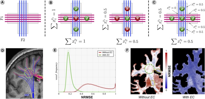
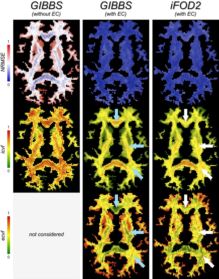
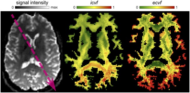
One of the major limitations of diffusion MRI tractography is that the fiber tracts recovered by existing algorithms are not truly quantitative. Local techniques for estimating more quantitative features of the tissue microstructure exist, but their combination with tractography has always been considered intractable. Recent advances in local and global modeling made it possible to fill this gap and a number of promising techniques for microstructure informed tractography have been suggested, opening new and exciting perspectives for the quantification of brain connectivity. The ease-of-use of the proposed solutions made it very attractive for researchers to include such advanced methods in their analyses; however, this apparent simplicity should not hide some critical open questions raised by the complexity of these very high-dimensional problems, otherwise some fundamental issues may be pushed into the background. The aim of this article is to raise awareness in the diffusion MRI community, notably researchers working on brain connectivity, about some potential pitfalls and modeling choices that make the interpretation of the outcomes from these novel techniques rather cumbersome. Through a series of experiments on synthetic and real data, we illustrate practical situations where erroneous and severely biased conclusions may be drawn about the connectivity if these pitfalls are overlooked, like the presence of partial/missing/duplicate fibers or the critical importance of the diffusion model adopted. Microstructure informed tractography is a young but very promising technology, and by acknowledging its current limitations as done in this paper, we hope our observations will trigger further research in this direction and new ideas for truly quantitative and biologically meaningful analyses of the connectivity.
Overview
The claims and statistics of this paper haven’t been checked yet.
Run the check — free, no account needed. It reads the open-access text and checks the sources it cites.
Integrity signals
Registry
Cites
Statistics
Figures
Verification
Claims the paper makes
The claims and statistics of this paper haven’t been checked yet.
Run the check — free, no account needed. It reads the open-access text and checks the sources it cites.
Claims other papers make about this one
Each is checked against this paper’s own text: does it say what the citing paper says it does?
The source backs part of the claim, not all of it — or backs it for a different population, condition or comparison.
In the citing paper
“Structural connectome analyses are most convincing when integrated with neuroanatomical knowledge, functional data, and careful control of tractography biases (Assaf et al., 2019; Daducci et al., 2016).”
From 40 Years of diffusion MRI in the brain: From history to emerging frontiers (2026) · cites this paper as Daducci et al., 2016
Experimental — accuracy not yet measuredThe source backs part of the claim, not all of it — or backs it for a different population, condition or comparison.
In the citing paper
“Higher angular-resolution methods, better distortion correction, and anatomically informed tractography improve matters, yet they do not eliminate the inverse problem (Daducci et al., 2016; Tournier et al., 2011).”
From 40 Years of diffusion MRI in the brain: From history to emerging frontiers (2026) · cites this paper as Daducci et al., 2016
Experimental — accuracy not yet measuredThe cited source says what the claim attributes to it.
In the citing paper
“despite the fact that white matter fibers synapse in the gray matter (Daducci et al., 2016)”
From Quantitative mapping of the brain’s structural connectivity using diffusion MRI tractography: A review (2022) · cites this paper as Daducci et al., 2016
The cited source says what the claim attributes to it.
In the citing paper
“Tractography, therefore, visualizes a model of anatomical connectivity constrained by diffusion data, not anatomy itself (Daducci et al., 2016; Tournier et al., 2011).”
From 40 Years of diffusion MRI in the brain: From history to emerging frontiers (2026) · cites this paper as Daducci et al., 2016
Self-check
Percentages, sums and reported differences re-computed from the paper’s own numbers.
The self-check needs the paper’s full text.
Citations
Cites
Donoho, D. L. (2006) · IEEE Transactions on Information Theory · doi:10.1109/tit.2006.871582 · Cited by 23,994
Donoho, D. L. (2004) · Cited by 16,703
Candes, E. J., Romberg, J., & Tao, T. (2006) · IEEE Transactions on Information Theory · doi:10.1109/tit.2005.862083 · Cited by 15,639
N4ITK: Improved N3 Bias Correction
Tustison, N. J., Avants, B., Cook, P. A., Zheng, Y., Egan, A., Yushkevich, P. A., & Gee, J. C. (2010) · IEEE Transactions on Medical Imaging · doi:10.1109/tmi.2010.2046908 · Cited by 6,413
The basis of anisotropic water diffusion in the nervous system—A technical review.
Beaulieu, C. (2002) · NMR in Biomedicine · doi:10.1002/nbm.782 · Cited by 4,628
Le Bihan, D., Breton, E., Lallemand, D., Grenier, P., Cabanis, E., & Laval-Jeantet, M. (1986) · Radiology · doi:10.1148/radiology.161.2.3763909 · Cited by 3,912
NODDI: Practical in vivo neurite orientation dispersion and density imaging of the human brain
Zhang, H., Schneider, T., Wheeler-Kingshott, C. A., & Alexander, D. C. (2012) · NeuroImage · doi:10.1016/j.neuroimage.2012.03.072 · Cited by 3,288
Characterization and propagation of uncertainty in diffusion-weighted MR imaging
Behrens, T. E. J., Woolrich, M. W., Jenkinson, M., Johansen‐Berg, H., Nunes, R. G., Clare, S., Matthews, P. M., Brady, J. M., & Smith, S. M. (2003) · Magnetic Resonance in Medicine · doi:10.1002/mrm.10609 · Cited by 3,038
The Human Connectome Project: A data acquisition perspective
Essen, D. C. V., Uǧurbil, K., Auerbach, E. J., Barch, D. M., Behrens, T. E. J., Bucholz, R. D., Chang, A., Chen, L., Corbetta, M., Curtiss, S. W., et al. (2012) · NeuroImage · doi:10.1016/j.neuroimage.2012.02.018 · Cited by 2,638
White matter integrity, fiber count, and other fallacies: The do's and don'ts of diffusion MRI
Jones, D. K., Knösche, T. R., & Turner, R. (2013) · NeuroImage · doi:10.1016/j.neuroimage.2012.06.081 · Cited by 2,402 · cited 6×
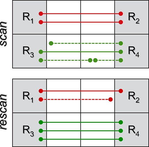
“…the connection strength between the four regions R1, . . . ,R4 is quantified by the fiber count (Jones et al., 2012), then a reduction inR1 ↔ R2 connectivity is observed between the two sessions (from 2 to 1), and an increase in R3 ↔ R4 (from 1 to 3), as the dashed tracts do not connect and so do…”
BackgroundInfluential
Cited by
Sorted by citations
Tractography-based connectomes are dominated by false-positive connections
Maier-Hein, K. H., Neher, P., Houde, J.-C., Côté, M.-A., Garyfallidis, E., Zhong, J., Chamberland, M., Yeh, F.-C., Lin, Y.-C., Ji, Q., et al. (2016) · doi:10.1101/084137 · Cited by 1,376
Self-citationDiffusion MRI fiber tractography of the brain
Jeurissen, B., Descoteaux, M., Mori, S., & Leemans, A. (2019) · NMR in Biomedicine · doi:10.1002/nbm.3785 · Cited by 574
Self-citationZhang, F., Daducci, A., He, Y., Schiavi, S., Seguin, C., Smith, R. E., Yeh, C.-H., Zhao, T., & O’Donnell, L. J. (2022) · NeuroImage · doi:10.1016/j.neuroimage.2021.118870 · Cited by 302
Self-citationBuilding connectomes using diffusion MRI: why, how and but
Sotiropoulos, S. N., & Zalesky, A. (2019) · NMR in Biomedicine · doi:10.1002/nbm.3752 · Cited by 290
Limits to anatomical accuracy of diffusion tractography using modern approaches
Schilling, K. G., Nath, V., Hansen, C., Parvathaneni, P., Blaber, J., Gao, Y., Neher, P., Aydogan, D. B., Shi, Y., Ocampo-Pineda, M., et al. (2019) · NeuroImage · doi:10.1016/j.neuroimage.2018.10.029 · Cited by 260
Self-citationEssayed, W. I., Zhang, F., Unadkat, P., Cosgrove, G. R., Golby, A. J., & O’Donnell, L. J. (2017) · NeuroImage Clinical · doi:10.1016/j.nicl.2017.06.011 · Cited by 249
Brain networks and their relevance for stroke rehabilitation
Guggisberg, A. G., Koch, P., Hummel, F. C., & Buetefisch, C. M. (2019) · Clinical Neurophysiology · doi:10.1016/j.clinph.2019.04.004 · Cited by 266
Design and Validation of Diffusion MRI Models of White Matter
Jelescu, I. O., & Budde, M. D. (2017) · Frontiers in Physics · doi:10.3389/fphy.2017.00061 · Cited by 240
Mapping Structural Connectivity Using Diffusion MRI : Challenges and Opportunities
Yeh, C., Jones, D. K., Liang, X., Descoteaux, M., & Connelly, A. (2021) · Journal of Magnetic Resonance Imaging · doi:10.1002/jmri.27188 · Cited by 187
Self-citationChallenges in diffusion MRI tractography – Lessons learned from international benchmark competitions
Schilling, K. G., Daducci, A., Maier-Hein, K. H., Poupon, C., Houde, J., Nath, V., Anderson, A. W., Landman, B. A., & Descoteaux, M. (2018) · Magnetic Resonance Imaging · doi:10.1016/j.mri.2018.11.014 · Cited by 148
Self-citationCommon misconceptions, hidden biases and modern challenges of dMRI tractography
Rheault, F., Poulin, P., Caron, A. V., St‐Onge, E., & Descoteaux, M. (2020) · Journal of Neural Engineering · doi:10.1088/1741-2552/ab6aad · Cited by 112
Self-citationSchiavi, S., Ocampo‐Pineda, M., Baraković, M., Petit, L., Descoteaux, M., Thiran, J., & Daducci, A. (2020) · Science Advances · doi:10.1126/sciadv.aba8245 · Cited by 107
Self-citationCalamante, F. (2019) · Diagnostics · doi:10.3390/diagnostics9030115 · Cited by 98
Quantitative streamlines tractography: methods and inter-subject normalisation
Smith, R. E., Raffelt, D. A., Tournier, J., & Connelly, A. (2022) · Aperture Neuro · doi:10.52294/apertureneuro.2022.2.neod9565 · Cited by 91
Bundle-specific tractography with incorporated anatomical and orientational priors
Rheault, F., St‐Onge, E., Sidhu, J., Maier-Hein, K. H., Tzourio‐Mazoyer, N., Petit, L., & Descoteaux, M. (2018) · NeuroImage · doi:10.1016/j.neuroimage.2018.11.018 · Cited by 92
Self-citationThe visual white matter: The application of diffusion MRI and fiber tractography to vision science
Rokem, A., Takemura, H., Bock, A. S., Scherf, K. S., Behrmann, M., Wandell, B. A., Fine, I., Bridge, H., & Pestilli, F. (2017) · Journal of Vision · doi:10.1167/17.2.4 · Cited by 92
Girard, G., Caminiti, R., Battaglia-Mayer, A., St-Onge, E., Ambrosen, K. S., Eskildsen, S. F., Krug, K., Dyrby, T. B., Descoteaux, M., Thiran, J.-P., et al. (2020) · NeuroImage · doi:10.1016/j.neuroimage.2020.117201 · Cited by 83
Self-citationGriffa, A., Ricaud, B., Benzi, K., Bresson, X., Daducci, A., Vandergheynst, P., Thiran, J., & Hagmann, P. (2017) · NeuroImage · doi:10.1016/j.neuroimage.2017.04.015 · Cited by 82
Self-citationYang, J. Y.-M., Yeh, C.-H., Poupon, C., & Calamante, F. (2021) · Physics in Medicine & Biology · doi:10.1088/1361-6560/ac0d90 · Cited by 80
Integrating across neuroimaging modalities boosts prediction accuracy of cognitive ability
Rasero, J., Sentis, A. I., Yeh, F.-C. F., & Verstynen, T. (2021) · PLoS Computational Biology · doi:10.1371/journal.pcbi.1008347 · Cited by 78
Models of Network Spread and Network Degeneration in Brain Disorders
Raj, A., & Powell, F. (2018) · Biological Psychiatry Cognitive Neuroscience and Neuroimaging · doi:10.1016/j.bpsc.2018.07.012 · Cited by 67
Attar, F. M., Kirilina, E. P., Haenelt, D., Pine, K., Trampel, R., Edwards, L., & Weiskopf, N. (2020) · Cerebral Cortex · doi:10.1093/cercor/bhaa049 · Cited by 64
Roine, T., Jeurissen, B., Perrone, D., Aelterman, J., Philips, W. R., Sijbers, J., & Leemans, A. (2018) · Medical Image Analysis · doi:10.1016/j.media.2018.10.009 · Cited by 68
AxTract: Toward microstructure informed tractography
Girard, G., Daducci, A., Petit, L., Thiran, J., Whittingstall, K., Deriche, R., Wassermann, D., & Descoteaux, M. (2017) · Human Brain Mapping · doi:10.1002/hbm.23741 · Cited by 62
Self-citationParallel Transport Tractography
Aydogan, D. B., & Shi, Y. (2020) · IEEE Transactions on Medical Imaging · doi:10.1109/tmi.2020.3034038 · Cited by 65
MEDUSA: A GPU-based tool to create realistic phantoms of the brain microstructure using tiny spheres
Ginsburger, K., Matuschke, F., Poupon, F., Mangin, J., Axer, M., & Poupon, C. (2019) · NeuroImage · doi:10.1016/j.neuroimage.2019.02.055 · Cited by 60
Larivière, S., de Wael, R. V., Paquola, C., Hong, S., Mišić, B., Bernasconi, N., Bernasconi, A., Bonilha, L., & Bernhardt, B. C. (2018) · Brain Connectivity · doi:10.1089/brain.2018.0587 · Cited by 51
Track-weighted imaging methods: extracting information from a streamlines tractogram
Calamante, F. (2017) · Magnetic Resonance Materials in Physics Biology and Medicine · doi:10.1007/s10334-017-0608-1 · Cited by 56
Brain connectivity alterations in early psychosis: from clinical to neuroimaging staging
Griffa, A., Baumann, P., Klauser, P., Mullier, E., Cleusix, M., Jenni, R., van den Heuvel, M. P., Kim, Q., Conus, P., & Hagmann, P. (2019) · Translational Psychiatry · doi:10.1038/s41398-019-0392-y · Cited by 54
Fan, Q., Eichner, C., Afzali, M., Mueller, L., Tax, C. M. W., Davids, M., Mahmutović, M., Keil, B. R., Bilgiç, B., Setsompop, K., et al. (2022) · NeuroImage · doi:10.1016/j.neuroimage.2022.118958 · Cited by 54
Sarubbo, S., Petit, L., BENEDICTIS, A. D., Chioffi, F., Ptito, M., & Dyrby, T. B. (2019) · Brain Structure and Function · doi:10.1007/s00429-019-01856-2 · Cited by 48
Sparse wars: A survey and comparative study of spherical deconvolution algorithms for diffusion MRI
Canales-Rodríguez, E. J., Legarreta, J. H., Pizzolato, M., Rensonnet, G., Girard, G., Rafael‐Patiño, J., Baraković, M., Romascano, D., Alemán‐Gómez, Y., Raduà, J., et al. (2018) · NeuroImage · doi:10.1016/j.neuroimage.2018.08.071 · Cited by 44
Self-citationMancini, M., Giulietti, G., Dowell, N., Spanò, B., Harrison, N., Bozzali, M., & Cercignani, M. (2018) · NeuroImage · doi:10.1016/j.neuroimage.2017.09.018 · Cited by 43
Evaluating arcuate fasciculus laterality measurements across dataset and tractography pipelines
Bain, J. S., Yeatman, J. D., Schurr, R., Rokem, A., & Mezer, A. (2019) · Human Brain Mapping · doi:10.1002/hbm.24626 · Cited by 43
Multidimensional encoding of brain connectomes
Caiafa, C. F., & Pestilli, F. (2017) · Scientific Reports · doi:10.1038/s41598-017-09250-w · Cited by 46
Validation of structural brain connectivity networks: The impact of scanning parameters
Ambrosen, K. S., Eskildsen, S. F., Hinne, M., Krug, K., Lundell, H., Schmidt, M. N., van Gerven, M. A. J., Mørup, M., & Dyrby, T. B. (2019) · NeuroImage · doi:10.1016/j.neuroimage.2019.116207 · Cited by 42
The role of diffusion tractography in refining glial tumor resection
Vanderweyen, D., Theaud, G., Sidhu, J., Rheault, F., Sarubbo, S., Descoteaux, M., & Fortin, D. (2020) · Brain Structure and Function · doi:10.1007/s00429-020-02056-z · Cited by 41
Self-citationSubtle white matter alterations in schizophrenia identified with a new measure of fiber density
Stämpfli, P., Sommer, S., Manoliu, A., Burrer, A., Schmidt, A., Herdener, M., Seifritz, E., Kaiser, S., & Kirschner, M. (2019) · Scientific Reports · doi:10.1038/s41598-019-40070-2 · Cited by 40
Maffei, C., Sarubbo, S., & Jovicich, J. (2019) · Frontiers in Neuroanatomy · doi:10.3389/fnana.2019.00027 · Cited by 41
Toward nonparametric diffusion‐ characterization of crossing fibers in the human brain
Reymbaut, A., Critchley, J., Durighel, G., Sprenger, T., Sughrue, M., Bryskhe, K., & Topgaard, D. (2020) · Magnetic Resonance in Medicine · doi:10.1002/mrm.28604 · Cited by 32
Modelling white matter in gyral blades as a continuous vector field
Cottaar, M., Bastiani, M., Boddu, N., Glasser, M. F., Haber, S. N., Essen, D. C. V., Sotiropoulos, S. N., & Jbabdi, S. (2020) · NeuroImage · doi:10.1016/j.neuroimage.2020.117693 · Cited by 36
Takemura, H., Pestilli, F., & Weiner, K. S. (2018) · Neuroscience Research · doi:10.1016/j.neures.2018.10.011 · Cited by 36
Tractography optimization using quantitative T1 mapping in the human optic radiation
Schurr, R., Duan, Y., Norcia, A. M., Ogawa, S., Yeatman, J. D., & Mezer, A. (2018) · NeuroImage · doi:10.1016/j.neuroimage.2018.06.060 · Cited by 35
Schiavi, S., Petracca, M., Battocchio, M., Mendili, M. M. E., Paduri, S., Fleysher, L., Inglese, M., & Daducci, A. (2020) · Human Brain Mapping · doi:10.1002/hbm.24989 · Cited by 34
Self-citationConnectome reorganization associated with temporal lobe pathology and its surgical resection
Larivière, S., Park, B., Royer, J., DeKraker, J., Ngo, A., Sahlas, E., Chen, J., Rodríguez‐Cruces, R., Weng, Y., Frauscher, B., et al. (2024) · Brain · doi:10.1093/brain/awae141 · Cited by 32
Advances in computational and statistical diffusion MRI
O’Donnell, L. J., Daducci, A., Wassermann, D., & Lenglet, C. (2017) · NMR in Biomedicine · doi:10.1002/nbm.3805 · Cited by 32
Self-citationGinsburger, K., Poupon, F., Beaujoin, J., Estournet, D., Matuschke, F., Mangin, J., Axer, M., & Poupon, C. (2018) · Frontiers in Physics · doi:10.3389/fphy.2018.00012 · Cited by 32
Subdividing the superior longitudinal fasciculus using local quantitative MRI
Schurr, R., Zelman, A., & Mezer, A. (2019) · NeuroImage · doi:10.1016/j.neuroimage.2019.116439 · Cited by 32
Intra– and inter–hemispheric network dynamics supporting object recognition and speech production
Kitazawa, Y., Sonoda, M., Sakakura, K., Mitsuhashi, T., Firestone, E., Ueda, R., Kambara, T., Iwaki, H., Luat, A. F., Marupudi, N. I., et al. (2023) · NeuroImage · doi:10.1016/j.neuroimage.2023.119954 · Cited by 29
Innocenti, G. M., Caminiti, R., Rouiller, E. M., Knott, G. W., Dyrby, T. B., Descoteaux, M., & Thiran, J. (2017) · Cerebral Cortex · doi:10.1093/cercor/bhx363 · Cited by 30
Self-citationGriffa, A., Ville, D. V. D., Herrmann, F. R., & Allali, G. (2020) · Neuroscience & Biobehavioral Reviews · doi:10.1016/j.neubiorev.2020.02.023 · Cited by 29
Tracking and validation techniques for topographically organized tractography
Aydogan, D. B., & Shi, Y. (2018) · NeuroImage · doi:10.1016/j.neuroimage.2018.06.071 · Cited by 27
Ultra-High Field Diffusion MRI Reveals Early Axonal Pathology in Spinal Cord of ALS mice
Gatto, R. G., Amin, M., Deyoung, D., Hey, M. T., Mareci, T. H., & Magin, R. L. (2018) · Translational Neurodegeneration · doi:10.1186/s40035-018-0122-z · Cited by 28
A multi-scale probabilistic atlas of the human connectome
Alemán‐Gómez, Y., Griffa, A., Houde, J., Najdenovska, E., Magon, S., Cuadra, M. B., Descoteaux, M., & Hagmann, P. (2022) · Scientific Data · doi:10.1038/s41597-022-01624-8 · Cited by 28
Self-citationBundle-Specific Axon Diameter Index as a New Contrast to Differentiate White Matter Tracts
Baraković, M., Girard, G., Schiavi, S., Romascano, D., Descoteaux, M., Granziera, C., Jones, D. K., Innocenti, G. M., Thiran, J., & Daducci, A. (2021) · Frontiers in Neuroscience · doi:10.3389/fnins.2021.646034 · Cited by 25
Self-citationSchiavi, S., Lu, P., Weigel, M., Lutti, A., Jones, D. K., Kappos, L., Granziera, C., & Daducci, A. (2022) · NeuroImage · doi:10.1016/j.neuroimage.2022.118922 · Cited by 24
Self-citationTractography delineation of the vertical occipital fasciculus using quantitative T1 mapping
Schurr, R., Filo, S., & Mezer, A. (2019) · NeuroImage · doi:10.1016/j.neuroimage.2019.116121 · Cited by 25
Gatto, R. G., Li, W., & Magin, R. L. (2017) · Brain Research · doi:10.1016/j.brainres.2017.11.017 · Cited by 24
The Hidden Control Architecture of Complex Brain Networks
Lee, B., Kang, U., Chang, H., & Cho, K. (2019) · iScience · doi:10.1016/j.isci.2019.02.017 · Cited by 24
Local connectome phenotypes predict social, health, and cognitive factors
Powell, M. A., Garcia, J. O., Yeh, F.-C. F., Vettel, J. M., & Verstynen, T. (2017) · Network Neuroscience · doi:10.1162/netn_a_00031 · Cited by 24
Topological principles and developmental algorithms might refine diffusion tractography
Innocenti, G. M., Dyrby, T. B., Girard, G., St‐Onge, E., Thiran, J., Daducci, A., & Descoteaux, M. (2018) · Brain Structure and Function · doi:10.1007/s00429-018-1759-1 · Cited by 20
Self-citationHe, J., Zhang, F., Pan, Y., Feng, Y., Rushmore, J., Torio, E., Yogesh Rathi, A., Makris, N., Kikinis, R., Golby, A. J., et al. (2023) · Human Brain Mapping · doi:10.1002/hbm.26497 · Cited by 20
Modelling the brain: Elementary components to explain ensemble functions
D'Angelo, E. U., & Wheeler‐Kingshott, C. A. M. G. (2017) · UCL Discovery (University College London) · doi:10.1393/ncr/i2017-10137-5 · Cited by 18
Dynamic spatiotemporal patterns of brain connectivity reorganize across development
Vohryzek, J., Griffa, A., Mullier, E., Friedrichs‐Maeder, C., Sandini, C., Schaer, M., Eliez, S., & Hagmann, P. (2019) · Network Neuroscience · doi:10.1162/netn_a_00111 · Cited by 19
Hierarchical microstructure informed tractography
Ocampo-Pineda, M., Schiavi, S., Rheault, F., Girard, G., Petit, L., Descoteaux, M., & Daducci, A. (2021) · Brain Connectivity · doi:10.1089/brain.2020.0907 · Cited by 19
Self-citationPhysical and digital phantoms for validating tractography and assessing artifacts
Drobnjak, I., Neher, P., Poupon, C., & Sarwar, T. (2021) · NeuroImage · doi:10.1016/j.neuroimage.2021.118704 · Cited by 18
Gatto, R. G., & Weissmann, C. (2018) · Current Medical Imaging Formerly Current Medical Imaging Reviews · doi:10.2174/1573405614666181115113400 · Cited by 18
Developmental Pathoconnectomics and Advanced Fetal MRI
András, J. (2019) · Topics in Magnetic Resonance Imaging · doi:10.1097/rmr.0000000000000220 · Cited by 19
Reducing false positives in tractography with microstructural and anatomical priorsPreprint
Schiavi, S., Baraković, M., Ocampo-Pineda, M., Descoteaux, M., Thiran, J.-P., & Daducci, A. (2019) · bioRxiv (Cold Spring Harbor Laboratory) · doi:10.1101/608349 · Cited by 18
Self-citationA Novel Anatomically-Constrained Global Tractography Approach to Monitor Sharp Turns in Gyri
Teillac, A., Beaujoin, J., Poupon, F., Mangin, J., & Poupon, C. (2017) · Lecture notes in computer science · doi:10.1007/978-3-319-66182-7_61 · Cited by 17
Asymmetric fiber trajectory distribution estimation using streamline differential equation
Feng, Y., & He, J. (2020) · Medical Image Analysis · doi:10.1016/j.media.2020.101686 · Cited by 15
Differences in topological progression profile among neurodegenerative diseases from imaging data
Garbarino, S., Lorenzi, M., Oxtoby, N. P., Vinke, E. J., Marinescu, R. V., Eshaghi, A., Ikram, M. A., Niessen, W. J., Ciccarelli, O., Barkhof, F., et al. (2019) · eLife · doi:10.7554/elife.49298 · Cited by 16
Fiber up‐sampling and quality assessment of tractograms – towards quantitative brain connectivity
Sommer, S., Kozerke, S., Seifritz, E., & Stämpfli, P. (2016) · Brain and Behavior · doi:10.1002/brb3.588 · Cited by 16
Differential Effects of Brain Disorders on Structural and Functional Connectivity
Vega-Pons, S., Olivetti, E., Avesani, P., Dodero, L., Gozzi, A., & Bifone, A. (2017) · Frontiers in Neuroscience · doi:10.3389/fnins.2016.00605 · Cited by 15
Real‐Time Tractography‐Assisted Neuronavigation for Transcranial Magnetic Stimulation
Aydogan, D. B., Souza, V. H., Matsuda, R. H., Lioumis, P., & Ilmoniemi, R. J. (2024) · Human Brain Mapping · doi:10.1002/hbm.70122 · Cited by 20
Cortical Surfaces Integration with Tractography for Structural Connectivity Analysis
St‐Onge, E., Al‐Sharif, N., Girard, G., Theaud, G., & Descoteaux, M. (2021) · Brain Connectivity · doi:10.1089/brain.2020.0930 · Cited by 15
Self-citationEvaluation of tractogram filtering methods using human-like connectome phantoms
Sarwar, T., Ramamohanarao, K., Daducci, A., Schiavi, S., Smith, R. E., & Zalesky, A. (2023) · NeuroImage · doi:10.1016/j.neuroimage.2023.120376 · Cited by 15
Self-citationThe impact of levamisole and alcohol on white matter microstructure in adult chronic cocaine users
Michels, L., Moisa, M., Stämpfli, P., Hirsiger, S., Baumgartner, M. R., Surbeck, W., Seifritz, E., & Quednow, B. B. (2022) · Addiction Biology · doi:10.1111/adb.13149 · Cited by 13
Bonet-Carné, E., Grussu, F., Ning, L., Sepehrband, F., & Tax, C. M. W. (2019) · Mathematics and visualization · doi:10.1007/978-3-030-05831-9 · Cited by 13
Jelescu, I., Grussu, F., Ianuș, A., Hansen, B., Barrett, R. L. C., Aggarwal, M., Michielse, S., Nasrallah, F. A., Syeda, W. T., Wang, N., et al. (2025) · Magnetic Resonance in Medicine · doi:10.1002/mrm.30429 · Cited by 21
Self-citationCerebro-Cerebellar Pathways for Verbal Working Memory
Sobczak‐Edmans, M., Lo, Y., Hsu, Y., Chen, Y.-J., Kwok, F. Y., Chuang, K., Tseng, W. I., & Chen, S. A. (2019) · Frontiers in Human Neuroscience · doi:10.3389/fnhum.2018.00530 · Cited by 13
Kumar, P. R., Jha, R. K., & Katti, A. P. (2023) · Acta Neurologica Belgica · doi:10.1007/s13760-023-02170-9 · Cited by 13
Papini, M. G., Avila, A. N., Fitzgerald, M., & Hellewell, S. C. (2024) · Journal of Neurotrauma · doi:10.1089/neu.2024.0039 · Cited by 10
Battocchio, M., Schiavi, S., Descoteaux, M., & Daducci, A. (2022) · NeuroImage · doi:10.1016/j.neuroimage.2022.119600 · Cited by 9
Self-citationNon-parametric graphnet-regularized representation of dMRI in space and time
Fick, R., Petiet, A., Santin, M. D., Philippe, A.-C., Lehéricy, S., Rachid Deriche, B., & Wassermann, D. (2017) · Medical Image Analysis · doi:10.1016/j.media.2017.09.002 · Cited by 9
Real-time tractography-assisted neuronavigation for TMSPreprint
Aydogan, D. B., Souza, V. H., Matsuda, R. H., Lioumis, P., & Ilmoniemi, R. J. (2023) · bioRxiv (Cold Spring Harbor Laboratory) · doi:10.1101/2023.03.09.531565 · Cited by 9
Mapping human brain pathways: challenges and opportunities in the integration of scales
Charvet, C. (2023) · Brain, Behavior and Evolution · doi:10.1159/000530317 · Cited by 8
Blurred streamlines: A novel representation to reduce redundancy in tractography
Gabusi, I., Battocchio, M., Bosticardo, S., Schiavi, S., & Daducci, A. (2024) · Medical Image Analysis · doi:10.1016/j.media.2024.103101 · Cited by 9
Self-citationWhite Matter Tractography and Diffusion‐Weighted Imaging
Vettel, J. M., Cooper, N., Garcia, J. O., Yeh, F.-C. F., & Verstynen, T. (2017) · Wiley eBooks · doi:10.1002/9780470015902.a0027162 · Cited by 9
Small brains but big challenges: white matter tractography in early life samples
Dubois, J., Grotheer, M., Yang, J. Y., Tournier, J., Beaulieu, C., & Lebel, C. A. (2025) · Brain Structure and Function · doi:10.1007/s00429-025-02922-8 · Cited by 6
Anisotropy Across Fields and Scales
Özarslan, E., Schultz, T., Zhang, E., & Fuster, A. (2021) · Mathematics and visualization · doi:10.1007/978-3-030-56215-1 · Cited by 6
Structural connectivity differs between males and females in the brain object manipulation network
Lee, D., & Son, T. (2021) · PLoS ONE · doi:10.1371/journal.pone.0253273 · Cited by 7
Diffusion MRI Fiber Tractography
Smith, R. E., Connelly, A., & Calamante, F. (2020) · Advances in magnetic resonance technology and applications · doi:10.1016/b978-0-12-817057-1.00023-8 · Cited by 10
Challenges for Tractogram Filtering
Jörgens, D., Descoteaux, M., & Moreno, R. (2021) · Mathematics and visualization · doi:10.1007/978-3-030-56215-1_7 · Cited by 9
Self-citationEvaluation of tractography-based myelin-weighted connectivity across the lifespan
Bosticardo, S., Schiavi, S., Schaedelin, S. A., Battocchio, M., Baraković, M., Lu, P., Weigel, M., Melie‐García, L., Granziera, C., & Daducci, A. (2024) · Frontiers in Neuroscience · doi:10.3389/fnins.2023.1228952 · Cited by 6
Self-citationDaducci, A., Schiavi, S., Christiaens, D., Smith, R. E., & Alexander, D. C. (2025) · Elsevier eBooks · doi:10.1016/b978-0-12-818894-1.00014-8 · Cited by 7
Self-citationA multi-compartment model for pathological connectomes
Bosticardo, S., Battocchio, M., Schiavi, S., Zalesky, A., Granziera, C., & Daducci, A. (2025) · Network Neuroscience · doi:10.1162/netn.a.30 · Cited by 4
Self-citationSommer, S., Kozerke, S., Seifritz, E., & Stämpfli, P. (2017) · Frontiers in Neuroscience · doi:10.3389/fnins.2017.00729 · Cited by 4
GPU-accelerated connectome discovery at scale
Sreenivasan, V., Kumar, S., Pestilli, F., Talukdar, P. P., & Sridharan, D. (2022) · Nature Computational Science · doi:10.1038/s43588-022-00250-z · Cited by 4
Improving Tractography Accuracy Using Dynamic Filtering
Battocchio, M., Schiavi, S., Descoteaux, M., & Daducci, A. (2021) · Mathematics and visualization · doi:10.1007/978-3-030-73018-5_4 · Cited by 4
Self-citation
Metadata
- Authors
- Daducci, A., Dal Palú, A., Descoteaux, M., & Thiran, J.-P.
- Affiliations
- Université de Sherbrooke (CA)
- Centre Hospitalier Universitaire Vaudois (CH)
- Corresponding author
- Daducci, Alessandro
- Venue
- Frontiers in Neuroscience
- Publisher
- Frontiers Media SA
- Published
- 2016-06-06
- Volume
- 10
- Pages
- 247-247
- ISSN
- 1662-453X (ISSN-L)
- PMC
- PMC4893481
- OpenAlex
- W2404527239
- Cited by
- 138
- References
- 51
- Citations by year
- 2026: 9 · 2025: 17 · 2024: 9 · 2023: 8 · 2022: 8 · 2021: 12 · 2020: 14 · 2019: 23 · 2018: 16 · 2017: 21 · 2016: 1
- FWCI
- 17.65
- Type
- journal article
- Language
- en
- Open access
- gold
- doi.org
- Licence
- cc-by
- Locations
- Frontiers in Neuroscience (publishedVersion, cc-by)
- Europe PMC (PubMed Central) (publishedVersion, cc-by)
- PubMed Central (publishedVersion, cc-by)
- PubMed (publishedVersion)
- DOAJ (DOAJ: Directory of Open Access Journals) (submittedVersion)
- Figshare (submittedVersion, cc-by)
- Infoscience (Ecole Polytechnique Fédérale de Lausanne) (submittedVersion)
- Infoscience (Ecole Polytechnique Fédérale de Lausanne) (submittedVersion, cc-by-nc-nd)
- Figshare (cc-by)
- Funders
- École Polytechnique Fédérale de Lausanne
- Centre d'Imagerie BioMédicale
- Topics
- Health Sciences › Medicine › Radiology, Nuclear Medicine and Imaging › Advanced Neuroimaging Techniques and Applications
- Life Sciences › Neuroscience › Cognitive Neuroscience › Functional Brain Connectivity Studies
- Health Sciences › Medicine › Radiology, Nuclear Medicine and Imaging › Advanced MRI Techniques and Applications
- Keywords
- diffusion MRI, brain connectivity
About these checks
Registry, retraction, Cites and statistics results are computed, not judged, with one exception: a printed reference that no index lists is read once by a model before it counts as a citation error. Claims are extracted by a model and kept only when their quote appears verbatim in the text; each cited source is then checked, with a model’s help, for whether it says what the claim relies on. Those verdicts can be wrong — read the claim’s quote and the cited source before relying on one.
Statistics and arithmetic are recomputed, not judged.
Confidence bands are shown only for fields calibrated on our annotated test set. None is calibrated yet, so no band is shown.
What counts as a claim ›How we measure confidence ›How we check this ›
Are you an author, or do you see a problem with this page? Request a review
