

The DOI matches its Crossref record; 63 of the 63 references it cites were found in a registry or an index.
Synaptic Tau Seeding Precedes Tau Pathology in Human Alzheimer’s Disease Brain
Open accessAbstract
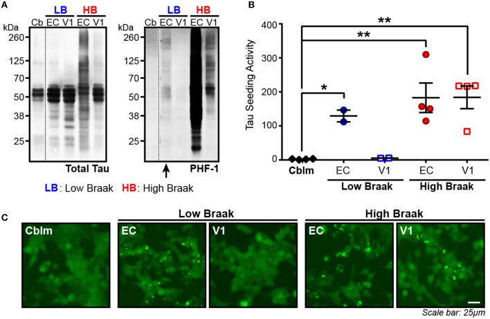
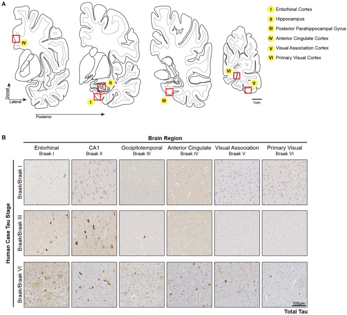
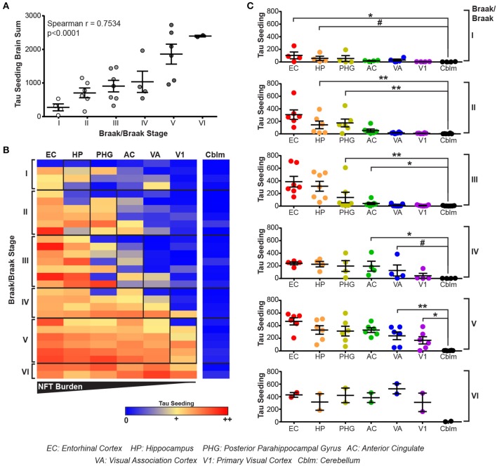
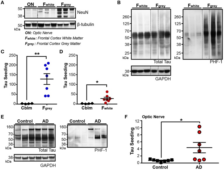
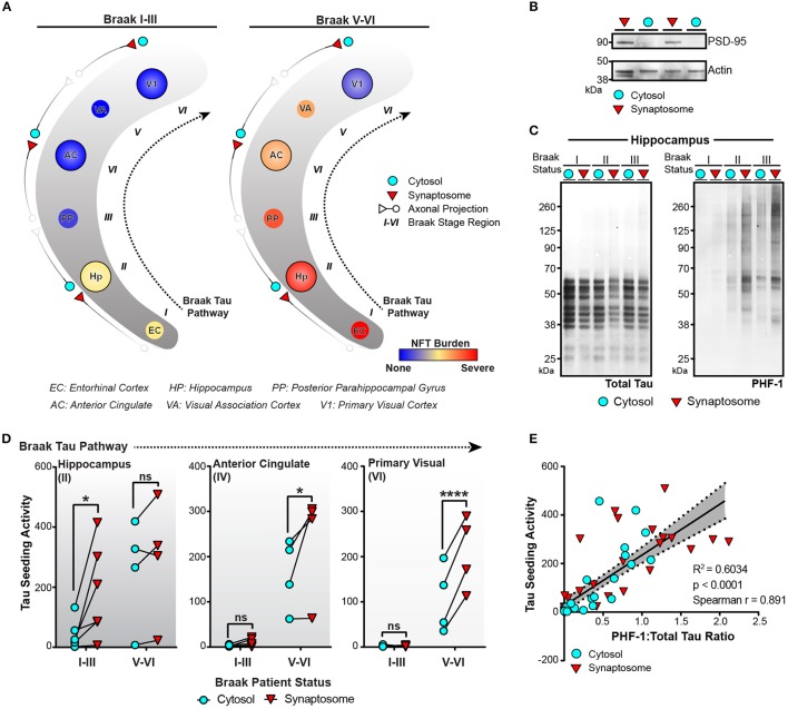
Alzheimer’s disease (AD) is defined by the presence of intraneuronal neurofibrillary tangles (NFTs) composed of hyperphosphorylated tau aggregates and extracellular amyloid-beta plaques. The presence and spread of tau pathology through the brain is classified by Braak stages and thought to correlate to the progression of AD. Several in vitro and in vivo studies have examined tau pathology moving from one neuron to the next, suggesting a “prion-like” spread of tau aggregates may be an underlying cause of Braak tau staging in AD. Using the HEK293 TauRD-P301S-CFP/YFP expressing biosensor cells as a highly sensitive and specific tool to identify the presence of seed competent aggregated tau in brain lysate – ie., tau aggregates that are capable of recruiting and misfolding monomeric tau – , we detected substantial tau seeding levels in the entorhinal cortex from human cases with only very rare NFTs, suggesting that soluble tau aggregates can exist prior to the development of overt tau pathology. We next looked at tau seeding levels in human brains of varying Braak stages along six regions of the Braak Tau Pathway. Tau seeding levels were detected not only in the brain regions impacted by pathology, but also in the subsequent non-pathology containing region along the Braak pathway. These data imply that pathogenic tau aggregates precede overt tau pathology in a manner that is consistent with transneuronal spread of tau aggregates. We then detected tau seeding in frontal white matter tracts and the optic nerve, two brain regions comprised of axons that contain little to no neuronal cell bodies, implying that tau aggregates can indeed traverse along axons. Finally, we isolated cytosolic and synaptosome fractions along the Braak Tau Pathway from brains of varying Braak stages. Phosphorylated and seed competent tau was significantly enriched in the synaptic fraction of brain regions that did not have extensive cellular tau pathology, further suggesting that aggregated tau seeds move through the human brain along synaptically connected neurons. Together, these data provide further evidence that the spread of tau aggregates through the human brain along synaptically connected networks results in the pathogenesis of human AD.
Overview
The claims and statistics of this paper haven’t been checked yet.
Run the check — free, no account needed. It reads the open-access text and checks the sources it cites.
Integrity signals
Registry
Cites
Statistics
Figures
Verification
Claims the paper makes
The claims and statistics of this paper haven’t been checked yet.
Run the check — free, no account needed. It reads the open-access text and checks the sources it cites.
Claims other papers make about this one
Each is checked against this paper’s own text: does it say what the citing paper says it does?
The source backs part of the claim, not all of it — or backs it for a different population, condition or comparison.
In the citing paper
“The progression of tau pathology has been correlated with memory dysfunction and cognitive impairments in AD (DeVos et al., 2018; Giannakopoulos et al., 2003).”
From Cholesterol 25-hydroxylase mediates neuroinflammation and neurodegeneration in a mouse model of tauopathy (2024) · cites this paper as DeVos et al., 2018
Experimental — accuracy not yet measuredThe cited source says what the claim attributes to it.
In the citing paper
“Several studies have shown that seeding precedes tau pathogenesis and can even occur in brain regions where tau pathology does not usually present (DeVos et al., 2018; Kaufman et al., 2018; Stopschinski et al., 2021).”
From Network analysis identifies strain-dependent response to tau and tau seeding-associated genes (2023) · cites this paper as DeVos et al., 2018
In the citing paper
“Thus, prion-like seeding and spreading may represent a mechanism whereby tau pathology propagates from the entorhinal cortex to the neocortex in AD”
From Alzheimer Disease: An Update on Pathobiology and Treatment Strategies (2019) · cites this paper as DeVos et al., 2018
Self-check
Percentages, sums and reported differences re-computed from the paper’s own numbers.
The self-check needs the paper’s full text.
Citations
Cites
Neuropathological stageing of Alzheimer-related changes.
Braak, H., & Braak, E. (1991) · Acta Neuropathologica · doi:10.1007/bf00308809 · Cited by 16,123 · cited 3×
“…disease (Morris et al., 1989), (2) the distribution of beta amyloid plaques (Thal score) (Thal et al., 2002), and (3) the Braak tau stage based on the location of neurofibrillary tau tangles as determined by a total tau immunostaining and Bielchowsky’s silver stain (Braak and Braak, 1991).”
BackgroundOverdependence on degraded gist memory in Alzheimer's disease.
Gallo, D. A., Shahid, K. R., Olson, M. A., Solomon, T. M., Schacter, D. L., & Budson, A. E. (2006) · Neuropsychology · doi:10.1037/0894-4105.20.6.625 · Cited by 4,757
Imaging brain amyloid in Alzheimer's disease with Pittsburgh Compound‐B
Klunk, W. E., Engler, H., Nordberg, A., Wang, Y., Blomqvist, G., Holt, D. P., Bergström, M., Savitcheva, I., Huang, G., Estrada, S., et al. (2004) · Annals of Neurology · doi:10.1002/ana.20009 · Cited by 4,496
Moms, J. C., Heyman, A., Mohs, R. C., Hughes, J. P., van Belle, G., Fillenbaum, G., Mellits, E. D., & Clark, C. (1989) · Neurology · doi:10.1212/wnl.39.9.1159 · Cited by 4,130
Phases of Aβ-deposition in the human brain and its relevance for the development of AD
Thal, D. R., Rüb, U., Orantes, M., & Braak, H. (2002) · Neurology · doi:10.1212/wnl.58.12.1791 · Cited by 3,490
Hyman, B. T., Phelps, C. H., Beach, T. G., Bigio, E. H., Cairns, N. J., Carrillo, M. C., Dickson, D. W., Duyckaerts, C., Frosch, M. P., Masliah, E., et al. (2012) · Alzheimer's & Dementia · doi:10.1016/j.jalz.2011.10.007 · Cited by 3,092
Self-citationNeurofibrillary tangles but not senile plaques parallel duration and severity of Alzheimer's disease
Arriagada, P. V., Growdon, J. H., Hedley-Whyte, E. T., & Hyman, B. T. (1992) · Neurology · doi:10.1212/wnl.42.3.631 · Cited by 2,668
Self-citationAlzheimer's disease: cell-specific pathology isolates the hippocampal formation.
Hyman, B. T., Van Hoesen, G. W., Damasio, A. R., & Barnes, C. L. (1984) · Science · doi:10.1126/science.6474172 · Cited by 2,268
Self-citationSynapse Loss and Microglial Activation Precede Tangles in a P301S Tauopathy Mouse Model
Yoshiyama, Y., Higuchi, M., Zhang, B., Huang, S.-M., Iwata, N., Saido, T. C., Maeda, J., Suhara, T., Trojanowski, J. Q., & Lee, V. M.-Y. (2007) · Neuron · doi:10.1016/j.neuron.2007.01.010 · Cited by 2,208
Transmission and spreading of tauopathy in transgenic mouse brain
Clavaguera, F., Bolmont, T., Crowther, R. A., Abramowski, D., Frank, S., Probst, A., Fraser, G., Stalder, A. K., Beibel, M., Staufenbiel, M., et al. (2009) · Nature Cell Biology · doi:10.1038/ncb1901 · Cited by 1,803
Cited by
Sorted by citations
Items 101–200 of 286
Chiu, M., Chen, T., Hu, C., Yan, S., Sun, Y., Liu, B.-H., Chang, Y.-T., Yang, C., & Yang, S. (2020) · Nanomedicine Nanotechnology Biology and Medicine · doi:10.1016/j.nano.2020.102182 · Cited by 28
Easton, A. E., Jensen, M. L., Wang, C., Hagedorn, P., Li, Y.-W., Weed, M. R., Meredith, J. E., Guss, V. L., Jones, K. M., Gill, M. B., et al. (2022) · Molecular Therapy — Nucleic Acids · doi:10.1016/j.omtn.2022.07.027 · Cited by 27
Retromer in Synaptic Function and Pathology
Brodin, L., & Shupliakov, O. (2018) · Frontiers in Synaptic Neuroscience · doi:10.3389/fnsyn.2018.00037 · Cited by 29
SIRT1 deficiency increases O-GlcNAcylation of tau, mediating synaptic tauopathy
Yin, X., Li, Y., Fan, X., Huang, F., Qiu, Y., Zhao, C., Zhou, Z., Gu, Q., Xia, L., Bao, J., et al. (2022) · Molecular Psychiatry · doi:10.1038/s41380-022-01689-2 · Cited by 28
Precision proteoform design for 4R tau isoform selective templated aggregation
Longhini, A. P., DuBose, A., Lobo, S., Vijayan, V., Bai, Y., Rivera, E. K., Sala‐Jarque, J., Nikitina, A. A., Carrettiero, D. C., Unger, M., et al. (2024) · Proceedings of the National Academy of Sciences · doi:10.1073/pnas.2320456121 · Cited by 26
Anatomic survey of seeding in Alzheimer’s disease brains reveals unexpected patterns
Stopschinski, B. E., Del Tredici, K., Estill-Terpack, S.-J., Ghebremedhin, E., Yu, F. F., Braak, H., & Diamond, M. I. (2021) · Acta Neuropathologica Communications · doi:10.1186/s40478-021-01255-x · Cited by 26
Tau seeding in cases of multiple sclerosis
LaCroix, M., Mirbaha, H., Shang, P., Zandee, S., Foong, C., Prat, A., White, C. L., Stüve, O., & Diamond, M. I. (2022) · Acta Neuropathologica Communications · doi:10.1186/s40478-022-01444-2 · Cited by 27
Frey, B., Holzinger, D., Taylor, K. C., Ehrnhoefer, D. E., Striebinger, A., Biesinger, S., Gasparini, L., O’Neill, M. J., Wegner, F., Barghorn, S., et al. (2023) · Acta Neuropathologica Communications · doi:10.1186/s40478-023-01676-w · Cited by 28
Zhang, H., Lin, A., Gong, P., Chen, Y., Ye, R. D., Qian, F., Zhang, Y., & Yu, Y. (2020) · The Journal of Neuroscience · doi:10.1523/jneurosci.0455-20.2020 · Cited by 24
SORL1 is a receptor for tau that promotes tau seeding
Cooper, J. M., Lathuiliere, A., Su, E. J., Song, Y., Torrente, D., Jo, Y., Weinrich, N., Sales, J. D., Migliorini, M., Sisson, T. H., et al. (2024) · Journal of Biological Chemistry · doi:10.1016/j.jbc.2024.107313 · Cited by 25
Self-citationTaddei, R. N., Sanchez-Mico, M. V., Bonnar, O., Connors, T. R., Gaona, A., Denbow, D., Frosch, M. P., & Gómez‐Isla, T. (2022) · Acta Neuropathologica Communications · doi:10.1186/s40478-022-01370-3 · Cited by 23
Self-citationHeparan Sulfate Proteoglycans in Tauopathy
Zhu, Y., Gandy, L. A., Zhang, F., Liu, J., Wang, C., Blair, L. J., Linhardt, R. J., & Wang, L. (2022) · Biomolecules · doi:10.3390/biom12121792 · Cited by 27
Tau-Mediated Dysregulation of Neuroplasticity and Glial Plasticity
Koller, E. J., & Chakrabarty, P. (2020) · Frontiers in Molecular Neuroscience · doi:10.3389/fnmol.2020.00151 · Cited by 23
Zhao, Y., Guo, Q., Tian, J., Liu, W., & Wang, X. (2024) · Ageing Research Reviews · doi:10.1016/j.arr.2024.102596 · Cited by 23
Braiding Braak and Braak: Staging patterns and model selection in network neurodegeneration
Putra, P. S., Thompson, T., Chaggar, P., Goriely, A., & Initiative, F. T. A. D. N. (2021) · Network Neuroscience · doi:10.1162/netn_a_00208 · Cited by 25
Pal, S., & Melnik, R. (2022) · Scientific Reports · doi:10.1038/s41598-022-11242-4 · Cited by 22
Hanger, D. P., Goniotaki, D., & Noble, W. J. (2019) · Advances in experimental medicine and biology · doi:10.1007/978-981-32-9358-8_9 · Cited by 23
Xue, Y., Zhang, Z., Wen, C., Liu, H., Wang, S., Li, J., Zhuge, Q., Chen, W., & Ye, Q. (2018) · Aging and Disease · doi:10.14336/ad.2018.1129 · Cited by 21
Longitudinal predictive modeling of tau progression along the structural connectome
Yang, F., Chowdhury, S. R., Jacobs, H. I. L., Sepulcre, J., Wedeen, V. J., Johnson, K. A., & Dutta, J. (2021) · NeuroImage · doi:10.1016/j.neuroimage.2021.118126 · Cited by 24
Chen, X., Zuo, X., Becker, A., Head, E., & Mobley, W. C. (2022) · Alzheimer s & Dementia · doi:10.1002/alz.12835 · Cited by 23
Alzheimer’s disease is an inherent, natural part of human brain aging: an integrated perspective
Ferrer, I. (2022) · Free Neuropathology · doi:10.17879/freeneuropathology-2022-3806 · Cited by 25
Hyperphosphorylated Tau Inflicts Intracellular Stress Responses that Are Mitigated by Apomorphine
Song, Z., Wang, K., Hagar, H. C., Chen, H., Kuan, C.-Y., Zhang, K., & Kuo, M. (2023) · Molecular Neurobiology · doi:10.1007/s12035-023-03689-x · Cited by 23
Lipid Rafts Act as a Common Platform for Amyloid-β Oligomer-Induced Alzheimer’s Disease Pathology
Kawarabayashi, T., Nakamura, T., Sato, K., Seino, Y., Ichii, S., Nakahata, N., Takatama, M., Westaway, D. A., George‐Hyslop, P. S., & Shoji, M. (2022) · Journal of Alzheimer s Disease · doi:10.3233/jad-215662 · Cited by 22
Perini, G., Ciasca, G., Minelli, E., Papi, M., Palmieri, V., Maulucci, G., Nardini, M., Latina, V., Corsetti, V., Florenzano, F., et al. (2019) · International Journal of Biological Macromolecules · doi:10.1016/j.ijbiomac.2019.08.220 · Cited by 21
Helboe, L., Rosenqvist, N., Volbracht, C., Pedersen, L. Ø., Pedersen, J. T., Christensen, S., Egebjerg, J., Christoffersen, C. T., Bang‐Andersen, B., Beach, T. G., et al. (2022) · Journal of Alzheimer s Disease · doi:10.3233/jad-220125 · Cited by 20
Tau R2 and R3 are essential regions for tau aggregation, seeding and propagation
Annadurai, N., Malina, L., Malohlava, J., Hajdúch, M., & Das, V. (2022) · Biochimie · doi:10.1016/j.biochi.2022.05.013 · Cited by 18
Kramarz, B., Roncaglia, P., Meldal, B., Huntley, R. P., Martin, M. J., Orchard, S. E., Parkinson, H., Brough, D., Bandopadhyay, R., Hooper, N. M., et al. (2018) · Genes · doi:10.3390/genes9120593 · Cited by 19
Wachter, A., Woodbury, M. E., Lombardo, S., Abdourahman, A., Wuest, C., McGlame, E. J., Pastika, T., Tamm, J. A., Romanul, N., Yanamandra, K., et al. (2024) · Acta Neuropathologica · doi:10.1007/s00401-024-02704-2 · Cited by 22
Self-citationRibeiro, J., Lopes, I., & Gomes, A. C. (2023) · Molecules · doi:10.3390/molecules28166015 · Cited by 21
Colom‐Cadena, M., Davies, C., Sirisi, S., Lee, J.-E., Simzer, E. M., Tzioras, M., Querol‐Vilaseca, M., Sánchez‐Aced, É., Chang, Y. Y., Holt, K., et al. (2023) · medRxiv · doi:10.1101/2023.03.15.23287268 · Cited by 19
Continuous Monitoring of Tau-Induced Neurotoxicity in Patient-Derived iPSC-Neurons
Oakley, D. H., Klickstein, N., Commins, C., Chung, M., Dujardin, S., Bennett, R. E., Hyman, B. T., & Frosch, M. P. (2021) · The Journal of Neuroscience · doi:10.1523/jneurosci.2590-20.2021 · Cited by 18
Self-citationInflammasome links traumatic brain injury, chronic traumatic encephalopathy, and Alzheimer’s disease
Seplovich, G., Bouchi, Y., Vaccari, J. P. D. R., Pareja, J. C. M., Reisner, A., Blackwell, L. S., Mechref, Y. S., Wang, K., Tyndall, J. A., Tharakan, B., et al. (2024) · Neural Regeneration Research · doi:10.4103/nrr.nrr-d-24-00107 · Cited by 21
Kinetics of tau aggregation reveals patient-specific tau characteristics among Alzheimer’s cases
Kamath, T. V., Klickstein, N., Commins, C., Fernandes, A. R., Oakley, D. H., Frosch, M. P., Hyman, B. T., & Dujardin, S. (2021) · Brain Communications · doi:10.1093/braincomms/fcab096 · Cited by 17
Self-citationLi, L., Miao, J., Chu, D., Jin, N., Tung, Y. C., Dai, C., Hu, W. H., Gong, C., Iqbal, K., & Liu, F. (2022) · CNS Neuroscience & Therapeutics · doi:10.1111/cns.13970 · Cited by 17
Xu, Y., Zheng, F., Zhong, Q., & Zhu, Y. (2023) · Journal of Alzheimer s Disease · doi:10.3233/jad-230002 · Cited by 20
The Role of Clearance in Neurodegenerative Diseases
Brennan, G. S., Thompson, T., Oliveri, H., Rognes, M. E., & Goriely, A. (2023) · SIAM Journal on Applied Mathematics · doi:10.1137/22m1487801 · Cited by 20
Shin, W. S., Di, J., Murray, K. A., Sun, C., Li, B., Bitan, G., & Jiang, L. (2019) · Frontiers in Molecular Neuroscience · doi:10.3389/fnmol.2019.00268 · Cited by 17
RetractedChi, H., Lee, S., Shiu, R.-H., Han, R., Hsieh, C.-P., Wei, T.-M., Lo, C., Chang, H.-Y., & Sang, T. (2020) · Scientific Reports · doi:10.1038/s41598-020-70423-1 · Cited by 17
Mammeri, N. E., Duan, P., & Hong, M. (2025) · Journal of the American Chemical Society · doi:10.1021/jacs.4c13772 · Cited by 16
Personalised regional modelling predicts tau progression in the human brain
Chaggar, P., Vogel, J. W., Binette, A. P., Thompson, T., Strandberg, O. T., Mattsson, N., Karlsson, L., Stomrud, E., Jbabdi, S., Magon, S., et al. (2025) · PLoS Biology · doi:10.1371/journal.pbio.3003241 · Cited by 19
Mathematical models on Alzheimer's disease and its treatment: A review
Maji, M., & Khajanchi, S. (2025) · Physics of Life Reviews · doi:10.1016/j.plrev.2025.01.004 · Cited by 17
Türker, F., Bharadwaj, R. A., Kleinman, J. E., Weinberger, D. R., Hyde, T. M., White, C. J., Williams, D. W., & Margolis, S. S. (2023) · Journal of Biological Chemistry · doi:10.1016/j.jbc.2023.104811 · Cited by 16
The reduction of astrocytic tau prevents amyloid-β-induced synaptotoxicity
Cisternas, P., Taylor, X., Martínez, P., Maldonado, O., Jury, N., & Lasagna‐Reeves, C. A. (2022) · Brain Communications · doi:10.1093/braincomms/fcac235 · Cited by 15
Tau trajectory in Alzheimer’s disease: Evidence from the connectome-based computational models
Bitra, V. R., Challa, S. R., Adiukwu, P. C., & Rapaka, D. (2023) · Brain Research Bulletin · doi:10.1016/j.brainresbull.2023.110777 · Cited by 15
Rather, M. A., Khan, A., Jahan, S., Siddiqui, A. J., & Wang, L. (2024) · Neuroscience · doi:10.1016/j.neuroscience.2024.05.042 · Cited by 18
Refining the interactions between microglia and astrocytes in Alzheimer’s disease pathology
Chen, J., Xu, S., Wang, L., Liu, X., Liu, G., Tan, Q., Li, W., Zhang, S., & Du, Y. (2025) · Neuroscience · doi:10.1016/j.neuroscience.2025.03.033 · Cited by 17
Macroscopic modelling of Alzheimer’s disease: difficulties and challenges
Bertsch, M., Franchi, B., Raj, A., & Tesi, M. C. (2021) · Brain Multiphysics · doi:10.1016/j.brain.2021.100040 · Cited by 14
Tai, H., Ma, H.-T., Huang, S.-C., Wu, M., Wu, C., Lai, Y., Li, Z., Margolin, R. A., Intorcia, A. J., Serrano, G. E., et al. (2022) · bioRxiv (Cold Spring Harbor Laboratory) · doi:10.1101/2022.06.24.497452 · Cited by 15
Jiang, S., Sydney, E. J., Runyan, A. M., Serpe, R., Srikanth, M., Figueroa, H. Y., Yang, M., & Myeku, N. (2024) · Frontiers in Cellular Neuroscience · doi:10.3389/fncel.2024.1338502 · Cited by 14
Annadurai, N., Malina, L., Salmona, M., Diomede, L., Bastone, A., Cagnotto, A., Romeo, M., Šrejber, M., Berka, K., Otyepka, M., et al. (2021) · FEBS Journal · doi:10.1111/febs.16270 · Cited by 14
Dysfunctional glia: contributors to neurodegenerative disorders
Sidoryk‐Wȩgrzynowicz, M., & Strużyńska, L. (2020) · Neural Regeneration Research · doi:10.4103/1673-5374.290877 · Cited by 14
Tok, S., Maurin, H., Delay, C., Crauwels, D., Manyakov, N. V., der Elst, W. V., Moechars, D., & Drinkenburg, W. H. I. M. (2022) · Acta Neuropathologica Communications · doi:10.1186/s40478-022-01393-w · Cited by 14
Ferrer‐Raventós, P., Puertollano‐Martín, D., Querol‐Vilaseca, M., Sánchez‐Aced, É., Valle‐Tamayo, N., González, A. C., Núñez‐Llaves, R., Pegueroles, J., Dols‐Icardo, O., Iulita, M. F., et al. (2023) · Neuropathology and Applied Neurobiology · doi:10.1111/nan.12879 · Cited by 13
Sárkány, B., Dávid, C., Hortobágyi, T., Gombás, P., Somogyi, P., Acsády, L., & Viney, T. J. (2024) · Acta Neuropathologica · doi:10.1007/s00401-024-02749-3 · Cited by 15
Front Propagation and Arrival Times in Networks with Application to Neurodegenerative Diseases
Putra, P. S., Oliveri, H., Thompson, T., & Goriely, A. (2023) · SIAM Journal on Applied Mathematics · doi:10.1137/21m1467547 · Cited by 13
Tau seeding in chronic traumatic encephalopathy parallels disease severity
Kaufman, S. K., Svirsky, S. E., Cherry, J. D., McKee, A. C., & Diamond, M. I. (2021) · Acta Neuropathologica · doi:10.1007/s00401-021-02373-5 · Cited by 14
Unknown roles of tau pathology in neurological disorders. Challenges and new perspectives
Olesen, M. A., Villavicencio-Tejo, F., Cuevas-Espinoza, V., & Quintanilla, R. A. (2024) · Ageing Research Reviews · doi:10.1016/j.arr.2024.102594 · Cited by 12
Anton, P. E., Maphis, N. M., Linsenbardt, D. N., & Coleman, L. G. (2025) · Advances in experimental medicine and biology · doi:10.1007/978-3-031-81908-7_10 · Cited by 12
Mohamed, A. Z., Cumming, P. D., Nasrallah, F. A., & Initiative, A. D. N. (2022) · Brain Sciences · doi:10.3390/brainsci12070876 · Cited by 12
Apolipoprotein E isoform does not influence trans-synaptic spread of tau pathology in a mouse model
Davies, C., Tulloch, J., Yip, E., Currie, L., Colom‐Cadena, M., Wegmann, S., Hyman, B. T., Wilkins, L., Hooley, M., Tzioras, M., et al. (2023) · Brain and Neuroscience Advances · doi:10.1177/23982128231191046 · Cited by 11
Self-citationValidation of Tau Antibodies for Use in Western Blotting and ImmunohistochemistryPreprint
Ellis, M. J., Lekka, C., Tulmin, H., O'Brien, D. P., Dhayal, S., Zeissler, M., Knudsen, J. G., Kessler, B. M., Morgan, N. G., Todd, J. A., et al. (2023) · bioRxiv (Cold Spring Harbor Laboratory) · doi:10.1101/2023.04.13.536711 · Cited by 15
Tau Acts in Concert With Kinase/Phosphatase Underlying Synaptic Dysfunction
Fan, X., Xia, L., Zhou, Z., Qiu, Y., Zhao, C., Yin, X., & Qian, W. (2022) · Frontiers in Aging Neuroscience · doi:10.3389/fnagi.2022.908881 · Cited by 14
Parekh, P. A., Mu, Q., Badachhape, A., Bhavane, R. C., Srivastava, M., Devkota, L., Sun, X., Bhandari, P., Eriksen, J. L., Tanifum, E. A., et al. (2022) · Theranostics · doi:10.7150/thno.72258 · Cited by 14
Dysfunction of the glutamatergic photoreceptor synapse in the P301S mouse model of tauopathy
Arouche-Delaperche, L., Cadoni, S., Joffrois, C., Labernède, G., Valet, M., Cesar, Q., Dégardin, J., Girardon, S., Gabriel, C., Krantic, S. M., et al. (2023) · Acta Neuropathologica Communications · doi:10.1186/s40478-022-01489-3 · Cited by 11
Mahady, L. J., Perez, S. E., Malek‐Ahmadi, M. H., & Mufson, E. J. (2023) · The Journal of Comparative Neurology · doi:10.1002/cne.25466 · Cited by 11
Lathuilière, A., Jo, Y., Perbet, R., Donahue, C., Commins, C., Quittot, N., Fan, Z., Bennett, R. E., & Hyman, B. T. (2023) · Molecular Neurodegeneration · doi:10.1186/s13024-023-00643-2 · Cited by 12
Self-citationSchmidt, S., Stapf, C. D., Schmutzler, S., Lachmann, I., Arendt, T., Holzer, M., Sonntag, M., & Morawski, M. (2020) · European Journal of Neuroscience · doi:10.1111/ejn.14923 · Cited by 11
Quantitative Methods for the Detection of Tau Seeding Activity in Human Biofluids
Lathuilière, A., & Hyman, B. T. (2021) · Frontiers in Neuroscience · doi:10.3389/fnins.2021.654176 · Cited by 13
Self-citationRole of Caffeine in the Age-related Neurodegenerative Diseases: A Review
Pohanka, M. (2022) · Mini-Reviews in Medicinal Chemistry · doi:10.2174/1389557522666220413103529 · Cited by 11
Eckhoff, K., Morris, R., Zuluaga, V., Polsky, R., & Cheng, F. (2021) · Brain Sciences · doi:10.3390/brainsci11070861 · Cited by 10
Alhadidy, M. M., & Kanaan, N. M. (2024) · Biochemical Society Transactions · doi:10.1042/bst20230596 · Cited by 10
Fukumoto, H., Kao, T., Tai, C., Jang, M., & Miyamoto, M. (2024) · The FASEB Journal · doi:10.1096/fj.202401704r · Cited by 9
Targeting tau in Alzheimer’s Disease: rationale, approach and challenges
Aslanyan, A., Foiani, M. S., Giovannucci, T. A., McDade, E., Duff, K., Mummery, C. J., Schöll, M., Wildsmith, K. R., & Paterson, R. W. (2026) · Molecular Neurodegeneration · doi:10.1186/s13024-026-00925-5 · Cited by 10
Tau‐mediated synaptic dysfunction is coupled with HCN channelopathy
Goniotaki, D., Tamagnini, F., Biasetti, L., Rumpf, S.-L., Troakes, C., Pollack, S. J., Ukwesa, S., Sun, H., Kraev, I., Serpell, L. C., et al. (2024) · Alzheimer s & Dementia · doi:10.1002/alz.14074 · Cited by 9
Arastoo, M., Penny, L. K., Lofthouse, R. A., Abdallah, A., Abrahamsson, A., Marini, P., Melis, V., Riedel, G., Harrington, C. R., Wischik, C. M., et al. (2024) · Alzheimer s Research & Therapy · doi:10.1186/s13195-024-01561-1 · Cited by 9
Will We Have a Drug for Alzheimer's Disease by 2030? The View From Pharma
Lovestone, S., & Manji, H. K. (2019) · Clinical Pharmacology & Therapeutics · doi:10.1002/cpt.1685 · Cited by 9
Symmetric and Asymmetric Synapses Driving Neurodegenerative Disorders
Romaus‐Sanjurjo, D., Custodia, A., Aramburu-Núñez, M., Posado-Fernández, A., Vázquez‐Vázquez, L., Camino-Castiñeiras, J., Leira, Y., Peleteiro, J. M. P., Aldrey, J. M., Ouro, A., et al. (2021) · Symmetry · doi:10.3390/sym13122333 · Cited by 8
[Metabolic syndrome and magnesium].
Kumeda, Y., & Inaba, M. (2005) · PubMed · Cited by 9
Diverse influences on tau aggregation and implications for disease progressionPreprint
Alstyne, M. V., Pratt, J., & Parker, R. (2025) · Genes & Development · doi:10.1101/gad.352551.124 · Cited by 11
Chronic Neuronal Hyperexcitation Exacerbates Tau Propagation in a Mouse Model of Tauopathy
Nishida, I., Yamada, K., Sakamoto, A., Wakabayashi, T., & Iwatsubo, T. (2024) · International Journal of Molecular Sciences · doi:10.3390/ijms25169004 · Cited by 8
pS396/pS404 (PHF1) tau vaccine outperforms pS199/pS202 (AT8) in rTg4510 tauopathy model
Hulse, J. P., Maphis, N. M., Peabody, J., Bondu, V., Chackerian, B., & Bhaskar, K. (2025) · npj Vaccines · doi:10.1038/s41541-025-01147-4 · Cited by 9
Ndunge, O. B. A., Shikani, H. J., Dai, M., Freeman, B. D., & Desruisseaux, M. S. (2023) · Journal of Neurochemistry · doi:10.1111/jnc.15972 · Cited by 7
Misfolded proteins as a therapeutic target in Alzheimer's disease
Liyanage, S. I., & Weaver, D. F. (2019) · Advances in protein chemistry and structural biology · doi:10.1016/bs.apcsb.2019.08.003 · Cited by 8
Age dependent trans-cellular propagation of human tau aggregates in Drosophila disease models
Aqsa, A., & Sarkar, S. (2020) · Brain Research · doi:10.1016/j.brainres.2020.147207 · Cited by 7
From particulates to pathways: environmental exposures and their impact on Alzheimer’s disease
Liu, B., Ahmad, M. A., Abbas, G., Ahmed, U., Javed, R., Ali, I., Ao, Q., & Deng, X. (2025) · Brain Research · doi:10.1016/j.brainres.2025.149880 · Cited by 6
Lin, N., Goh, A., Lin, S., Chuang, K., Chang, C.-H., Li, M., Lu, C.-H., Chen, W., Wei, P.-H., Pan, I.-H., et al. (2023) · Journal of Alzheimer s Disease Reports · doi:10.3233/adr-220056 · Cited by 6
DnaJs are enriched in tau regulators
Esquivel, A. R., Hill, S. E., & Blair, L. J. (2023) · International Journal of Biological Macromolecules · doi:10.1016/j.ijbiomac.2023.127486 · Cited by 6
Qiu, N., Hou, H.-M., Guo, T.-Y., Lv, Y.-L., zhou, Y., Zhang, F., Zhang, F., Wang, X., Chen, W., Gao, Y., et al. (2024) · Neurochemical Research · doi:10.1007/s11064-024-04156-2 · Cited by 5
Smith, E. D., Torrellas, J., McKenna, R., Vo, Q., Mietzsch, M., Borchelt, D., Prokop, S., & Chakrabarty, P. (2025) · Journal of Neurochemistry · doi:10.1111/jnc.70221 · Cited by 6
Wu, J., Shahid, S. S., Lin, Q., Hone‐Blanchet, A., Smith, J. L., Risk, B. B., Bisht, A. S., Loring, D. W., Goldstein, F. C., Levey, A. I., et al. (2022) · Frontiers in Aging Neuroscience · doi:10.3389/fnagi.2022.901140 · Cited by 6
Li, L., Miao, J., Jiang, Y., Dai, C., Iqbal, K., Liu, F., & Chu, D. (2023) · Experimental Neurology · doi:10.1016/j.expneurol.2023.114337 · Cited by 6
Schwab, K., Lauer, D., Magbagbeolu, M., Theuring, F., Gąsiorowska, A., Zadrożny, M., Harrington, C. R., Wischik, C. M., Niewiadomska, G., & Riedel, G. (2024) · Brain Research Bulletin · doi:10.1016/j.brainresbull.2024.110955 · Cited by 6
Seeding-Competent Tau in Gray Matter Versus White Matter of Alzheimer’s Disease Brain
Wu, R., Gu, J., Zhou, D., Tung, Y. C., Jin, N., Chu, D., Hu, W. H., Węgiel, J., Gong, C., Iqbal, K., et al. (2021) · Journal of Alzheimer s Disease · doi:10.3233/jad-201290 · Cited by 5
SIRT1: A Novel Way to Target Tau?
Futch, H. S., & Croft, C. L. (2018) · Journal of Neuroscience · doi:10.1523/jneurosci.1201-18.2018 · Cited by 6
LRP1 and SORL1 regulate tau internalization and degradation and enhance tau seedingPreprint
Cooper, J. M., Lathuilière, A., Migliorini, M. M., Arai, A. L., Wani, M. M., Dujardin, S., Muratoglu, S. C., Hyman, B. T., & Strickland, D. K. (2020) · bioRxiv (Cold Spring Harbor Laboratory) · doi:10.1101/2020.11.17.386581 · Cited by 6
Self-citationXiao, Y., Spotorno, N., An, L., Bazinet, V., Hansen, J. Y., Strandberg, O. T., Shafiei, G., Behjat, H., Funck, T., Salvadó, G., et al. (2025) · bioRxiv (Cold Spring Harbor Laboratory) · doi:10.1101/2025.04.17.648358 · Cited by 6
Neurodegeneration emerges at a cellular tipping point between aggregate accumulation and removalPreprint
Cotton, M. W., Venkatesan, S., Beckwith, J. S., Böken, D., Xu, C. K., Fertan, E., Breiter, J. C., Berkowicz, L. E., Salazar, L. S., Schulze, A. T. V., et al. (2025) · bioRxiv (Cold Spring Harbor Laboratory) · doi:10.1101/2025.09.08.674880 · Cited by 7
Goud, A. C., Kozlov, I., Skoupilová, P., Malina, L., Roy, S., & Das, V. (2025) · Protein Science · doi:10.1002/pro.70240 · Cited by 5
Malik, N., Miah, M.-U., Galgani, A., McAleese, K. E., Walker, L., LeBeau, F. E. N., Attems, J., Outeiro, T. F., Thomas, A. J., & Koss, D. J. (2024) · Acta Neuropathologica · doi:10.1007/s00401-024-02691-4 · Cited by 5
Segura, L., Santos, N., Flores, R., Sikazwe, D., McGibbon, M., Blay, V., & Cheng, K. H. (2024) · Molecules · doi:10.3390/molecules29122818 · Cited by 5
Metadata
- Authors
- DeVos, S. L., Corjuc, B. T., Oakley, D. H., Nobuhara, C. K., Bannon, R. N., Chase, A., Commins, C., Gonzalez, J. A., Dooley, P. M., Frosch, M. P., & Hyman, B. T.
- Affiliations
- Harvard University (US)
- Corresponding author
- Hyman, Bradley T.
- ORCID
- Venue
- Frontiers in Neuroscience
- Publisher
- Frontiers Media SA
- Published
- 2018-04-24
- Volume
- 12
- Pages
- 267-267
- ISSN
- 1662-453X (ISSN-L)
- PMC
- PMC5928393
- OpenAlex
- W2800998714
- Cited by
- 285
- References
- 63
- Citations by year
- 2026: 13 · 2025: 28 · 2024: 47 · 2023: 41 · 2022: 34 · 2021: 50 · 2020: 36 · 2019: 23 · 2018: 11
- FWCI
- 20.63
- Type
- journal article
- Language
- en
- Open access
- gold
- doi.org
- Locations
- Frontiers in Neuroscience (publishedVersion, cc-by)
- Europe PMC (PubMed Central) (publishedVersion, cc-by)
- PubMed Central (publishedVersion, cc-by)
- PubMed (publishedVersion)
- Digital Access to Scholarship at Harvard (DASH) (Harvard University) (submittedVersion, cc-by)
- DOAJ (DOAJ: Directory of Open Access Journals) (submittedVersion, cc-by-sa)
- Funders
- National Institutes of Health (AG000222, AG026249)
- BrightFocus Foundation
- Topics
- Life Sciences › Neuroscience › Cellular and Molecular Neuroscience › Alzheimer's disease research and treatments
- Health Sciences › Medicine › Psychiatry and Mental health › Dementia and Cognitive Impairment Research
- Life Sciences › Neuroscience › Neurology › Neuroinflammation and Neurodegeneration Mechanisms
- Keywords
- Alzheimer's disease, tau seeding, tau pathology, neurofibrillary tangles, Braak staging, prion-like spread, transneuronal spread, entorhinal cortex, optic nerve, synaptosomes, tau phosphorylation, P301S tau, human brain
About these checks
Registry, retraction, Cites and statistics results are computed, not judged, with one exception: a printed reference that no index lists is read once by a model before it counts as a citation error. Claims are extracted by a model and kept only when their quote appears verbatim in the text; each cited source is then checked, with a model’s help, for whether it says what the claim relies on. Those verdicts can be wrong — read the claim’s quote and the cited source before relying on one.
Statistics and arithmetic are recomputed, not judged.
Confidence bands are shown only for fields calibrated on our annotated test set. None is calibrated yet, so no band is shown.
What counts as a claim ›How we measure confidence ›How we check this ›
Are you an author, or do you see a problem with this page? Request a review
